

Micromelic dysplasia-like syndrome in a captive colony of common marmosets (Callithrix jacchus)
- PMID: 25402180
- PMCID: PMC4236788
Micromelic dysplasia-like syndrome in a captive colony of common marmosets (Callithrix jacchus)
Abstract
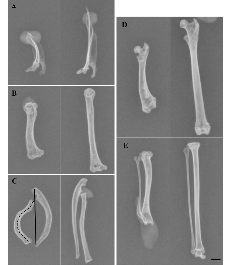
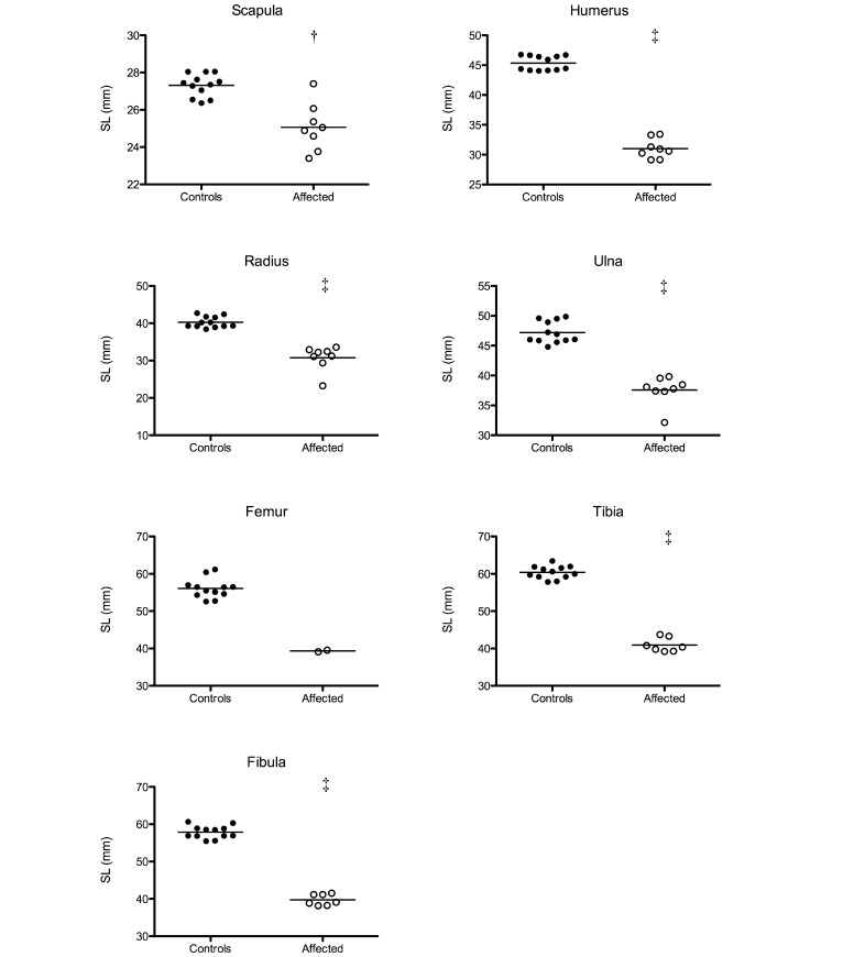
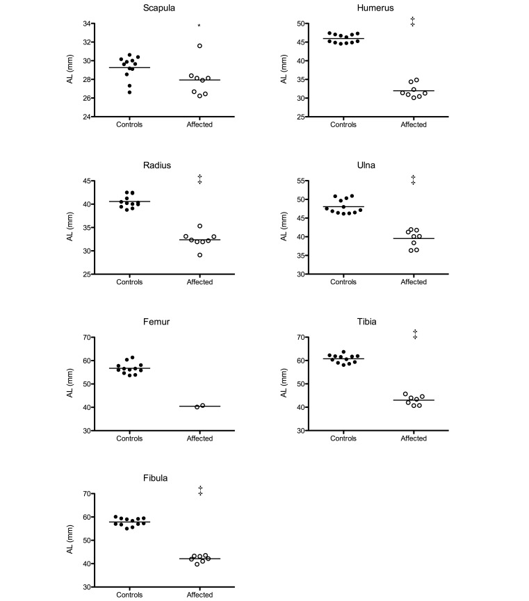
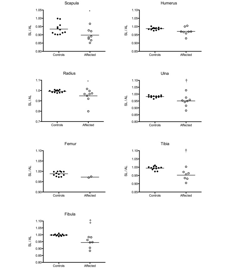
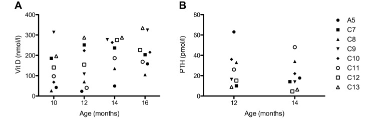
Over several years, 0% to 5% of adolescent animals in a captive colony of common marmosets (Callithrix jacchus) showed severely bended arms and legs over several years. The animals showed no pain, discomfort, or altered behavior but were unable to stretch their distal limbs to their full extent. To characterize the lesion morphologically, the bones of 4 affected marmosets were compared macroscopically and radiographically with those of 6 unaffected animals. The deformities were characterized by mid- to distal diaphyseal bending and pronounced shortening of long bones. The morphology and density of other bones including the skull and vertebrae were unaffected. Although vitamin D values were low in a fifth affected marmoset during 10 to 16 mo of age, lesions associated with rickets were not observed. To our knowledge, this report is the first to describe a micromelic dysplasia-like syndrome comprising severe, idiopathic bending and shortening of long bones in a colony of marmosets.
Figures
References
-
- Al-Gazali LI, Bakir M, Hamid Z, Nath D, Haas D. 2001. Micromelic dwarfism—humerus, femur and tibia type. Clin Dysmorphol 10:24–28. - PubMed
-
- Allen JM, Cross AJ, Yeats JC, Ghatei MA, McGregor GP, Close SP, Pay S, Marriott AS, Tyers MB, Crow TJ, Bloom SR. 1986. Neuropeptides and dopamine in the marmoset. Effect of treatment with 1-methyl-4-phenyl-1, 2, 3, 6 tetrahydropyridine (MPTP): an animal model for Parkinson's disease? Brain 109:143–157. - PubMed
-
- Casteleyn C, Bakker J, Breugelmans S, Kondova I, Saunders J, Langermans JA, Cornillie P, Van den Broeck W, Van Loo D, Van Hoorebeke L, Bosseler L, Chiers K, Decostere A. 2012. Anatomical description and morphometry of the skeleton of the common marmoset (Callithrix jacchus). Lab Anim 46:152–163. - PubMed
-
- Chun RF, Chen H, Boldrick L, Sweet C, Adams JS. 2001. Cloning, sequencing, and functional characterization of the vitamin D receptor in vitamin D-resistant New World primates. Am J Primatol 54:107–118. - PubMed
-
- Clark L. 1970. The effect of excess vitamin A on longbone growth in kittens. J Comp Pathol 80:625–634. - PubMed
MeSH terms
Substances
LinkOut - more resources
Full Text Sources