. 2014 Nov 13;15(11):20913-26.
doi: 10.3390/ijms151120913.

Chrysin protects against focal cerebral ischemia/reperfusion injury in mice through attenuation of oxidative stress and inflammation

Affiliations

Chrysin protects against focal cerebral ischemia/reperfusion injury in mice through attenuation of oxidative stress and inflammation

Yang Yao et al. Int J Mol Sci. .

Abstract

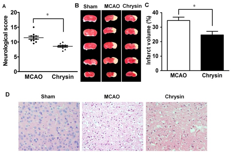
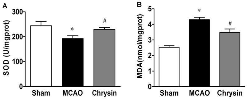
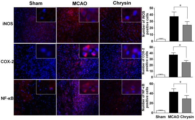
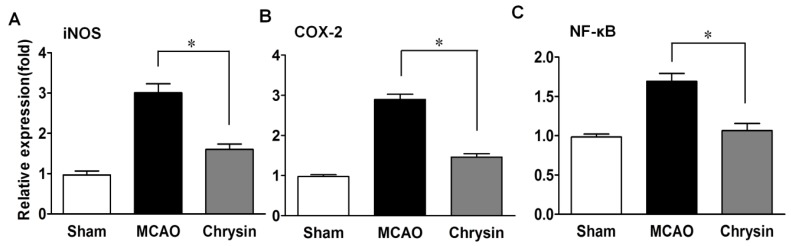
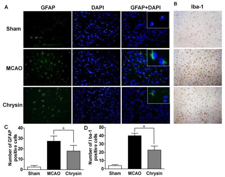
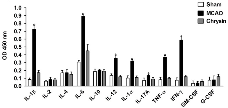
Inflammation and oxidative stress play an important part in the pathogenesis of focal cerebral ischemia/reperfusion (I/R) injury, resulting in neuronal death. The signaling pathways involved and the underlying mechanisms of these events are not fully understood. Chrysin, which is a naturally occurring flavonoid, exhibits various biological activities. In this study, we investigated the neuroprotective properties of chrysin in a mouse model of middle cerebral artery occlusion (MCAO). To this end, male C57/BL6 mice were pretreated with chrysin once a day for seven days and were then subjected to 1 h of middle cerebral artery occlusion followed by reperfusion for 24 h. Our data show that chrysin successfully decreased neurological deficit scores and infarct volumes, compared with the vehicle group. The increases in glial cell numbers and proinflammatory cytokine secretion usually caused by ischemia/reperfusion were significantly ameliorated by chrysin pretreatment. Moreover, chrysin also inhibited the MCAO-induced up-regulation of nuclear factor-kappa B (NF-κB), cyclooxygenase-2 (COX-2), and inducible nitric oxide synthase (iNOS), compared with the vehicle. These results suggest that chrysin could be a potential prophylactic agent for cerebral ischemia/reperfusion (I/R) injury mediated by its anti-inflammatory and anti-oxidative effects.


Figures

Figure 1
Figure 1
Effect of chrysin on neurological deficits, brain infarct volume and pathomorphological changes in mice with cerebral I/R injury. (A) Neurological score was measured in mice that underwent 1 h of ischemia followed by 24 h of reperfusion. Chrysin decreased the neurological score compared to the middle cerebral artery occlusion (MCAO) group; (B) Representative coronal brain sections stained with 1% TTC. Mice were subjected to 1 h of ischemia followed by 24 h of reperfusion; the infarct area is white; (C) Quantitative analysis of the percentage of brain infarct volume. Chrysin pretreatment diminished the percentage of brain infarct volume and there is a statistical difference compared with the MCAO group; (D) H&E staining showed that the normal morphologic features of neurons were present in the sham group. The MCAO group showed the loss of neurons and the presence of multiple vacuolated interspaces. Chrysin significantly ameliorated the damage of neurons that is associated with ischemia in the MCAO group (Magnified 20×). Data are expressed as means ± SEM, * p < 0.05, vs. MCAO group, n = 6.
Figure 2
Figure 2
Effect of chrysin on the activity of SOD and the contents of MDA. (A) SOD activity were significantly increased in the MCAO group as compared to the sham group, Compared with the MCAO group, the activities of SOD were significantly increased in the chrysin pretreatment group; (B) The contents of MDA were significantly increased in the MCAO group as compared to the sham group, while it was significantly decreased in the chrysin pretreatment group. Data are expressed as means ± SEM, * p < 0.05, MCAO vs. sham group, # p < 0.05, chrysin pretreatment group vs. MCAO group, n = 6.
Figure 3
Figure 3
Chrysin pretreatment suppresses the production of iNOS, COX-2 and NF-κB in the I/R injured mouse brain. Representative coronal brain sections of the sham group, the MCAO group and the chrysin pretreatment group were stained using immunofluorescence for iNOS, COX-2 and NF-κB. The sham group showed negligible staining. However, the expression of iNOS, COX-2 and NF-κB was prominent in the MCAO group compared with the sham group, and the chrysin pretreatment group showed moderate staining for iNOS, COX-2 and NF-κB (Magnified 20×). Data are expressed as means ± SEM, * p < 0.05, vs. MCAO group, n = 6.
Figure 4
Figure 4
Chrysin pretreatment suppresses the mRNA expression of iNOS (A), COX-2 (B) and NF-κB (C) in the MCAO model. Representative mRNA expression of iNOS, COX-2 and NF-κB detected by RT-PCR. Compared to the MCAO group, chrysin pretreatment also inhibited the mRNA expression of iNOS, COX-2 and NF-κB. Data are expressed as means ± SEM, * p < 0.05, vs. MCAO group, n = 6.
Figure 5
Figure 5
Chrysin suppressed the expression of GFAP and Iba-1. (A) Immunofluorescence staining in cortical brain sections for the sham group, the MCAO group and the chrysin pretreated group show the expression of GFAP; (A,C) The sham group showed negligible GFAP-positive cells. However, the expression level of GFAP was prominent for the MCAO group compared with the sham group, while the chrysin pretreatment group showed moderate expression of GFAP levels. GFAP-positive cells and DAPI-positive nuclei were co-localized (Magnification 40×); (B,D) The expression of Iba-1 was observed in the all three treatment groups. The MCAO group showed increased gliosis compared to the sham group, while the chrysin pretreatment group showed a moderate expression of Iba-1 (Magnification 20×). Data are expressed as means ± SEM, * p < 0.05, vs. MCAO group, n = 6.
Figure 6
Figure 6
Chrysin reduced the secretion of proinflammatory cytokines. The brain tissue supernatants in mice that underwent 1 h of ischemia followed by 24 h of reperfusion were measured by ELISA. IL-1β, IL-6, IL-12, IL-1α, IL-17A, IFN-γ and TNF-α were inhibited, however, IL-2, IL-4, IL-10, G-CSF and GM-CSF showed no significant changes. The results are presented as mean OD ± SEM. * p < 0.05, vs. MCAO group, n = 6.

References

    1. Nagakannan P., Shivasharan B.D., Thippeswamy B.S., Veerapur V.P., Bansal P. Protective effect of hydroalcoholic extract of Mimusops elengi Linn. flowers against middle cerebral artery occlusion induced brain injury in rats. J. Ethnopharmacol. 2012;140:247–254. - PubMed
    1. Yen T.L., Hsu C.K., Lu W.J., Hsieh C.Y., Hsiao G., Chou D.S., Wu G.J., Sheu J.R. Neuroprotective effects of xanthohumol, a prenylated flavonoid from hops (Humulus lupulus), in ischemic stroke of rats. J. Agric. Food Chem. 2012;60:1937–1944. doi: 10.1021/jf204909p. - DOI - PubMed
    1. Ning N., Dang X., Bai C., Zhang C., Wang K. Panax notoginsenoside produces neuroprotective effects in rat model of acute spinal cord ischemia-reperfusion injury. J. Ethnopharmacol. 2012;139:504–512. doi: 10.1016/j.jep.2011.11.040. - DOI - PubMed
    1. Pichichero E., Cicconi R., Mattei M., Muzi M.G., Canini A. Acacia honey and chrysin reduce proliferation of melanoma cells through alterations in cell cycle progression. Int. J. Oncol. 2010;37:973–981. - PubMed
    1. Barbaric M., Miskovic K., Bojic M., Loncar M.B., Smolcic-Bubalo A., Debeljak Z., Medic-Saric M. Chemical composition of the ethanolic propolis extracts and its effect on HeLa cells. J. Ethnopharmacol. 2011;135:772–778. doi: 10.1016/j.jep.2011.04.015. - DOI - PubMed

Publication types

MeSH terms

LinkOut - more resources