. 2014 Nov 15;28(22):2464-76.
doi: 10.1101/gad.251041.114.

Telomere position effect: regulation of gene expression with progressive telomere shortening over long distances

Affiliations

Telomere position effect: regulation of gene expression with progressive telomere shortening over long distances

Jérôme D Robin et al. Genes Dev. .

Abstract

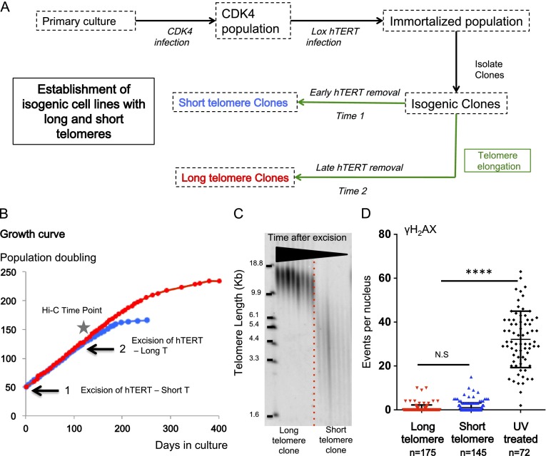
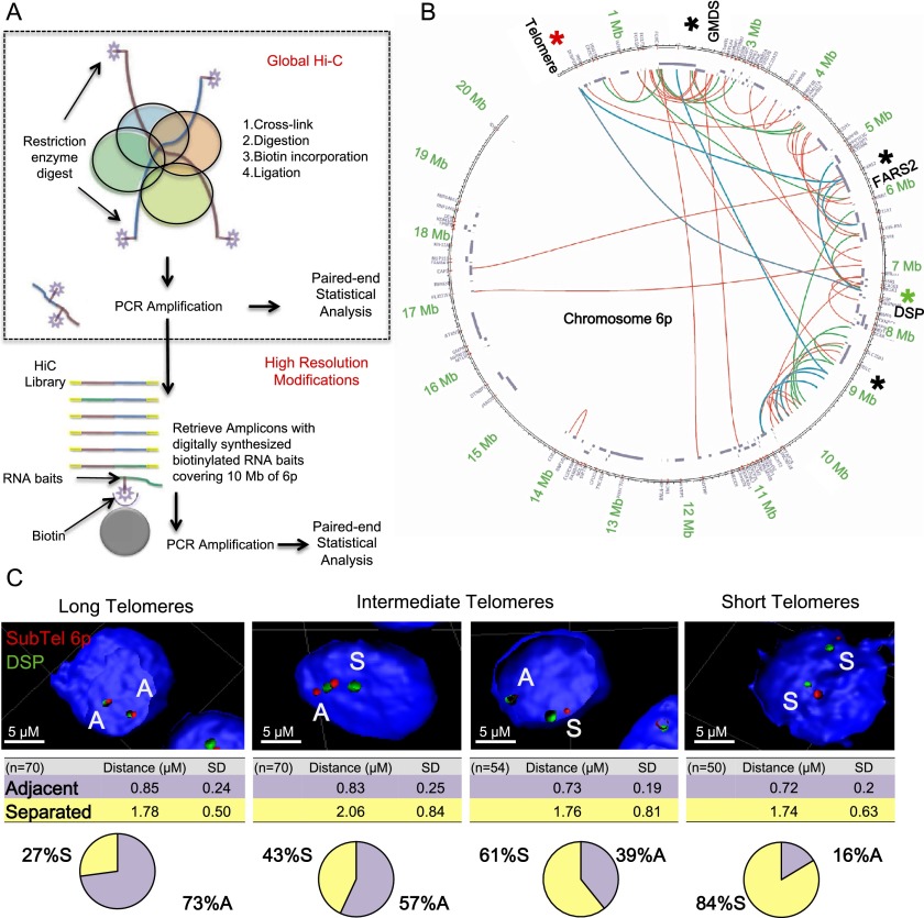
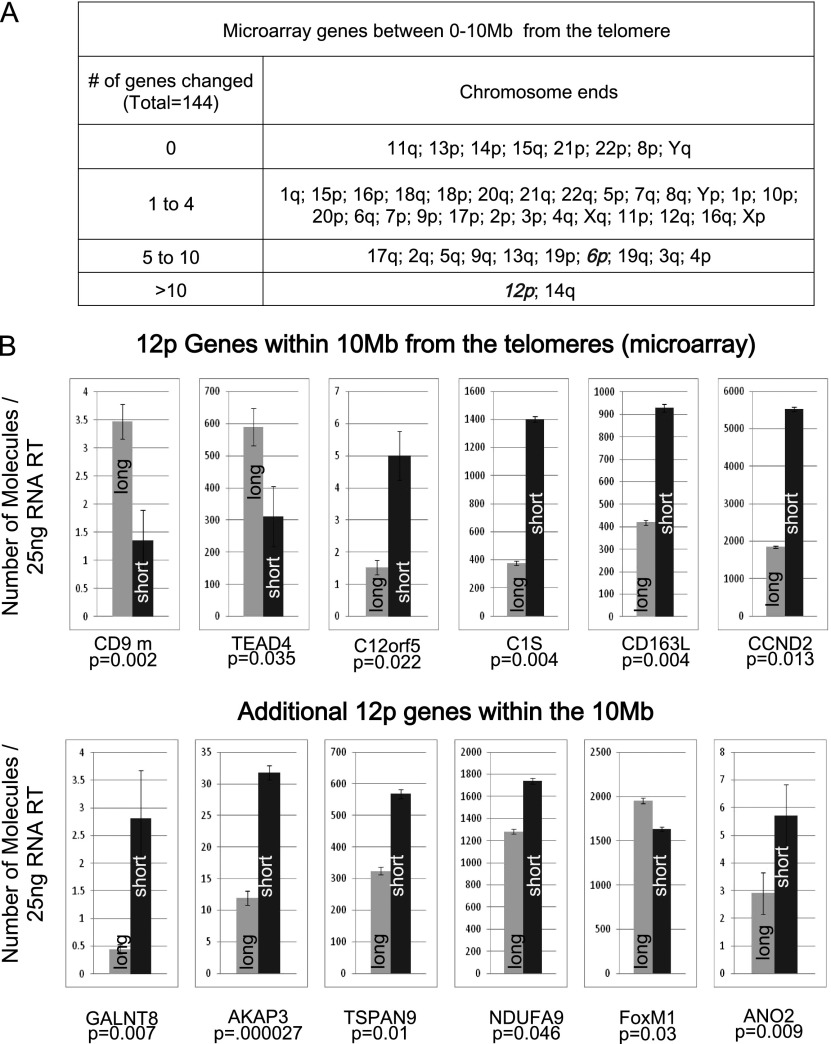
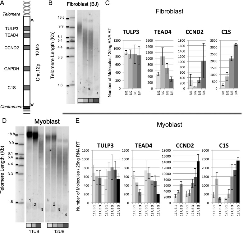
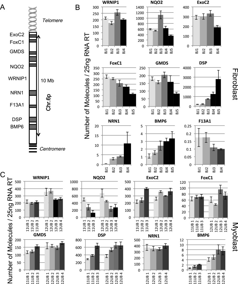
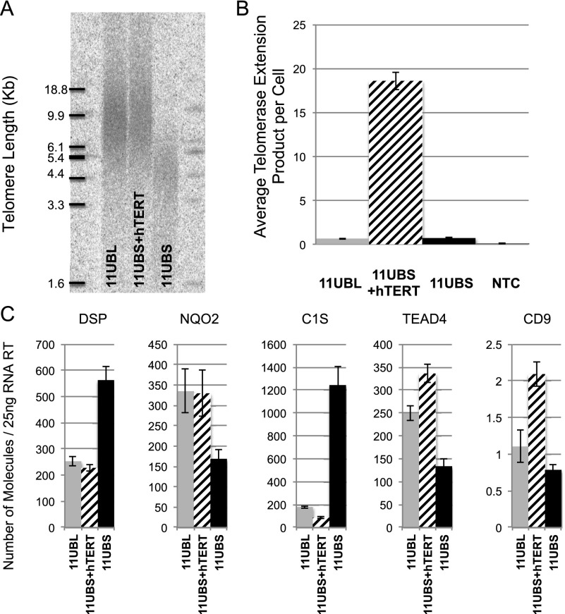
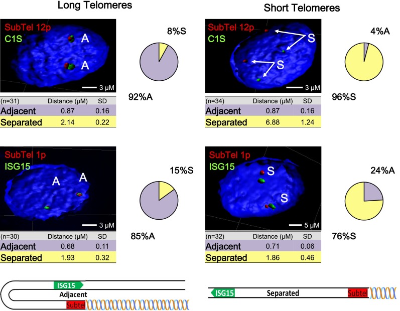
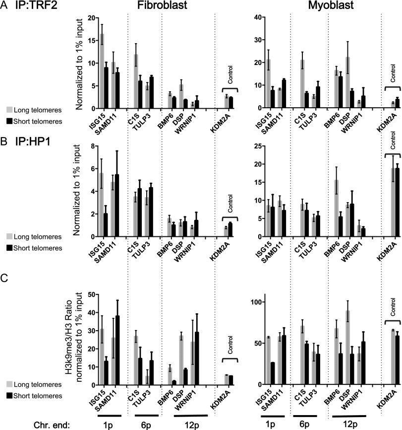
While global chromatin conformation studies are emerging, very little is known about the chromatin conformation of human telomeres. Most studies have focused on the role of telomeres as a tumor suppressor mechanism. Here we describe how telomere length regulates gene expression long before telomeres become short enough to produce a DNA damage response (senescence). We directly mapped the interactions adjacent to specific telomere ends using a Hi-C (chromosome capture followed by high-throughput sequencing) technique modified to enrich for specific genomic regions. We demonstrate that chromosome looping brings the telomere close to genes up to 10 Mb away from the telomere when telomeres are long and that the same loci become separated when telomeres are short. Furthermore, expression array analysis reveals that many loci, including noncoding RNAs, may be regulated by telomere length. We report three genes (ISG15 [interferon-stimulated gene 15 kd], DSP [Desmoplakin], and C1S [complement component 1s subcomplement]) located at three different subtelomeric ends (1p, 6p, and 12p) whose expressions are altered with telomere length. Additionally, we confirmed by in situ analysis (3D-FISH [three-dimensional fluorescence in situ hybridization]) that chromosomal looping occurs between the loci of those genes and their respective telomere ends. We term this process TPE-OLD for "telomere position effect over long distances." Our results suggest a potential novel mechanism for how telomere shortening could contribute to aging and disease initiation/progression in human cells long before the induction of a critical DNA damage response.

Keywords: age-dependent gene expression; cancer; chromatin; chromosome looping; replicative aging; senescence; telomerase.


Figures

Figure 1.
Figure 1.
Establishment of isogenic clones with long and short telomeres. Isogenic clones with long and short telomeres were produced as described (Stadler et al. 2013). (A) CDK4 is required for myoblasts to prevent growth arrest due to inadequate culture conditions. Telomerase expression preferentially elongates the shortest telomeres so that all telomeres within one cell become normalized to have approximately the same length. Thus, the confounding effects of inheritance (whether a long or short telomere was inherited from one parent) are eliminated, and the specific effects of telomere length can be directly analyzed. An early excision of hTERT produced the “short” telomere subclones, and later excisions yielded subclones with long telomeres. The population doublings for both were similar at the time of experiments. (B) One example of growth curves of isogenic clones. No differences in growth rates were detectable at the time points chosen for the analysis. (C) Telomere length was measured by TRF. The TRF shown has a series of relatively early time points for the long telomere clone and includes several samples from a short telomere clone when cells are entering senescence (cells were used for in situ or expression analysis at least 30 population doublings prior to senescence, when the growth rate had not begun to slow). (D) γH2AX staining. We report the number of events (DNA damage) per nucleus in cells with long and short telomeres along with cells subjected to UV radiation (positive control). No increase in DNA damage was seen in the short telomere cells.
Figure 2.
Figure 2.
Telomere shortening causes a change of chromatin organization involving a 7-Mb loop in myoblasts. (A) Schematic of high-resolution Hi-C: A chromatin looping interaction brings two distant parts of a chromosome (brown and blue lines, respectively) close together (colored circles represent proteins). After cross-linking, restriction digest, and fill-in incorporation of biotin, ligation under dilute conditions will connect the two distant DNA fragments (brown and blue lines). The incorporation of biotin during the ligation step allows these interactions to be purified. Because short-range (enhancer/repressor) interactions are very abundant, in global Hi-C, they dominate the paired-end sequencing results, and long-range interactions cannot be identified. In the modified approach, PCR amplification removes the biotin used in global Hi-C. This then allows biotinylated probes specific to the 6p terminus to enrich for interactions involving 6p. (B) Organization of the 6p locus in myoblasts with long telomeres, shown as a Circos representation of the 6p-specific Hi-C experiment. The first 20 Mb of chromosome 6p is shown as a circle, and the locations of known genes are shown as segments in a purple inner circle. Interactions are color-coded ([red] P < 0.05; [green] P < 0.01; [blue] P < 0.001); areas involved in interactions confirmed through further experiments are marked with an asterisk. (C) In situ validation. Confocal images of 3D-FISH ([green] fosmid targeting the gene DSP 7.5 Mb from the telomere of 6p; [red] BAC targeting subtelomeric 6p 200 kb from the telomere of 6p) were processed with IMARIS. n = ∼50 nuclei. Adjacent (A) and separated (S) events are labeled. Isogenic clones with progressively shorter telomeres, corresponding to the four lanes of the 12UB myoblast in Figure 4, were tested. Myoblasts with short telomeres show significant looping differences (independent test between all conditions for binary choice, P < 10 × −7; t-test of the mean distance between centers, P = 0.01).
Figure 3.
Figure 3.
Telomere shortening causes a change in transcription of genes located up to 10 Mb from a telomere in myoblasts. (A) Microarray results comparing transcription in myoblasts with long versus short telomeres. Changes in expression of genes 0–10 Mb from each telomere are grouped by chromosome ends. A microarray was performed using a biological triplicate. (B) ddPCR validation of six of the microarray hits at chromosome 12p and six genes not on the array within the same 10-Mb region. Expression in myoblasts with long telomeres (gray) is compared with an isogenic clone with short telomeres (black). Results are expressed as the number of molecules detected in a 25-ng RT input. P-values indicate significance of the difference for assays done in duplicate using biological triplicates (total of six measurements). All six array genes were validated as significant, and all additional genes tested showed significant differences (P < 0.05, and two-thirds of these showed a >50% change in expression). Many subtelomeric genes were not on the microarray; thus, the table in A provides a significant underestimate of the number of genes whose expression is influenced by telomere length.
Figure 4.
Figure 4.
TPE-OLD at chromosome 12p. The same procedure described for myoblasts in Figure 1 was used for four isogenic fibroblast clones (BJ1, BJ2, BJ3, and BJ4, each indicated by a different shade of gray) and two series of isogenic myoblast clones (11UB and 12UB). All clones were analyzed during logarithmic growth at least 30 population doublings prior to senescence. The location of each gene is shown in A in a schematic representation of the short arm (p) of chromosome 12 and different genes present in the 10-Mb subtelomeric region. (B) TRF analysis of fibroblast isogenic clones. (C) Expression in fibroblasts with long telomeres (light gray) is compared with those with shorter telomeres (grayscale). ddPCR analysis of four genes on chromosome 12p. Results are expressed as the number of molecules detected in a 25-ng RT input. (D) TRF analysis of myoblast isogenic clones. (E) The same genes in the myoblasts are changing as were seen in fibroblast (C1S and TEAD4). This region contains three genes with observed changes (TEAD4, CCND2, and C1S), a housekeeping gene (GAPDH) (Supplemental Fig. S6), and a gene with no changes (TULP3). Each assay was performed in biological triplicate and technical duplicate.
Figure 5.
Figure 5.
TPE-OLD at chromosome 6p. Gene expression of four isogenic fibroblast clones (BJ1, BJ2, BJ3, and BJ4, each indicated by a different shade of gray) and two series of isogenic myoblast clones (11UB and 12UB). All clones were analyzed during logarithmic growth at least 30 population doublings prior to senescence. The location of each gene is shown in A in a schematic representation of the short arm (p) of chromosome 6 and different genes present in the 10-Mb subtelomeric region. Nine genes located within the first 10 Mb of chromosome 6 were examined in fibroblasts (B) and myoblasts (C). Expression with long telomeres is compared with a series of isogenic clones with shorter telomeres. Results are expressed as the number of molecules detected in a 25-ng RT input. Each assay was performed in biological triplicate and technical duplicate.
Figure 6.
Figure 6.
Reverted expression of TPE-OLD genes by telomerase. (A) TRF analysis of isogenic myoblast clones with long (11UBL), re-elongated (11UBS+hTERT), and short (11UBS) telomeres. (B) hTERT overexpression was assessed by measuring telomerase activity, as reported by the ddTRAP assay. Results are expressed as average telomerase extension products per cell from biological duplicates and technical triplicates. (NTC) Nontemplate control. (C) Gene expression analysis of TPE-OLD genes in myoblasts from A. All clones were analyzed during logarithmic growth at least 30 population doublings prior to senescence. Results are expressed as the number of molecules detected in a 25-ng RT input. Each assay was performed in biological duplicate and technical duplicate.
Figure 7.
Figure 7.
Telomere shortening causes a change of chromatin organization. Confocal images of 3D-FISH after IMARIS processing. (Top) Telomere shortening causes a change in chromatin organization at chromosome 12p. A Bac containing the C1S probe (7.5 Mb from the telomere; green) and a fosmid containing the most unique subtelomeric region of 12p (∼200 kb from the telomeres; red) showed a dramatic change from >90% within 2 μm of each other to >95% separated by almost 7 μm. The loop identified by 3D-FISH is 7.5 Mb long. (Bottom) Chromatin organization at chromosome 1p using a fosmid ISG15 probe (1 Mb from the telomere; green) and another (∼20 kb from the telomere; red) containing the most unique subtelomeric region of chromosome 1p. An average of 30 nuclear z-stacks per condition were processed. Adjacent (A) and separated (S) are labeled. A test of independence confirms the binary observation based on the distance associated (P < 0.00001). No significant differences were seen in the amount of separation with telomere length, only in the fraction of adjacent or separated locations. A schematic representation of the loop is attached.
Figure 8.
Figure 8.
Effect of telomere shortening on histone modifications and TRF2. Antibodies against H3, trimethylated H3K9, HP1α, and TRF2 were used in ChIP. Immunoprecipitated DNA was analyzed by ddPCR using primers designed against the promoter region of TPE-OLD genes (ISG15, C1S, BMP6, and DSP) of three chromosome ends (1p, 6p, and 12p). For each chromosome end, a control gene (located in between the telomere and the TPE-OLD gene: SAMD11 for 1p, WRNIP1 for 6p, and TULP3 for 12p) whose expression was not affected by telomere length was assayed. An external gene located far from the telomeres was added as an additional control (KDM2A; 70 Mb away from 11q); no telomere length-dependent variation in the control genes was observed. Each assay was performed in biological duplicate and technical triplicate. (A,B) A decrease of TRF2 enrichment was seen in cells with short telomeres (loss of the loop). HP1 showed little change, suggesting that this marker of constitutive heterochromatin may not be involved in TPE-OLD. (C) Effect of telomere length on the heterochromatic signature of TPE-OLD genes. TPE-OLD genes exhibit a significant decrease of H3K9me3 in cells with short telomeres, underlying a loss of this repressive transcription marker.

Comment in

Similar articles

Cited by

References

    1. Apostolou E, Thanos D. 2008. Virus infection induces NF-κB-dependent interchromosomal associations mediating monoallelic IFN-β gene expression. Cell 134: 85–96. - PubMed
    1. Arnoult N, Schluth-Bolard C, Letessier A, Drascovic I, Bouarich-Bourimi R, Campisi J, Kim SH, Boussouar A, Ottaviani A, Magdinier F, et al. . 2010. Replication timing of human telomeres is chromosome arm-specific, influenced by subtelomeric structures and connected to nuclear localization. PLoS Genet 6: e1000920. - PMC - PubMed
    1. Azzalin CM, Reichenbach P, Khoriauli L, Giulotto E, Lingner J. 2007. Telomeric repeat containing RNA and RNA surveillance factors at mammalian chromosome ends. Science 318: 798–801. - PubMed
    1. Barbieri M, Paolisso G, Kimura M, Gardner JP, Boccardi V, Papa M, Hjelmborg JV, Christensen K, Brimacombe M, Nawrot TS, et al. . 2009. Higher circulating levels of IGF-1 are associated with longer leukocyte telomere length in healthy subjects. Mech Ageing Dev 130: 771–776. - PubMed
    1. Bau D, Sanyal A, Lajoie BR, Capriotti E, Byron M, Lawrence JB, Dekker J, Marti-Renom MA. 2011. The three-dimensional folding of the α-globin gene domain reveals formation of chromatin globules. Nat Struct Mol Biol 18: 107–114. - PMC - PubMed

Publication types