. 2015 Jan;31(1):98-106.
doi: 10.1089/AID.2014.0121.

Designed transcription activator-like effector proteins efficiently induced the expression of latent HIV-1 in latently infected cells

Affiliations

Designed transcription activator-like effector proteins efficiently induced the expression of latent HIV-1 in latently infected cells

Xiaohui Wang et al. AIDS Res Hum Retroviruses. 2015 Jan.

Abstract

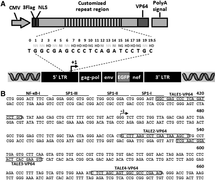
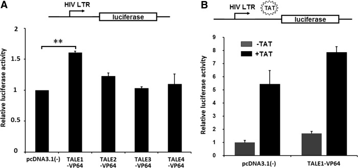
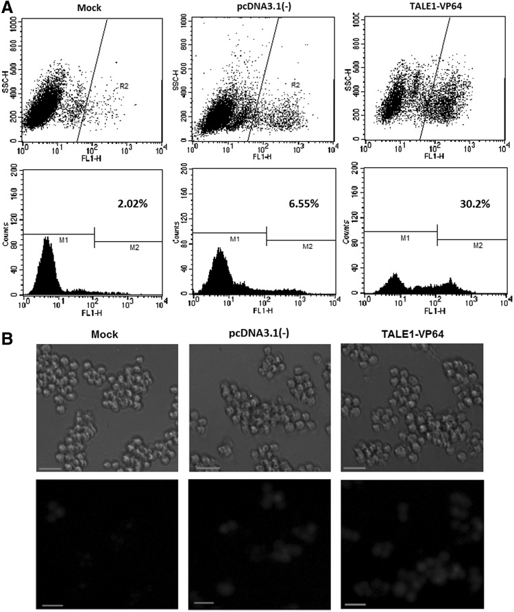
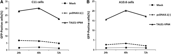
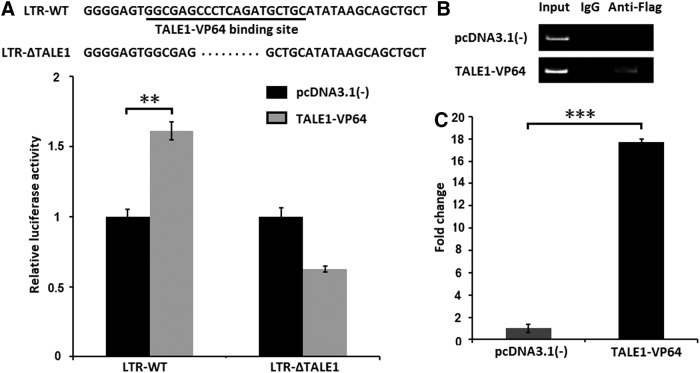
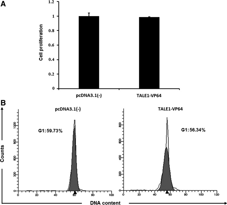
HIV latency is the foremost barrier to clearing HIV infection from patients. Reactivation of latent HIV-1 represents a promising strategy to deplete these viral reservoirs. Here, we report a novel approach to reactivate latent HIV-1 provirus using artificially designed transcription activator-like effector (TALE) fusion proteins containing a DNA-binding domain specifically targeting the HIV-1 promoter and the herpes simplex virus-based transcriptional activator VP64 domain. We engineered four TALE genes (TALE1-4) encoding TALE proteins, each specifically targeting different 20-bp DNA sequences within the HIV-1 promoter, and we constructed four TALE-VP64 expression vectors corresponding to TALE1-4. We found that TALE1-VP64 effectively reactivated HIV-1 gene expression in latently infected C11 and A10.6 cells. We further confirmed that TALE1-VP64 reactivated latent HIV-1 via specific binding to the HIV-LTR promoter. Moreover, we also found that TALE1-VP64 did not affect cell proliferation or cell cycle distribution. Taken together, our data demonstrated that TALE1-VP64 can specifically and effectively reactivate latent HIV-1 transcription, suggesting that this strategy may provide a novel approach for anti-HIV-1 latency therapy in the future.


Figures

<b>FIG. 1.</b>
FIG. 1.
Basic structure and target sites of transcription activator-like effector (TALE)-VP64. (A) Schematic representation of TALE-VP64-mediated activation of HIV-1 expression. The diagram illustrates the composition of the tandem repeat of a TALE-VP64 and its corresponding 20-bp DNA binding target site on the 5′-LTR of the HIV-1 genome. NLS, nuclear localization signal; VP64, synthetic transcription activation domain. (B) The DNA sequence of the HIV-1 (HXB2 strain) 5′-LTR. Several features of the HIV-1 promoter [binding sites for NF-κB, Sp1, and the transcription initiation site (arrow)] are indicated. The sites targeted by TALE-VP64 are overlaid on the promoter sequence.
<b>FIG. 2.</b>
FIG. 2.
Luciferase assays of TALE-VP64-mediated HIV-1 5′-LTR activity. (A) Transient reporter assays comparing the activation potential of various designer TALE-VP64 transcription factors. HEK-293 cells were transfected with an HIV LTR-luciferase reporter plasmid and a plasmid expressing the chimeric protein indicated; the empty vector pcDNA3.1(-) was used as a control. Then, 48 h posttransfection, the relative luciferase activity was measured. The data represent the mean±standard deviation of three independent experiments. *p<0.05, **p<0.01, ***p<0.001; paired t test. (B) Synergistic activation of HIV-1 by TALE1-VP64 and Tat. HEK-293 cells were transfected with the TALE1-VP64 expression plasmid in the presence or absence of Tat; the empty vector pcDNA3.1(-) was used as a control. Then, 48 h posttransfection, the relative luciferase activity was measured. The data represent the mean±standard deviation of three independent experiments.
<b>FIG. 3.</b>
FIG. 3.
Reactivation of latent HIV-1 in latently infected C11 cells by TALE1-VP64. (A) C11 cells were untransfected (mock) or transfected with pcDNA3.1(-) (2 μg) or TALE1-VP64 (2 μg). Then, 48 h posttreatment, the percentage of cells expressing green fluorescent protein (GFP) was measured via flow cytometry to determine the extent of reactivation. The results are presented as fluorescence histograms. (B) Fluorescence microscopic images of TALE1-VP64-induced GFP expression in C11 cells 48 h posttransfection. Scale bar, 100 μm.
<b>FIG. 4.</b>
FIG. 4.
Time dependence of the effect of TALE1-VP64 on C11 and A10.6 cells. The cells were untransfected (mock) or transfected with pcDNA3.1(-) (2 μg) or TALE1-VP64 (2 μg) for the indicated periods. The level of GFP expression in (A) C11 cells or (B) A10.6 cells was measured at 24, 48, and 72 h using standard flow cytometric techniques. The data are presented as the means±standard deviations of three independent experiments.
<b>FIG. 5.</b>
FIG. 5.
TALE1-VP64 activates latent HIV-1 replication by binding to the HIV-1 LTR promoter. (A) HEK-293 cells were transfected with the HIV-1 LTR-Luc or HIV-1 LTR(ΔTALE1)-Luc reporter plasmid and the TALE1-VP64 expression plasmid; the empty vector pcDNA3.1(-) was used as a control. Then, 48 h posttransfection, the relative luciferase activity was measured. The data represent the mean±standard deviation of three independent experiments. *p<0.05, **p<0.01, ***p<0.001; paired t test. (B) C11 cells were treated with pcDNA3.1(-) (2 μg) or TALE1-VP64 (2 μg). Then, 48 h posttreatment, ChIP assays were performed using an anti-FLAG antibody or normal mouse IgG. PCR primers corresponding to the LTR promoter were used to amplify the DNA isolated from the immunoprecipitated chromatin as described in the Materials and Methods section. (C) ChIP analysis data are presented as fold change (mean value±standard error). Each ChIP experiment was repeated three times to confirm the reproducibility of the results. *p<0.05, **p<0.01, ***p<0.001; paired t test.
<b>FIG. 6.</b>
FIG. 6.
Effects of TALE1-VP64 on cell proliferation and cell cycle distribution in human peripheral blood mononuclear cells (PBMCs). (A) PBMCs were transfected with pcDNA3.1(-) or TALE1-VP64. Then, 48 h posttransfection, the cell viability was measured using a CCK-8 kit (Dojindo). The cell viability of the transfected groups was normalized to that of the untransfected group. The data are presented as the means±standard deviations of three independent experiments. (B) PBMCs were transfected with pcDNA3.1(-) or TALE1-VP64. Then, 48 h posttransfection, the distribution of the cells in each phase of the cell cycle was determined based on their DNA content via propidium iodide staining and FACS analysis.

References

    1. Katlama C, Deeks SG, Autran B, et al. : Barriers to a cure for HIV: New ways to target and eradicate HIV-1 reservoirs. Lancet 2013;381(9883):2109–2117 - PMC - PubMed
    1. Tyagi M. and Bukrinsky M: Human immunodeficiency virus (HIV) latency: The major hurdle in HIV eradication. Mol Med 2012;18(1):1096. - PMC - PubMed
    1. Iglesias-Ussel MD. and Romerio F: HIV reservoirs: The new frontier. AIDS Rev 2011;13(1):13–29 - PubMed
    1. Finzi D, Hermankova M, Pierson T, et al. : Identification of a reservoir for HIV-1 in patients on highly active antiretroviral therapy. Science 1997;278(5341):1295–1300 - PubMed
    1. Siliciano JD, Kajdas J, Finzi D, et al. : Long-term follow-up studies confirm the stability of the latent reservoir for HIV-1 in resting CD4+ T cells. Nat Med 2003;9(6):727–728 - PubMed

Publication types

MeSH terms

Substances

LinkOut - more resources