. 2014 Nov 18:14:281.
doi: 10.1186/s12866-014-0281-8.

Extracellular excystation and development of Cryptosporidium: tracing the fate of oocysts within Pseudomonas aquatic biofilm systems

Affiliations

Extracellular excystation and development of Cryptosporidium: tracing the fate of oocysts within Pseudomonas aquatic biofilm systems

Wan Koh et al. BMC Microbiol. .

Abstract

Background: Aquatic biofilms often serve as environmental reservoirs for microorganisms and provide them with a nutrient-rich growth environment under harsh conditions. With regard to Cryptosporidium, biofilms can serve as environmental reservoirs for oocysts, but may also support the growth of additional Cryptosporidium stages.

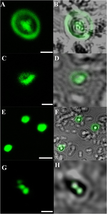
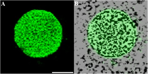
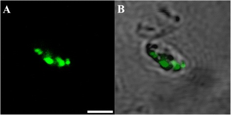
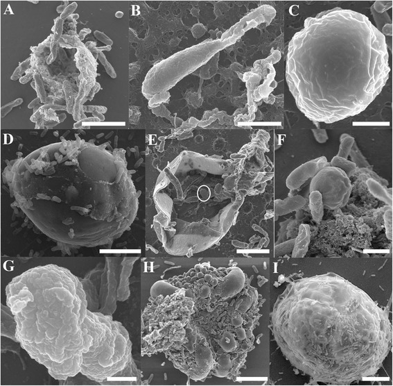
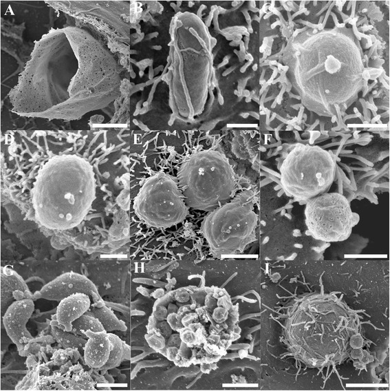
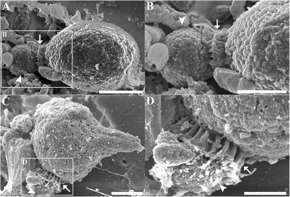
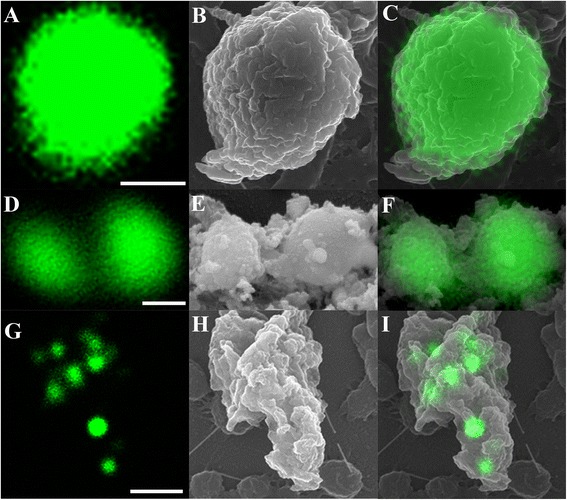
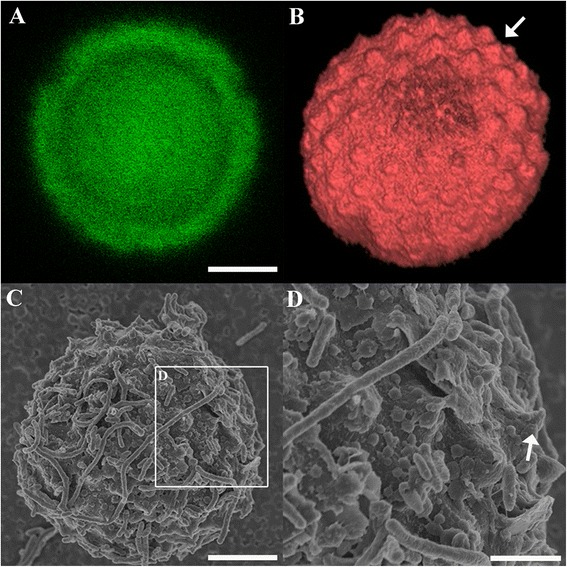
Results: Here we used confocal laser scanning microscopy, scanning electron microscopy (SEM), and flow cytometry to identify and describe various Cryptosporidium developmental stages present within aquatic biofilm systems, and to directly compare these to stages produced in cell culture. We also show that Cryptosporidium has the ability to form a parasitophorous vacuole independently, in a host-free biofilm environment, potentially allowing them to complete an extracellular life cycle. Correlative data from confocal and SEM imaging of the same cells confirmed that the observed developmental stages (including trophozoites, meronts, and merozoites) were Cryptosporidium. These microscopy observations were further supported by flow cytometric analyses, where excysted oocyst populations were detected in 1, 3 and 6 day-old Cryptosporidium-exposed biofilms, but not in biofilm-free controls.

Conclusions: These observations not only highlight the risk that aquatic biofilms pose in regards to Cryptosporidium outbreaks from water distribution systems, but further indicate that even simple biofilms are able to stimulate oocyst excystation and support the extracellular multiplication and development of Cryptosporidium within aquatic environments.


Figures

Figure 1
Figure 1
Micrographs of Cryptosporidium within Cryptosporidium- exposed biofilms. (A & B) Oocyst containing sporozoites; (C & D) Free sporozoites; (E & F) Individual trophozoites; (G & H) Aggregated trophozoites. (A, C, E, G) Confocal images; (B, D, F, H) Superimposed confocal and brightfield images. Scale bars = A & G: 3 μm; C: 1 μm; E: 4 μm.
Figure 2
Figure 2
Micrographs of a Cryptosporidium microgamont. (A) Confocal image; (B) Superimposed confocal and brightfield images. Scale bar = 2 μm.
Figure 3
Figure 3
Cryptosporidium gamont-like cell identified within 6 day-old Cryptosporidium-exposed biofilms. (A) Confocal image; (B) Superimposed confocal and brightfield images. Scale bars = 2.5 μm.
Figure 4
Figure 4
Scanning electron micrographs of Cryptosporidium within Cryptosporidium-exposed biofilms. (A) Empty oocysts with a rough membrane appearance; (B) Free sporozoite; (C) Trophozoite; (D) Large gamont cells (meronts) identified within 6 day-old biofilms; (E) type II meront containing type II merozoites within (circled); (F) Free type I merozoites; (G) Free type II merozoites; (H) Microgamont; (I) Extra-large gamont. Scale bars = A & B: 2.5 μm; C & F: 1 μm; D & E: 3 μm; G: 500 nm; H: 5 μm; I: 8 μm.
Figure 5
Figure 5
Scanning electron micrographs of various Cryptosporidium stages identified from Cryptosporidium infected in-vitro cell culture showing complementary stages to that seen in biofilms. (A) Empty oocyst; (B-D) Various shaped individual trophozoites; (E) Aggregated trophozoites; (F) Type I merozoites; (G) Type II merozoites; (H) Microgamonts; (I) Large gamont-like cell. Scale bars = A: 2 μm; B: 0.5 μm; C-D & F-G: 1 μm; E: 2 μm; H: 1.5 μm; I: 4 μm.
Figure 6
Figure 6
Parasitophorous vacuole formation. (A) Scanning electron micrograph showing evidence of parasitophorous vacuole formation by Cryptosporidium in a biofilm environment. (B) Magnified region depicted in A. (C) Parasitophorous vacuole formation by Cryptosporidium in HCT8 cell culture, included for comparison. (D) Magnified area depicted in C. Arrows indicate radial fold and arrowheads indicate dense band area. Scale bars = A: 1.5 μm; B & D: 1 μm; C: 2μm.
Figure 7
Figure 7
Direct correlation of Cryptosporidium stages using confocal microscopy (A, D, G) and SEM (B, E, H). (C, F, I) Superimposed confocal and SEM images; (A-C) Single trophozoite; (D-F) Free type II merozoites; (G-I) Type I meront containing type I merozoites. Scale bars = A: 1 μm; D: 0.5 μm; G: 2 μm.
Figure 8
Figure 8
Correlation of a meront completely enveloped by a PV (arrowhead) under confocal microscopy (A, B) and SEM (C-D). B) The three dimensional Z-stack images of image A. D) Magnified area depicted in image C showing undulating membrane peaks (arrow). Scale bars = A & C: 5 μm; D: 2 μm.

References

    1. Thompson RCA, Olson ME, Zhu G, Enomoto S, Abrahamsen MS, Hijjawi NS. Cryptosporidium and cryptosporidiosis. Adv Parasitol. 2005;59:77–158. doi: 10.1016/S0065-308X(05)59002-X. - DOI - PubMed
    1. Howe AD, Forster S, Morton S, Marshall R, Osborn KS, Wright P, Hunter PR. Cryptosporidium oocysts in a water supply associated with a cryptosporidiosis outbreak. Emerg Infect Dis. 2002;8(6):619–624. doi: 10.3201/eid0806.010271. - DOI - PMC - PubMed
    1. Wingender J, Flemming HC. Biofilms in drinking water and their role as reservoir for pathogens. Int J Hyg Environ Health. 2011;214(6):417–423. doi: 10.1016/j.ijheh.2011.05.009. - DOI - PubMed
    1. Rogers J, Keevil C. Survival of Cryptosporidium parvum oocysts in biofilm and planktonic samples in a model system. In: Betts W, Casemore D, Fricker C, Smith H, Watkins J, editors. Protozoan parasites and water. Cambridge: The Royal Society of Chemistry; 1995.
    1. Wolyniak EA, Hargreaves BR, Jellison KL. Retention and release of Cryptosporidium parvum oocysts by experimental biofilms composed of a natural stream microbial community. Appl Environ Microbiol. 2009;75(13):4624–4626. doi: 10.1128/AEM.02916-08. - DOI - PMC - PubMed

Publication types

MeSH terms

LinkOut - more resources