. 2015 Jan;15(2-3):340-55.
doi: 10.1002/pmic.201400315.

Identifying novel targets of oncogenic EGF receptor signaling in lung cancer through global phosphoproteomics

Affiliations

Identifying novel targets of oncogenic EGF receptor signaling in lung cancer through global phosphoproteomics

Xu Zhang et al. Proteomics. 2015 Jan.

Abstract

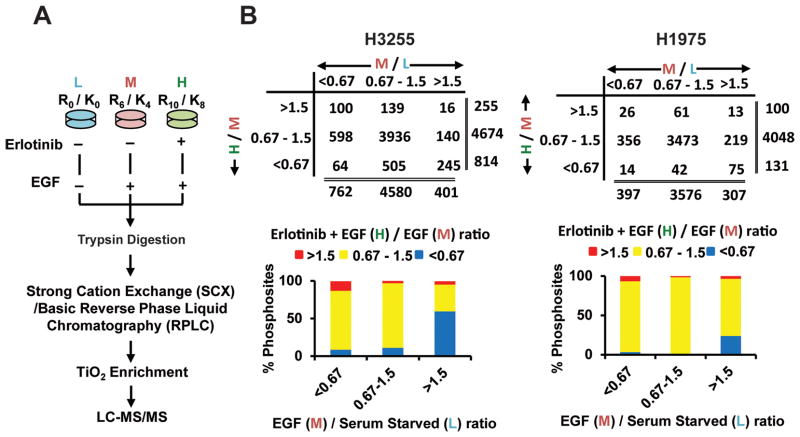
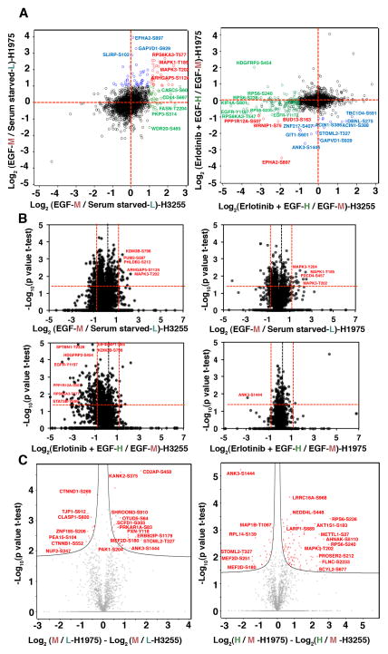
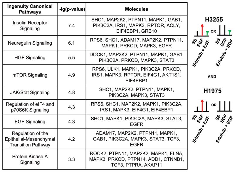
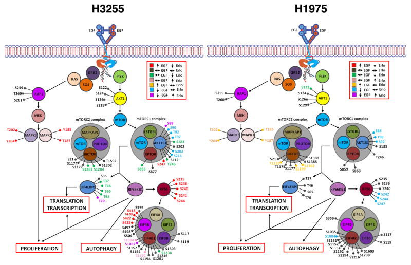
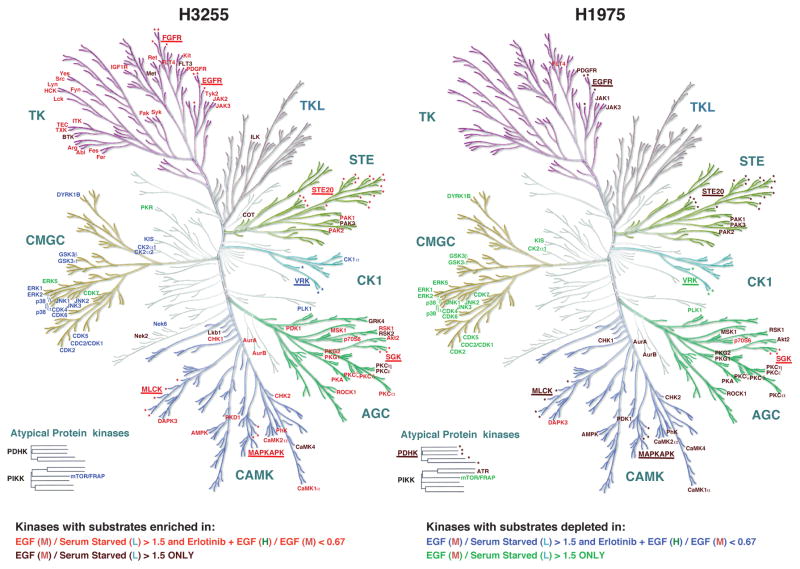
Mutations in the epidermal growth factor receptor (EGFR) kinase domain occur in 10-30% of lung adenocarcinoma and are associated with tyrosine kinase inhibitor (TKI) sensitivity. We sought to identify the immediate direct and indirect phosphorylation targets of mutant EGFRs in lung adenocarcinoma. We undertook SILAC strategy, phosphopeptide enrichment, and quantitative MS to identify dynamic changes of phosphorylation downstream of mutant EGFRs in lung adenocarcinoma cells harboring EGFR(L858R) and EGFR(L858R/T790M) , the TKI-sensitive, and TKI-resistant mutations, respectively. Top canonical pathways that were inhibited upon erlotinib treatment in sensitive cells, but not in the resistant cells include EGFR, insulin receptor, hepatocyte growth factor, mitogen-activated protein kinase, mechanistic target of rapamycin, ribosomal protein S6 kinase beta 1, and Janus kinase/signal transducer and activator of transcription signaling. We identified phosphosites in proteins of the autophagy network, such as ULK1 (S623) that is constitutively phosphorylated in these lung adenocarcinoma cells; phosphorylation is inhibited upon erlotinib treatment in sensitive cells, but not in resistant cells. Finally, kinase-substrate prediction analysis from our data indicated that substrates of basophilic kinases from, AGC and Calcium and calmodulin-dependent kinase groups, as well as STE group kinases were significantly enriched and those of proline-directed kinases from, CMGC and Casein kinase groups were significantly depleted among substrates that exhibited increased phosphorylation upon EGF stimulation and reduced phosphorylation upon TKI inhibition. This is the first study to date to examine global phosphorylation changes upon erlotinib treatment of lung adenocarcinoma cells and results from this study provide new insights into signaling downstream of mutant EGFRs in lung adenocarcinoma. All MS data have been deposited in the ProteomeXchange with identifier PXD001101 (http://proteomecentral.proteomexchange.org/dataset/PXD001101).

Keywords: Autophagy; EGFR; Erlotinib; Mass spectrometry; NSCLC; Phosphoproteomics; SILAC.


Conflict of interest statement

Conflict of interest: The authors declare no financial or commercial conflict of interest.

Figures

Figure 1
Figure 1
SILAC-based quantitative mass spectrometry and phosphopeptides identified in groups of specific SILAC ratios. A. Flowchart showing biological treatment of SILAC-labelled cells, enrichment of phosphopeptides, and detection by tandem mass spectrometry. B. Number of class I phosphosites (localization probability >0.75) identified with SILAC ratios >1.5 (increased), 0.67–1.5 (unchanged) and <0.67 (decreased) (upper panel) and percentage of phosphosites in each of those groups (lower panel) in H3255 (left panel) and H1975 (right panel) cells. Ratio M/L is EGF/Serum starved and ratio H/M is erlotinib + EGF / EGF states.
Figure 2
Figure 2
Distribution of quantified class I phosphosites and volcano plots showing significant SILAC ratios of phosphopeptides in H3255 and H1975. (A) Scatter plot showing distribution of SILAC ratios of EGF stimulation or erlotinib inhibition. Left panel is a plot against the Log 2 base ratio of EGF stimulated and serum starved cells (M/L). Right panel is a plot against the Log 2 base ratio of erlotinib inhibited before EGF stimulation and EGF stimulated cells (H/M). Each circle is a phosphorylation site. Annotated circles represent specific phosphosites with 2 fold-changes. The phosphosites that change in both H3255 and H1975, H3255 only, H1975 only are depicted in red, green, and blue circles, respectively; unaltered phosphosites between H3255 and H1975 are depicted in black. (B) Volcano plots showing significant SILAC ratios (M/L-upper panel; H/M-lower panel) of phosphosites that show hyperphosphorylation, no change or dephosphorylation with EGF and erlotinib treatment. Log2 ratios of the median of biological replicates were plotted versus −log10 of the p-values derived from a t-test. Proteins with a minimum 2-fold change combined with a p<0.05 are considered significant (red lines). Left panels are the changes in H3255; and right panels are the changes in H1975. Paired t-test was used for significance analysis from five biological replicates for H3255 and three biological replicates of H1975. (C) Volcano plots representing significant alterations of phosphorylation changes upon EGF stimulation or TKI inhibition between H3255 and H1975 cells. Differences in log2 ratios of M/L (left panel) and H/M (right panel) of H1975 and H3255 were plotted against −log10 of the p-values derived from a t-test. Hyperbolic curves separate the phosphosites significantly different between the two cell lines (red circles) from the ones unaltered between the two cell lines (grey circles).
Figure 3
Figure 3
Ingenuity pathway analysis (IPA) shows top canonical pathways represented by proteins with phosphosites that are hypophosphorylated upon erlotinib inhibition in the sensitive cell line H3255, but remain unchanged in the resistant cell line H1975. The p value is a measure of the likelihood that the association between the set of phosphosites with the given pathway is due to random chance.
Figure 4
Figure 4
Phosphosites identified in proteins of the RAS-RAF-MAPK and PI3K-AKT-mTOR signaling pathway in H3255 (left panel) and H1975 (right panel). Colors represent specific alterations in M/L (EGF/serum starved) and H/M (erlotinib+EGF/EGF) ratios as indicated.
Figure 5
Figure 5
Validation of phosphorylation changes of specific phosphosites in proteins using Western blotting. Phosphorylation specific and total protein antibody blots of key proteins that are regulated upon EGF stimulation or erlotinib treatment in H3255 and H1975 cell lines. pEGFR, pAKT, pMEK, pERK and pULK1 levels were increased or not changed upon EGF stimulation and decreased upon erlotinib treatment in H3255, but not upon erlotinib treatment in H1975.
Figure 6
Figure 6
Group-based Prediction System (GPS) analysis reveals predicted kinases for groups of phosphosites with specific alteration upon EGF or erlotinib treatment in H3255 (left panel) and H1975 (right panel) cells. Color-coding of kinases is based on the statistically significant enrichment or depletion of kinase prediction among phosphosites in specific groups of SILAC ratios as shown. CAMK, AGC, STE and TK protein kinase groups were statistically enriched and CMGC group of kinases were depleted among phosphosites that had enhanced phosphorylation upon EGF stimulation and also among phosphosites that had reduced phosphorylation upon erlotinib inhibition in H3255 cells. In H1975 the same groups of kinases were statistically enriched or depleted, but only among phosphosites that had increased phosphorylation upon EGF stimulation. Names of kinase families are underlined and the protein kinase members are marked by asterisks for those predictions where the enrichment of a single kinase within a family did not reach significance, but the family as a whole reached statistical significance.

References

    1. Siegel R, Naishadham D, Jemal A. Cancer statistics, 2013. CA Cancer J Clin. 2013;63:11–30. - PubMed
    1. Lynch TJ, Bell DW, Sordella R, Gurubhagavatula S, et al. Activating mutations in the epidermal growth factor receptor underlying responsiveness of non-small-cell lung cancer to gefitinib. N Engl J Med. 2004;350:2129–2139. - PubMed
    1. Paez JG, Janne PA, Lee JC, Tracy S, et al. EGFR mutations in lung cancer: correlation with clinical response to gefitinib therapy. Science. 2004;304:1497–1500. - PubMed
    1. Pao W, Miller V, Zakowski M, Doherty J, et al. EGF receptor gene mutations are common in lung cancers from “never smokers” and are associated with sensitivity of tumors to gefitinib and erlotinib. Proc Natl Acad Sci U S A. 2004;101:13306–13311. - PMC - PubMed
    1. Pao W, Miller VA, Politi KA, Riely GJ, et al. Acquired resistance of lung adenocarcinomas to gefitinib or erlotinib is associated with a second mutation in the EGFR kinase domain. PLoS Med. 2005;2:e73. - PMC - PubMed

Publication types

MeSH terms