Review
. 2015 Jan;16(1):465.
doi: 10.1007/s11934-014-0465-7.

Recent advances in ureteral tissue engineering

Affiliations
Review

Recent advances in ureteral tissue engineering

Paul K J D de Jonge et al. Curr Urol Rep. 2015 Jan.

Abstract

Reconstruction of long ureteral defects often warrants the use of graft tissue and extensive surgical procedures to maintain the safe transport of urine from the kidneys to the urinary bladder. Complication risks, graft failure-related morbidity, and the lack of suitable tissue are major concerns. Tissue engineering might offer an alternative treatment approach in these cases, but ureteral tissue engineering is still an underreported topic in current literature. In this review, the most recent published data regarding ureteral tissue engineering are presented and evaluated, with a focus on cell sources, implantation strategies, and (bio)materials.


Conflict of interest statement

Paul K.J.D. de Jonge, Vasileios Simaioforidis, Dr. Paul J. Geutjes, Dr. Egbert Oosterwijk, and Dr. Wout F.J. Feitz each declare no potential conflicts of interest.

Figures
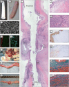
Fig. 1
Fig. 1
Implantation strategy and outcome after ureteral reconstruction using tubular collagen scaffolds. a, b Macroscopic overview of a tubular 0.5 % type-I collagen scaffold (length = 6 cm, Ø = 6 mm). c SEM overview of the scaffold surface, which was highly porous, facilitating cell penetration into the scaffold (scale bar = 500 μm) d Immunofluorescent staining for collagen (green), nuclei (blue), and RCK103 (red) of a cell-seeded scaffold. Urothelial cells (RCK103 positive) were lining the scaffold (scale bar = 400 μm). e Immunofluorescent staining for collagen (green), nuclei (blue), and α smooth muscle actin (red) of a cell-seeded scaffold. Smooth muscle cells (α smooth muscle actin positive) were found throughout the scaffold (scale bar = 400 μm). f The scaffolds were implanted by end-to-end anastomosis. g, h Ureteral regeneration was evaluated after 1 month. Four animals showed intact ureters (g), while seven animals presented with defects or dissections (h). i Histological overview of a regenerating ureter (scale bar = 5 mm). Black dotted lines indicate the anastomosis sites. Specific locations are highlighted (JO). j Hematoxylin and eosin (HE) staining of the native ureter (scale bar = 400 μm). k Inflammatory response in the regenerating tissue near scaffold remnants (red arrows). Mostly lymphocytes, a few granulocytes, and some multinucleated giant cells (black arrows) were observed (scale bar = 200 μm). l Pancytokeratin staining in the middle of the neo-ureter, indicating the presence of (multilayered) urothelium (scale bar = 400 μm). m Smoothelin staining near the anastomosis site, indicating ingrowth and maturation of smooth muscle tissue into the neo-ureter (scale bar = 400 μm). n Masson’s trichrome staining of the native ureter (scale bar = 400 μm). o Masson’s trichrome staining near the anastomosis site, indicating the ingrowth of new muscle tissue (scale bar = 400 μm)

References

    1. Vakili B, et al. The incidence of urinary tract injury during hysterectomy: a prospective analysis based on universal cystoscopy. Am J Obstet Gynecol. 2005;192(5):1599–1604. doi: 10.1016/j.ajog.2004.11.016. - DOI - PubMed
    1. Dandolu V, et al. Accuracy of cystoscopy in the diagnosis of ureteral injury in benign gynecologic surgery. Int Urogynecol J Pelvic Floor Dysfunct. 2003;14(6):427–431. doi: 10.1007/s00192-003-1095-7. - DOI - PubMed
    1. Dowling RA, Corriere JN, Jr, Sandler CM. Iatrogenic ureteral injury. J Urol. 1986;135(5):912–915. - PubMed
    1. Johnson EB, et al. Obstetric complications of ureteroscopy during pregnancy. J Urol. 2012;188(1):151–154. doi: 10.1016/j.juro.2012.02.2566. - DOI - PubMed
    1. Adamowicz J, et al. Urine is a highly cytotoxic agent: does it influence stem cell therapies in urology? Transplant Proc. 2012;44(5):1439–1441. doi: 10.1016/j.transproceed.2012.01.128. - DOI - PubMed

LinkOut - more resources