. 2015 Feb;248(1):145-55.
doi: 10.1007/s00232-014-9752-y. Epub 2014 Nov 18.

The connexin46 mutant, Cx46T19M, causes loss of gap junction function and alters hemi-channel gating

Affiliations

The connexin46 mutant, Cx46T19M, causes loss of gap junction function and alters hemi-channel gating

Jun-Jie Tong et al. J Membr Biol. 2015 Feb.

Abstract

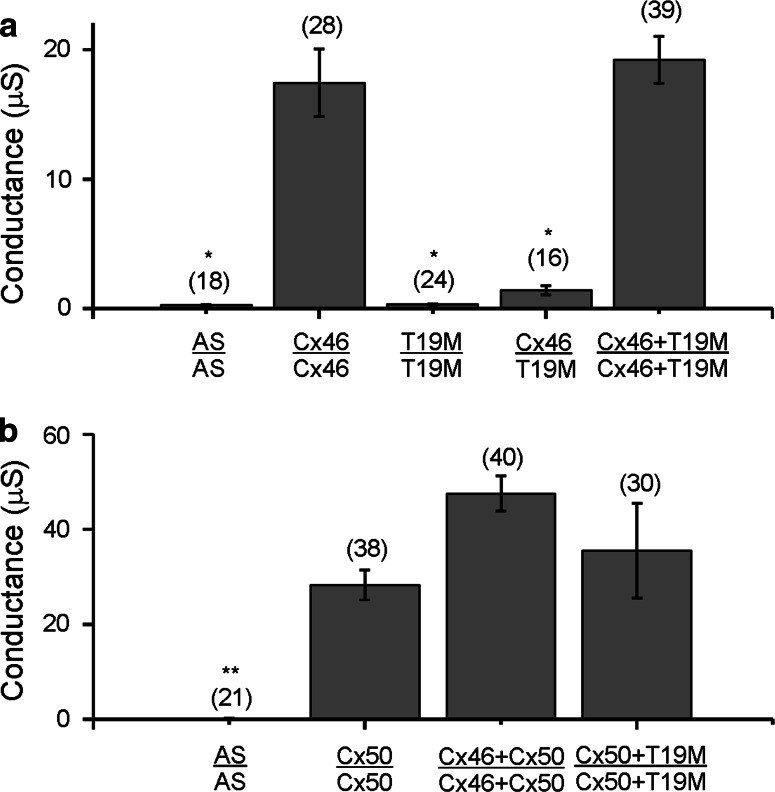
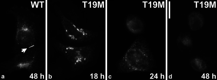
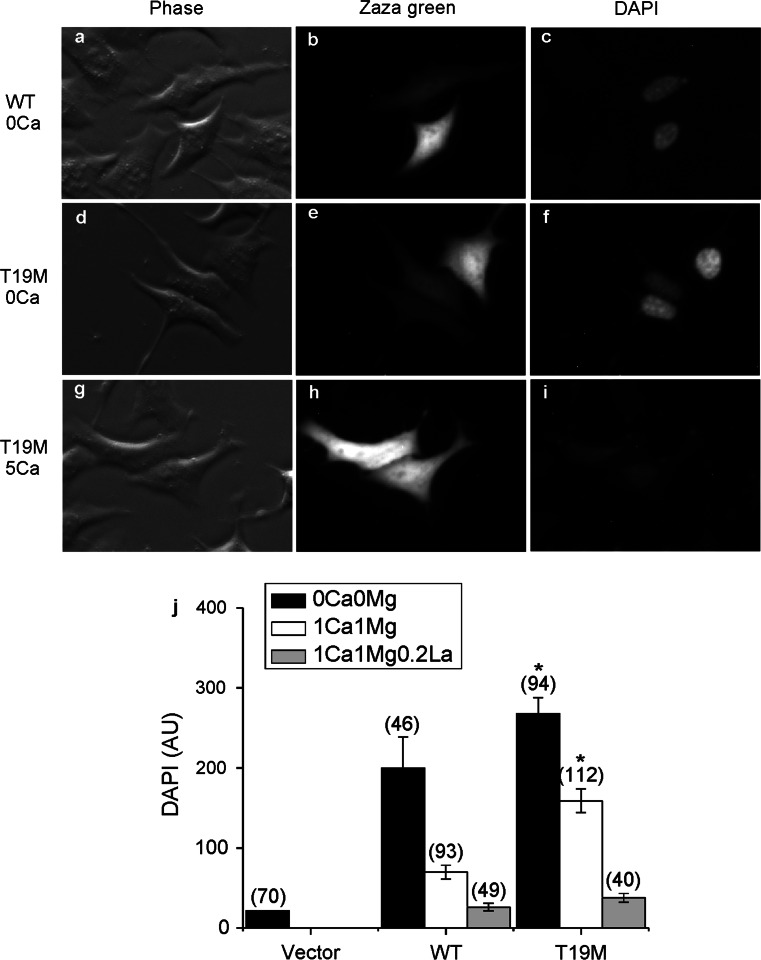
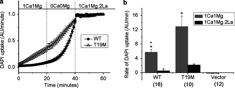
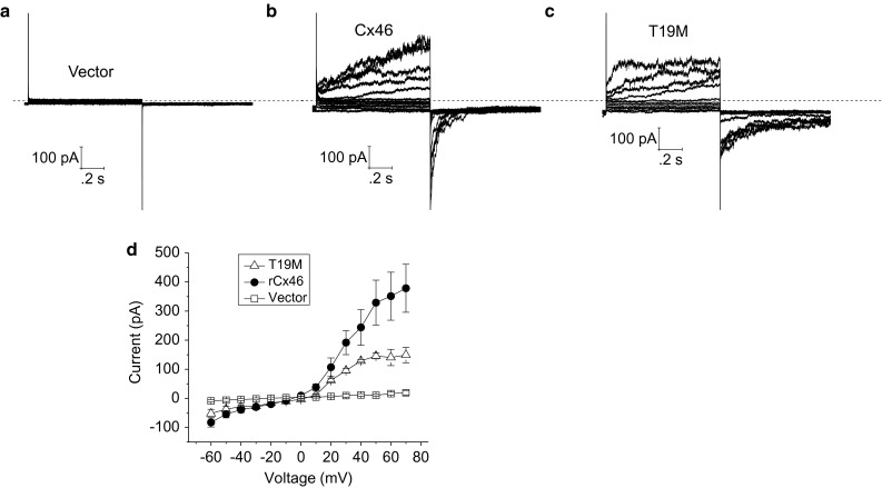
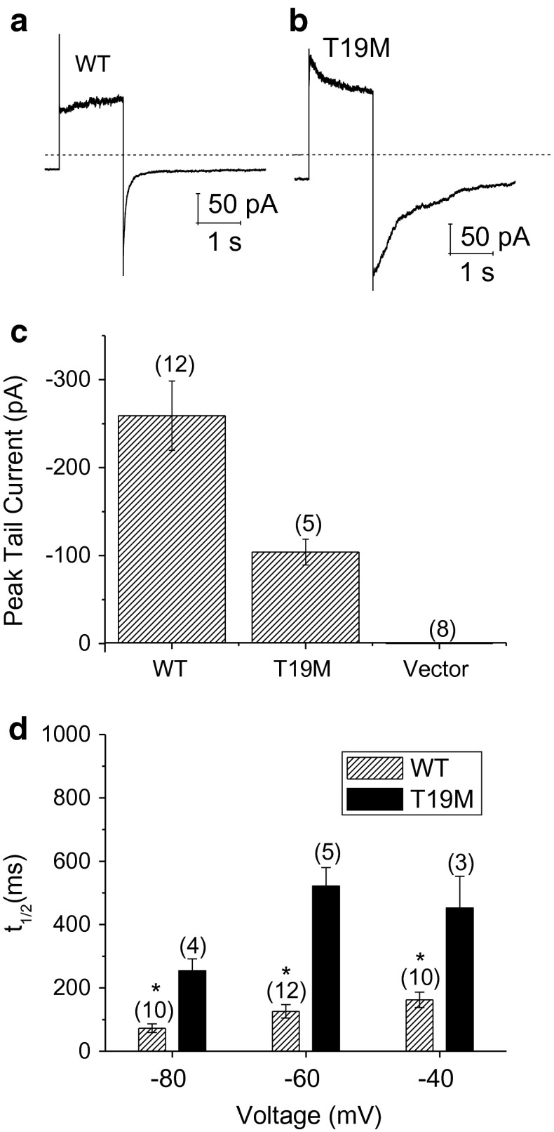
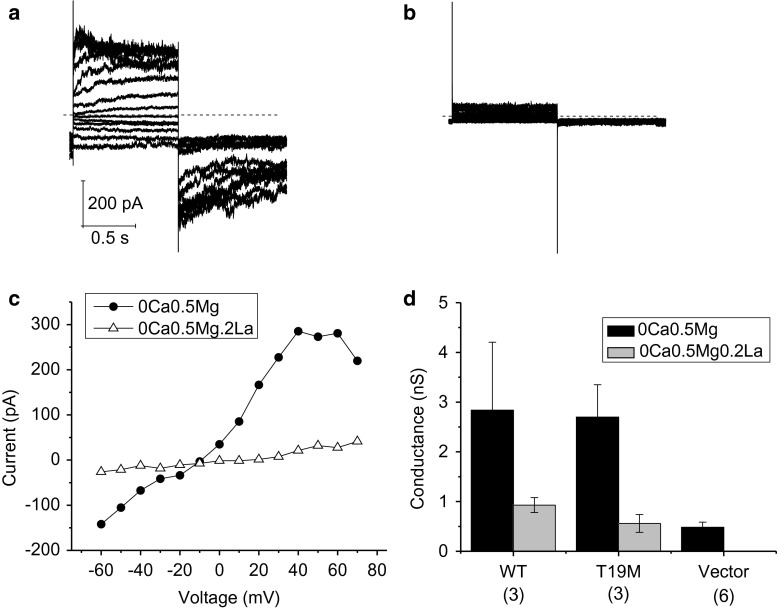
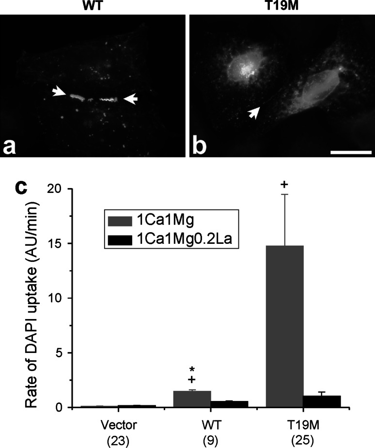
An N-terminal mutant of connexin46 (T19M) alters a highly conserved threonine and has been linked to autosomal dominant cataracts. To study the cellular and functional consequences of substitution of this amino acid, T19M was expressed in Xenopus oocytes and in HeLa cells. Unlike wild-type Cx46, T19M did not induce intercellular conductances in Xenopus oocytes. In transfected HeLa cells, T19M was largely localized within the cytoplasm, with drastically reduced formation of gap junction plaques. Expression of rat T19M was cytotoxic, as evidenced by an almost complete loss of viable cells expressing the mutant protein by 48-72 h following transfection. When incubated in medium containing physiological concentrations of divalent cations, T19M-expressing cells showed increased uptake of DAPI as compared with cells expressing wild-type Cx46, suggesting aberrant connexin hemi-channel activity. Time-lapse and dye uptake studies suggested that T19M hemi-channels had reduced sensitivity to Ca(2+). Whole cell patch clamp studies of single transfected HeLa cells demonstrated that rat T19M formed functional hemi-channels with altered voltage-dependent gating. These data suggest that T19M causes cataracts by loss of gap junctional channel function and abnormally increased hemi-channel activity. Furthermore, they implicate this conserved threonine in both gap junction plaque formation and channel/hemi-channel gating in Cx46.


Figures

Fig. 1
Fig. 1
T19M does not induce gap junctional coupling when expressed by itself, and it acts as a loss-of-function mutation without dominant-negative inhibition when co-expressed with wild-type lens connexins. Bar graphs show mean gap junctional conductances in pairs of oocytes expressing different combinations of wild-type and mutant lens connexins as determined using the double two-electrode voltage clamp technique. a Rat Cx46 or T19M were expressed alone or in combination with each other. b Mouse Cx50 was expressed alone or in combination with either rat Cx46 or T19M. AS indicates oocytes that were injected with no cRNA (i.e., Xenopus Cx38 antisense oligonucleotide alone). The number of pairs tested is indicated within parentheses. *p < 0.001 (Student’s t test compared with Cx46-injected oocyte pairs); **p < 0.001 (Student’s t test compared with Cx50-injected oocyte pairs)
Fig. 2
Fig. 2
T19M is inefficient at forming gap junction plaques. Photomicrographs show the distribution of wild-type rat Cx46 (a) and T19M (bd) at the indicated times following transient transfection of HeLa cells. Bar 30 μM
Fig. 3
Fig. 3
T19M causes increased uptake of connexon-permeant dyes. Photomicrographs show examples of HeLa cells that were transfected with wild-type rat Cx46 (ac) or T19M (d–i) (using the vector PBI-CMV3 which also drives expression of Zaza green) and incubated a day later with DAPI in Na gluconate Ringer’s solution containing 0 mM Ca2+ (af) or 5 mM Ca+2 (gi) for 20 min. Phase contrast images (a, d, g). Zaza-green fluorescence (b, e, h). DAPI fluorescence (c, f, i). After a 20-min incubation in control solution containing 0 mM Ca2+, cells expressing T19M showed DAPI uptake (e, f) that was mostly inhibited by 5 mM Ca2+ (h, i). Bar graph summarizes the quantification of the DAPI uptake data (j). Data are graphed as mean ± SEM. The number of cells tested is indicated within parentheses. *p < 0.001 (Mann–Whitney rank sum test compared with wild-type rat Cx46-transfected cells)
Fig. 4
Fig. 4
The rate of DAPI uptake is increased by lowering divalent cations and inhibited by La3+. Average time course of DAPI uptake by transfected HeLa cells in control solution (1 mM Ca2+, 1 mM Mg2+), in external solutions with no added divalent cations and in control solution plus 200 µM La3+. a Wild-type rat Cx46 (closed circles); T19M (open triangles). To measure changes in the rate of dye uptake over time, the mean DAPI fluorescence intensity per pixel from ROI’s located in the nuclei of Zaza-green positive cells were normalized to mean DAPI fluorescence intensity of the ROI’s at 60 min, averaged and plotted as a function of time. The cells were initially bathed in control solution (containing 1 mM Ca2+, 1 mM Mg2+). Then, the cells were exposed to a solution containing no added divalent cations followed by reperfusion with control solution containing 200 μM La3+. All the solutions contained 4 μM DAPI. b Bar graph shows the rates of DAPI uptake in cells expressing wild-type Cx46, T19M, or vector alone in the presence of 1 mM Ca2+, 1 mM Mg2+ (gray bar); or 1 mM Ca2+, 1 mM Mg2+, 0.2 mM La3+ (black bar). Data are presented as the mean ± SEM. *p < 0.002 (Mann–Whitney rank sum test compared with T19M-transfected cells); + p < 0.001 (Mann–Whitney rank sum test compared with vector-transfected cells). The number of cells analyzed is indicated within parentheses
Fig. 5
Fig. 5
Representative families of current traces recorded from single HeLa cells transfected with vector alone (a), wild-type rat Cx46 (b), or T19M (c). Families of current traces were recorded in response to a series of voltage clamp steps between −60 and 50 mV in increments of 10 mV from a holding potential of −60 mV. Dashed line indicates zero current level. d Average steady-state I–V relationships for vector alone (open squares, n = 5), wild-type (solid circles, n = 4), and T19M (open triangles, n = 3)
Fig. 6
Fig. 6
T19M hemi-channels show alterations in voltage gating. Ensemble averaged current traces recorded from cells expressing wild-type rat Cx46 (a) or T19M (b) in response to a 2-s voltage clamp step to 80 mV followed by a hyperpolarizing step to −60 mV. The holding potential was −60 mV. Dashed line indicates zero current level. c Averaged peak tail currents at −60 mV. The number of cells tested is indicated within parentheses. d Bar graph summarizes the t 1/2’s of deactivation of peak tail currents for wild-type rat Cx46 (hatched bars) and T19M (black bars) at −80, −60, and −40 mV. Data are graphed as mean ± SEM. *p < 0.01 (Student’s t test or Mann–Whitney rank sum test compared with T19M-transfected cells). The number of cells analyzed is indicated within parentheses
Fig. 7
Fig. 7
Effect of lanthanum ions. Currents before (a) and after the application of La3+ (b; 200 µM) recorded from a HeLa cell expressing T19M. Families of current traces were recorded in response to a series of voltage clamp steps between −60 and 70 mV in increments of 10 mV from a holding potential of −60 mV. Dashed line indicates zero current level. c I–V relations obtained from the data shown in (a, b). The current was measured at the end of the 1-s pulse and plotted as a function of voltage. The concentrations of divalent cations in the bath solution were reduced to zero added Ca2+ and 0.5 mM Mg2+ to augment the size of the hemi-channel currents. d Bar graph summarizes the input conductance measured at −60 mV in HeLa cells expressing wild-type Cx46, T19M, or vector (alone) when exposed to extracellular solutions containing 0 mM Ca2+, 0.5 mM Mg2+ (black bars) or 0 mM Ca2+, 0.5 mM Mg2+, 0.2 mM La3+ (gray bars). The number of cells analyzed is indicated within parentheses
Fig. 8
Fig. 8
Effects of T19M mutation on human Cx46. Photomicrographs showing the distribution of human wild-type Cx46 (a) and T19M (b) in transfected HeLa cells. Bar 30 µm. c Bar graph summarizes the DAPI uptake data for vector alone, human wild-type Cx46 and T19M obtained in transfected HeLa cells when exposed to extracellular solutions containing 1 mM Ca2+, 1 mM Mg2+ (gray bars) or 1 mM Ca2+, 1 mM Mg2+, 0.2 mM La3+ (black bars). Data are graphed as mean ± SEM. *p < 0.004 (Mann–Whitney rank sum test compared with T19M-transfected cells in the same external solution); + p < 0.001 (Mann–Whitney rank sum test compared with vector-transfected cells). The number of cells analyzed is indicated within parentheses

Similar articles

Cited by

References

    1. Arora A, Minogue PJ, Liu X, Reddy MA, Ainsworth JR, Bhattacharya SS, Webster AR, Hunt DM, Ebihara L, Moore AT, Beyer EC, Berthoud VM. A novel GJA8 mutation is associated with autosomal dominant lamellar pulverulent cataract: further evidence for gap junction dysfunction in human cataract. J Med Genet. 2006;43:e2. doi: 10.1136/jmg.2005.034108. - DOI - PMC - PubMed
    1. Arora A, Minogue PJ, Liu X, Addison PK, Russel-Eggitt I, Webster AR, Hunt DM, Ebihara L, Beyer EC, Berthoud VM, Moore AT. A novel connexin50 mutation associated with congenital nuclear pulverulent cataracts. J Med Genet. 2008;45:155–160. doi: 10.1136/jmg.2007.051029. - DOI - PMC - PubMed
    1. Berthoud VM, Minogue PJ, Guo J, Williamson EK, Xu X, Ebihara L, Beyer EC. Loss of function and impaired degradation of a cataract-associated mutant connexin50. Eur J Cell Biol. 2003;82:209–221. doi: 10.1078/0171-9335-00316. - DOI - PMC - PubMed
    1. Beyer EC, Berthoud VM. The family of connexin genes. In: Harris AL, Locke D, editors. Connexins: a guide. New York: Humana Press; 2009. pp. 3–26.
    1. Beyer EC, Ebihara L, Berthoud VM. Connexin mutants and cataracts. Front Pharmacol. 2013;4:43. - PMC - PubMed

Publication types

Supplementary concepts

LinkOut - more resources