. 2014 Dec 9;111(49):17612-7.
doi: 10.1073/pnas.1414053111. Epub 2014 Nov 17.

Young capillary vessels rejuvenate aged pancreatic islets

Affiliations

Young capillary vessels rejuvenate aged pancreatic islets

Joana Almaça et al. Proc Natl Acad Sci U S A. .

Abstract

Pancreatic islets secrete hormones that play a key role in regulating blood glucose levels (glycemia). Age-dependent impairment of islet function and concomitant dysregulation of glycemia are major health threats in aged populations. However, the major causes of the age-dependent decline of islet function are still disputed. Here we demonstrate that aging of pancreatic islets in mice and humans is notably associated with inflammation and fibrosis of islet blood vessels but does not affect glucose sensing and the insulin secretory capacity of islet beta cells. Accordingly, when transplanted into the anterior chamber of the eye of young mice with diabetes, islets from old mice are revascularized with healthy blood vessels, show strong islet cell proliferation, and fully restore control of glycemia. Our results indicate that beta cell function does not decline with age and suggest that islet function is threatened by an age-dependent impairment of islet vascular function. Strategies to mitigate age-dependent dysregulation in glycemia should therefore target systemic and/or local inflammation and fibrosis of the aged islet vasculature.

Keywords: aging; glucose metabolism; insulin secretion; pancreatic islets; vasculature.


Conflict of interest statement

Conflict of interest statement: P.-O.B. is one of the founders of the biotech company Biocrine, which is going to use the anterior chamber of the eye as a commercial servicing platform. A.C. holds a patent on this servicing platform. M.H.A. is a consultant of Biocrine.

Figures
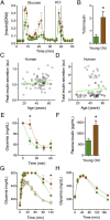
Fig. 1.
Fig. 1.
Beta cells in aged mice and humans are functionally robust, but glucose tolerance is fragile. (A) Insulin secretion from islets isolated from young (2 mo, green) and aged (18 mo, brown) C57BL/6 mice, stimulated with 11 mM glucose and KCl (25 mM; n = 4; insulin levels normalized to DNA concentration). (B) Total amount of insulin released during high glucose (area under the curve). (C and D) Insulin secretion in response to high glucose (11 mM) from human islets from 82 cadaveric donors (ages, 17–65 y). Peak insulin secretion (C) denotes the peak of first-phase insulin secretion, and total insulin secretion (D) denotes the total amount of insulin released during 20 min glucose (expressed relative to responses to KCl). (E) Insulin tolerance tests performed with young and old mice (0.75 units insulin/kg body weight, i.p.; n = 10 mice; glycemia normalized to value at t0). (F) Fed plasma insulin concentration (n = 10 mice). (G and H) Glucose tolerance tests with young and aged mice with different glucose loads (G: 2 g/kg, n =10 mice, open symbols; 4 g/kg, n = 5 mice, filled symbols; one-way ANOVA, * P < 0.05) or in restrained mice (H: 2 g/kg, n = 7–8 mice, area under the curve, 28,010 ± 581 for young mice versus 36,350 ± 3399; *P = 0.02). Mean ± SEM are shown.
Fig. 2.
Fig. 2.
Aged pancreatic mouse and human islets have inflamed and fibrotic blood vessels. (A) Maximal projection of confocal images showing macrophages (green) and endothelial cells expressing the adhesion molecule ICAM-1 (red) in an old islet. (Scale bar, 20 μm.) (B) Quantification of the number of macrophages per islet area (mm2) in islets in pancreatic sections from young and old mice and humans. (C) Quantification of the fractional area of ICAM-1 immunostaining inside islets in young and aged mice. (D) Transcript levels of macrophage colony-stimulating factor receptor gene (CSFR1) and genes involved in the recruitment of immune cells (ICAM1, VCAM1). (E) Expression levels of the extracellular matrix remodeling genes MMP2 and MMP9 in old islets, relative to values in young islets (*P < 0.05, unpaired t test). 18S RNA was used as an internal reference (n = 200 young or old islets, three replicates). (F and G) Maximal projections of confocal images of islets labeled for laminin (green) and the endothelial cell marker PECAM (red) in pancreatic sections from young (F) and aged (G) mice. (Scale bars, 50 μm.) (H) Quantification of the fractional area of laminin immunostaining inside young and old islets.
Fig. 3.
Fig. 3.
Aged islet grafts functionally recover after a prolonged period in young recipient mice. (A) Illustration of islet transplantation into the anterior chamber of the eye. (B) In the experimental strategy, young and old islets were transplanted into the eye of young mice with diabetes and followed for 11 mo (gray arrow). Green and brown arrows, respectively, indicate the actual aging of young and old islet grafts. Dashed line represents the survival curve of C57BL/6 mice and shows that aged islets were studied beyond the 50% survival age [28 mo old (36)]. (C) Fed glycemia in streptozotocin (STZ)-induced young mice with diabetes after transplantation of young (green; n = 5 mice) and old (brown; n = 6 mice) islets (enucleation = removal of the islet graft-bearing eye). (D) Percentage of young mice with diabetes (fed glycemia > 200 mg/dL) transplanted with young (green) or old (brown) islets and of old mice with diabetes transplanted with old islets (black). (E and F) Average glycemia during glucose tolerance tests performed at 3, 4.5, 7, and 10 mo after transplantation in young mice with young islets (E; n = 5 mice) or old islets (F; n = 5 mice). (G) Glycemia at 120 min (t120) after glucose injection in mice with young (green) and old islets [brown; one-way ANOVA, *P < 0.05; dashed line, glycemia (t120) in young donor mice]. (H) Fed plasma insulin concentration in young and old islet recipient mice.
Fig. 4.
Fig. 4.
Old islet grafts in young recipient mice show strong proliferative activity. (A) Photograph of a mouse eye (*, pupil) showing two old islet grafts 3 and 9 mo after transplantation. A marked increase in islet graft size can be seen. (Scale bar, 200 μm.) (B) Quantification of the fraction of proliferating beta cells (*P < 0.05; n = 6 young or old islet grafts). BrdU (1 mg/mL) was added for 21 d to the drinking water at 11 mo after transplantation. (C and D) Confocal images of a young (C) and an aged (D) islet graft showing BrdU-labeled, proliferating cells (green, BrdU; red, insulin; blue, DNA). Arrows point at beta cells, arrowhead indicates a nonbeta cell. (Scale bars, 50 μm.)
Fig. 5.
Fig. 5.
Revascularization of old islet grafts. (A) Longitudinal in vivo images of blood vessels in an old islet graft. Capillaries with smaller diameters appear in newly formed regions. (Scale bars, 100 μm.) (B) Distribution curves of the diameters of blood vessels in young and old islets grafts at 2 wk and 2 and 10 mo after transplantation. (C and D) Quantification of the fractional area of laminin immunostaining (C) and of the number of macrophages per islet area (mm2, D) in young and old islet grafts 11 mo after transplantation (n = 3 islet grafts/eye; n = 2 recipient mice/age).

References

    1. Chang AM, Halter JB. Aging and insulin secretion. Am J Physiol Endocrinol Metab. 2003;284(1):E7–E12. - PubMed
    1. Gu Z, et al. Effect of aging on islet beta-cell function and its mechanisms in Wistar rats. Age (Dordr) 2012;34(6):1393–1403. - PMC - PubMed
    1. Gumbiner B, et al. Effects of aging on insulin secretion. Diabetes. 1989;38(12):1549–1556. - PubMed
    1. Ihm SH, et al. Effect of aging on insulin secretory function and expression of beta cell function-related genes of islets. Diabetes Res Clin Pract. 2007;77(Suppl 1):S150–S154. - PubMed
    1. Leiter EH, Premdas F, Harrison DE, Lipson LG. Aging and glucose homeostasis in C57BL/6J male mice. FASEB J. 1988;2(12):2807–2811. - PubMed

Publication types

MeSH terms