. 2014 Dec 2;111(48):17104-9.
doi: 10.1073/pnas.1418637111. Epub 2014 Nov 17.

NanoFlares for the detection, isolation, and culture of live tumor cells from human blood

Affiliations

NanoFlares for the detection, isolation, and culture of live tumor cells from human blood

Tiffany L Halo et al. Proc Natl Acad Sci U S A. .

Abstract

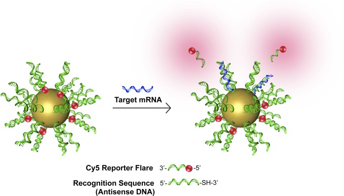
Metastasis portends a poor prognosis for cancer patients. Primary tumor cells disseminate through the bloodstream before the appearance of detectable metastatic lesions. The analysis of cancer cells in blood—so-called circulating tumor cells (CTCs)—may provide unprecedented opportunities for metastatic risk assessment and investigation. NanoFlares are nanoconstructs that enable live-cell detection of intracellular mRNA. NanoFlares, when coupled with flow cytometry, can be used to fluorescently detect genetic markers of CTCs in the context of whole blood. They allow one to detect as few as 100 live cancer cells per mL of blood and subsequently culture those cells. This technique can also be used to detect CTCs in a murine model of metastatic breast cancer. As such, NanoFlares provide, to our knowledge, the first genetic-based approach for detecting, isolating, and characterizing live cancer cells from blood and may provide new opportunities for cancer diagnosis, prognosis, and personalized therapy.

Keywords: NanoFlares; cancer metastasis; diagnostic; mRNA; nanotechnology.


Conflict of interest statement

Conflict of interest statement: C.A.M. and C.S.T. are cofounders of AuraSense, LLC, a start-up biotechnology company that licensed the NanoFlare technology from Northwestern University.

Figures

Fig. 1.
Fig. 1.
Schematic of NanoFlare structure and function. The NanoFlare contains a monolayer of antisense DNA (recognition sequence) adsorbed to the surface of a 13-nm spherical gold nanoparticle. A reporter flare sequence is hybridized to the recognition sequence, which contains a fluorophore (red). The dye is quenched in close proximity to the gold surface. The reporter flare is displaced when complementary mRNA (blue) binds the recognition sequence, providing a fluorescent signal.
Fig. 2.
Fig. 2.
NanoFlares detect mesenchymal markers in EMT model cell lines. (A) NanoFlares detect E-cadherin and Twist mRNA in model mammary cell lines, HMLE, and HMLE-Twist. Results represent flow-cytometric analysis of cells treated with NanoFlares (100 pM) for 16 h. (B) NanoFlares detect EMT markers in metastatic breast cancer cell line MDA-MB-231. NanoFlares were designed for mesenchymal markers (Vimentin and Fibronectin), an epithelial marker (E-cadherin), and a nontargeting Scrambled Control NanoFlare. Results represent flow-cytometric analysis of cells treated with NanoFlares (100 pM) for 8 h.
Fig. 3.
Fig. 3.
NanoFlares isolate metastatic breast cancer cells (MDA-MB-231) spiked into human whole blood. (A) Recovery of mCherry MDA-MB-231 cells from whole blood upon isolation with NanoFlares. (B) Relative cell-associated fluorescence of Vimentin (Vim), Fibronectin (HFN), or nontargeted Scrambled (Scr) Control NanoFlares in 100–100,000 MDA-MB-231 added to whole blood. (C) Scatter plots showing ability of Vimentin NanoFlare to detect small numbers of cells added to whole blood, with a range of 10,000, 1000, and 100 cells. ***P < 0.0005, **P < 0.005, *P < 0.05.
Fig. 4.
Fig. 4.
NanoFlares detect circulating breast cancer cells from whole blood in a murine model of metastatic breast cancer. (A) Representative scatter plots (n = 1 per scatter plot) show mCherry MDA-MB-231 cells recovered from whole blood by Vimentin NanoFlares from two example tumor-bearing mice (Experimental 1 and 2) (n = 12 per group) or non–tumor-bearing (Control) mice (n = 12 per group). (B) Relative cell associated fluorescence of isolated blood samples treated with Vimentin (Vim), Fibronectin (HFN), or Scrambled (Scr) Control NanoFlares added 6 h before flow-cytometric analysis. (C) Efficiency of mCherry-MDA-MB-231 cell detection by NanoFlares; values were calculated by correlating mCherry fluorescence to NanoFlare fluorescence. Untreated population detection indicates channel bleed-through of mCherry to NanoFlare channel. Samples were collected after 6.5-wk tumor inoculation. Cy5 represents NanoFlare signal. *P < 0.05.
Fig. 5.
Fig. 5.
Cell isolation and mammosphere formation post NanoFlare treatment and flow cytometry analysis. Representative scatter plots show Cy5 fluorescence (NanoFlare) of GFP recurrent cells spiked into (A) untreated human whole blood or (B) Vimentin NanoFlare-treated blood. Upon treatment with NanoFlares, Cy5 fluorescence of GFP-positive cells increases 5.4-fold. Cells in the red gate in the Vimentin sample were sorted for mammosphere culture. Cells retrieved from blood form mammospheres (C) untreated or (D) Vimentin NanoFlare-treated.

References

    1. Liotta LA, Kohn EC. The microenvironment of the tumour-host interface. Nature. 2001;411(6835):375–379. - PubMed
    1. Chiang AC, Massagué J. Molecular basis of metastasis. N Engl J Med. 2008;359(26):2814–2823. - PMC - PubMed
    1. Cristofanilli M, et al. Circulating tumor cells: A novel prognostic factor for newly diagnosed metastatic breast cancer. J Clin Oncol. 2005;23(7):1420–1430. - PubMed
    1. Budd GT, et al. Circulating tumor cells versus imaging: Predicting overall survival in metastatic breast cancer. Clin Cancer Res. 2006;12(21):6403–6409. - PubMed
    1. Hayes DF, et al. Circulating tumor cells at each follow-up time point during therapy of metastatic breast cancer patients predict progression-free and overall survival. Clin Cancer Res. 2006;12(14 Pt 1):4218–4224. - PubMed

Publication types

MeSH terms