. 2014 Dec 16;111(50):17977-82.
doi: 10.1073/pnas.1420258111. Epub 2014 Nov 17.

Melanopsin mediates light-dependent relaxation in blood vessels

Affiliations

Melanopsin mediates light-dependent relaxation in blood vessels

Gautam Sikka et al. Proc Natl Acad Sci U S A. .

Abstract

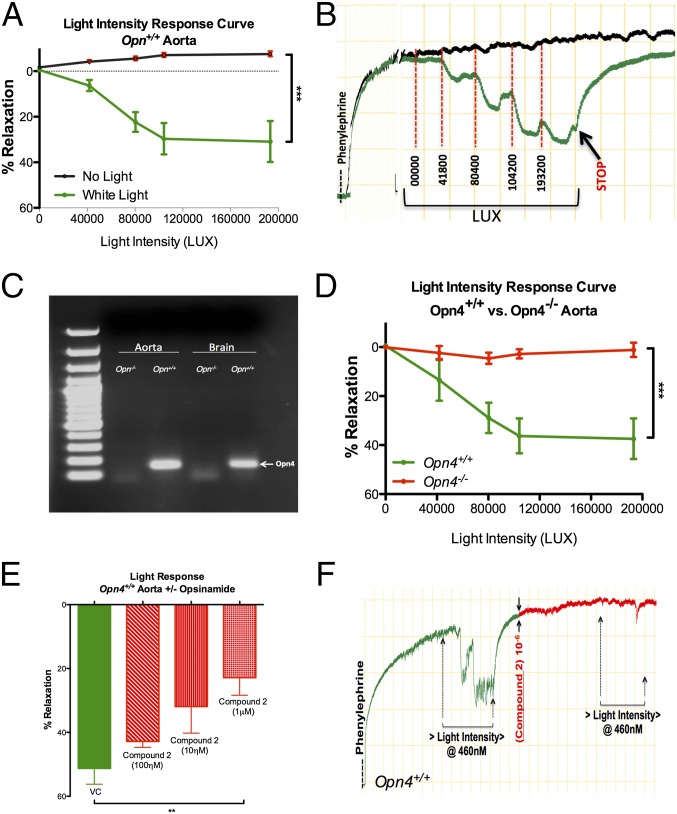
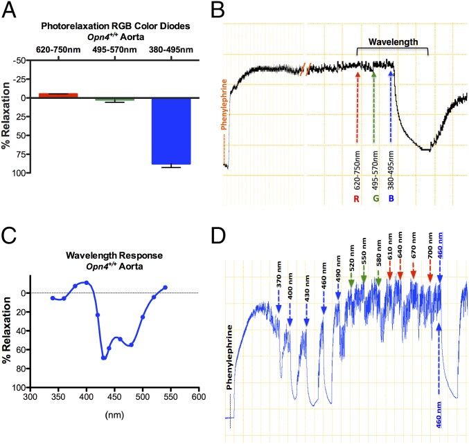
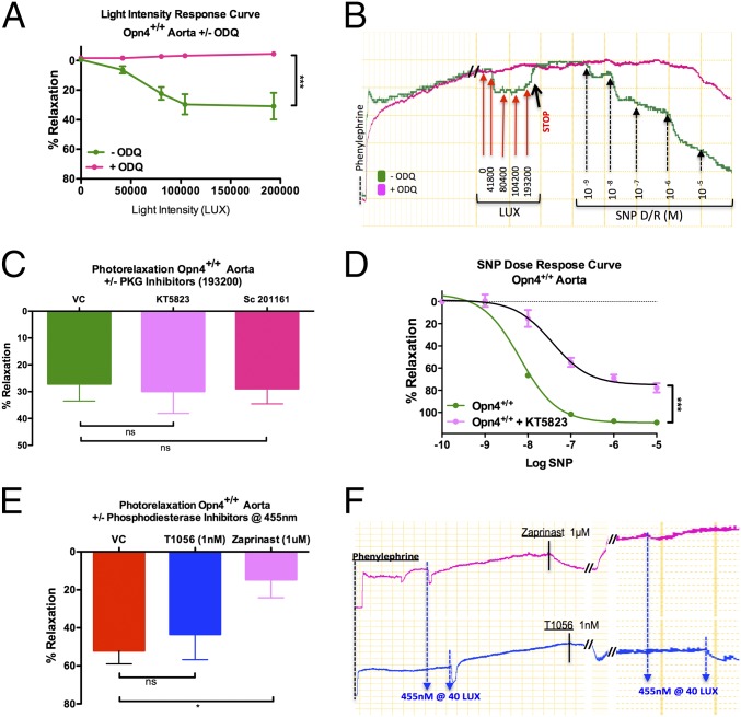
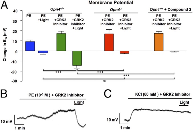
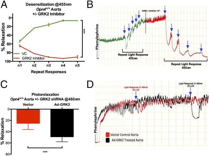
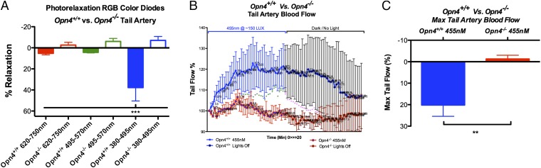
Melanopsin (opsin4; Opn4), a non-image-forming opsin, has been linked to a number of behavioral responses to light, including circadian photo-entrainment, light suppression of activity in nocturnal animals, and alertness in diurnal animals. We report a physiological role for Opn4 in regulating blood vessel function, particularly in the context of photorelaxation. Using PCR, we demonstrate that Opn4 (a classic G protein-coupled receptor) is expressed in blood vessels. Force-tension myography demonstrates that vessels from Opn4(-/-) mice fail to display photorelaxation, which is also inhibited by an Opn4-specific small-molecule inhibitor. The vasorelaxation is wavelength-specific, with a maximal response at ∼430-460 nm. Photorelaxation does not involve endothelial-, nitric oxide-, carbon monoxide-, or cytochrome p450-derived vasoactive prostanoid signaling but is associated with vascular hyperpolarization, as shown by intracellular membrane potential measurements. Signaling is both soluble guanylyl cyclase- and phosphodiesterase 6-dependent but protein kinase G-independent. β-Adrenergic receptor kinase 1 (βARK 1 or GRK2) mediates desensitization of photorelaxation, which is greatly reduced by GRK2 inhibitors. Blue light (455 nM) regulates tail artery vasoreactivity ex vivo and tail blood blood flow in vivo, supporting a potential physiological role for this signaling system. This endogenous opsin-mediated, light-activated molecular switch for vasorelaxation might be harnessed for therapy in diseases in which altered vasoreactivity is a significant pathophysiologic contributor.

Keywords: GRK2; melanopsin; opsin; photorelaxation; signal transduction.


Conflict of interest statement

The authors declare no conflict of interest.

Figures

Fig. 1.
Fig. 1.
Opsin 4 expression in blood vessels and its role in photorelaxation. (A) Summary data and (B) representative trace of ex vivo vasoreactivity demonstrating dose-dependent effects of cold white light on Opn4+/+ mouse aorta compared with no light exposure. Error bars denote SEM, n = 6, ***P < 0.001. (C) RT-PCR analysis of mouse aorta and brain demonstrates Opn4 mRNA expression in Opn4+/+ mice but not Opn4−/− mice. n = 6. (D) Vasoreactivity to cold white light demonstrates absence of photorelaxation in aortas from Opn4−/− mice compared with Opn4+/+ mice. Error bars denote SEM, n = 6, ***P < 0.001. (E) Dose-dependent inhibition of the photorelaxation response in presence of opsinamide, Compound 2 (an Opn4 inhibitor). Error bars denote SEM, n = 4, **P < 0.005. (F) Representative trace: vasoreactivity data showing photorelaxation response before and after pharmacological inhibition of Opn4 with 1 μM concentration of Compound 2 in Opn4+/+ aorta.
Fig. 2.
Fig. 2.
Determination of optimal wavelength within the visible electromagnetic spectrum mediating the photorelaxation response. (A) Vasoreactivity of Opn4+/+ mouse aorta to red (620–750 nm) to green (495–570 nm) to blue (380–495 nm) (RGB) wavelengths of the visible spectrum using light-emitting diodes. n = 4. (B) Representative trace demonstrates no vasorelaxation in Opn4+/+ aorta in the R to G spectrum but maximum vasorelaxation in the presence of B spectrum light. (C) Summary data of vasoreactivity of Opn4+/+ aorta to precise wavelength changes within blue light spectrum using monochromator (precise peak wavelength spectral changes at 30-nm intervals). n = 3. (D) Representative trace demonstrating vasoreactivity of Opn4+/+ aorta to precise 30-nm wavelength increments from 370 nm to 700 nm, followed by repeat 460-nm exposure to confirm this optimal wavelength and ensure lack of desensitization.
Fig. 3.
Fig. 3.
Signal transduction mechanism involved in photorelaxation. (A) Vasoreactivity in Opn4+/+ mouse aorta to cold white light demonstrates marked attenuation of photorelaxation in the vessels preincubated with the sGC inhibitor ODQ (∼−4% relaxation), whereas untreated vessels relaxed ∼31%. Error bars denote SEM, ***P < 0.001. (B) Representative trace: vasoreactivity of Opn4+/+ mouse aorta to cold white light in the presence and absence of ODQ, followed by dose–response curve with nitric oxide donor SNP to confirm sGC inhibition. (C) Vasoreactivity of Opn4+/+ mouse aorta to cold white light demonstrates no significant difference in photorelaxation in vessels preincubated with the PKG inhibitor KT5823 (0.5 μM), (n = 3) or sc-201161 (10 μM) (n = 4) compared with untreated vessels. n = 7. (D) Significant impairment in vasorelaxation to the NO donor SNP in Opn4+/+ mouse aorta preincubated with KT5823 compared with VC. n = 3, ***P < 0.001. (E) Maximum photorelaxation response to 455 nm blue light in untreated Opn4+/+ aortas compared with aorta preincubated with 1 nM T1056 (selective PDE5 inhibitor) and 1 μM Zaprinast (nonselective PDE5 and PDE6 inhibitor). Only Zaprinast-treated vessels had attenuated photorelaxation (∼52% vs. ∼14%). Error bars denote SEM, *P < 0.05. (F) Representative trace comparing photorelaxation in vessels treated with 1 nM T1056 and 1 μM Zaprinast.
Fig. 4.
Fig. 4.
Sharp electrode intracellular recordings of in situ smooth muscle Em in endothelium-denuded segments of mouse thoracic aorta. n = 5–12. (A) Bar graph shows composite data (mean ± SEM) for change in Em under various conditions in aortas from Opn4+/+ mice, Opn4−/− mice, and after pretreatment with opsinamide, Compound 2 for 30 min (1 μM). Depolarizing stimuli with and without the GPK2 inhibitor methyl 5-[2-(5-nitro-2-furyl)vinyl]-2-furoate were applied for 5 min in dark conditions before subjecting the tissue to light. n = 5–12. There was a significant decrease in Em when blue light (380–495 nm) was applied to GRK2 pretreated vessels (with PE) from Opn4+/+ mice compared with vessels from Opn4−/− and after pretreatment of Opn4+/+ vessels with Compound 2. n = 5–12; ***P < 0.001. (B and C) Representative traces show the effect of blue light (380–495 nm) on Em in cells depolarized with (B) PE or (C) potassium chloride (KCl).
Fig. 5.
Fig. 5.
Stimulus-dependent desensitization, photorelaxation, and its modulation. (A) Vasoreactivity: Opn4+/+ aorta demonstrate desensitization/diminished vasodilatory responses to iso-intensity repeat light stimulation (455 nm at 40 lux; ∼2-min interval) and attenuation of desensitization/enhanced vasodilatory responses to the same iso-intensity repeat light stimulation after inhibition with GPK2 inhibitor. Error bars denote SEM, n = 5, ***P < 0.001. (B) Representative trace demonstrating desensitization resulting in loss of photorelaxation, followed by resensitization of receptors with use of GPK2 inhibitor, resulting in exaggerated vasodilatory responses with blue light (455 nm at 40 lux). (C) Vasoreactivity: Opn4+/+ aortic rings, preincubated with the GRK2 shRNA adenovirus (Ad-GRK2), demonstrating enhanced wavelength-specific photorelaxation (∼49%) compared with the vector-treated vessels (∼25%). Error bars denote SEM, n = 4, *P < 0.05. (D) Representative trace demonstrating enhanced photorelaxation in Opn4+/+ aortas treated with Ad-GRK2 compared with vector control.
Fig. 6.
Fig. 6.
Opsin4 in vivo peripheral blood flow and vasoreactivity. (A) Vasoreactivity of mouse-tail artery to RGB wavelength spectrum demonstrates that only blue light causes vasorelaxation in Opn4+/+ but no response in Opn4−/− tail arteries. Error bars denote SEM, n = 4, ***P < 0.001. (B) In vivo laser Doppler flowmetry of mouse tail demonstrates a significant increase in tail blood flow after 10 min of cumulative blue light (455 nm at 40 lux) exposure in Opn4+/+ but not in Opn4−/− mice. This is reversible on reexposure to dark. Error bars denote SD, *P < 0.05, **P < 0.01. (C) Maximum change in tail flow in Opn4+/+ and Opn4−/− mice. Error bars denote SEM, n = 4, **P < 0.01.

Comment in

  • PDE and sGC hand in hand to see the light.
    Vanhoutte PM. Vanhoutte PM. Proc Natl Acad Sci U S A. 2014 Dec 16;111(50):17704-5. doi: 10.1073/pnas.1421161111. Epub 2014 Dec 2. Proc Natl Acad Sci U S A. 2014. PMID: 25468979 Free PMC article. No abstract available.

References

    1. Furchgott RF, Sleator WJ, Mccaman MW, Elchlepp J. Relaxation of arterial strips by light and the influence of drugs on this photodynamic effect. J Pharmacol Exp Ther. 1955;113:22.
    1. Matsunaga K, Furchgott RF. Interactions of light and sodium nitrite in producing relaxation of rabbit aorta. J Pharmacol Exp Ther. 1989;248(2):687–695. - PubMed
    1. Chen X, Gillis CN. Enhanced photorelaxation in aorta, pulmonary artery and corpus cavernosum produced by BAY K 8644 or N-nitro-L-arginine. Biochem Biophys Res Commun. 1992;186(3):1522–1527. - PubMed
    1. Andrews KL, McGuire JJ, Triggle CR. A photosensitive vascular smooth muscle store of nitric oxide in mouse aorta: No dependence on expression of endothelial nitric oxide synthase. Br J Pharmacol. 2003;138(5):932–940. - PMC - PubMed
    1. Andrews KL, Triggle CR, Ellis A. NO and the vasculature: Where does it come from and what does it do? Heart Fail Rev. 2002;7(4):423–445. - PubMed

Publication types