. 2014 Dec 2;111(48):17182-7.
doi: 10.1073/pnas.1414164111. Epub 2014 Nov 17.

Structures of protective antibodies reveal sites of vulnerability on Ebola virus

Affiliations

Structures of protective antibodies reveal sites of vulnerability on Ebola virus

Charles D Murin et al. Proc Natl Acad Sci U S A. .

Abstract

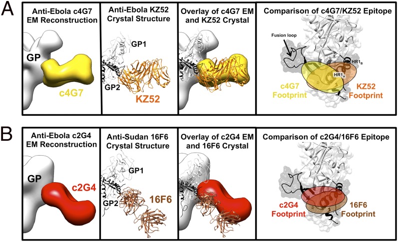
Ebola virus (EBOV) and related filoviruses cause severe hemorrhagic fever, with up to 90% lethality, and no treatments are approved for human use. Multiple recent outbreaks of EBOV and the likelihood of future human exposure highlight the need for pre- and postexposure treatments. Monoclonal antibody (mAb) cocktails are particularly attractive candidates due to their proven postexposure efficacy in nonhuman primate models of EBOV infection. Two candidate cocktails, MB-003 and ZMAb, have been extensively evaluated in both in vitro and in vivo studies. Recently, these two therapeutics have been combined into a new cocktail named ZMapp, which showed increased efficacy and has been given compassionately to some human patients. Epitope information and mechanism of action are currently unknown for most of the component mAbs. Here we provide single-particle EM reconstructions of every mAb in the ZMapp cocktail, as well as additional antibodies from MB-003 and ZMAb. Our results illuminate key and recurring sites of vulnerability on the EBOV glycoprotein and provide a structural rationale for the efficacy of ZMapp. Interestingly, two of its components recognize overlapping epitopes and compete with each other for binding. Going forward, this work now provides a basis for strategic selection of next-generation antibody cocktails against Ebola and related viruses and a model for predicting the impact of ZMapp on potential escape mutations in ongoing or future Ebola outbreaks.

Keywords: EM; Ebola; ZMapp; antibodies.


Conflict of interest statement

The authors declare no conflict of interest.

Figures

Fig. 1.
Fig. 1.
Competition binding assays. Antibodies from the anti-Ebola cocktails MB-003 and ZMAb were compared for their binding to Ebola GPΔTM to determine if there were any overlapping binding sites within or between mAb cocktails. The percent binding of the competing mAb in the presence of the first mAb was determined by comparing the maximal signal of competing mAb applied after the first mAb complex to the maximal signal of competing mAb alone. MAbs were considered competing for the same site if maximum binding of antibody 2 was reduced to <10% of its binding to GP alone (black boxes with white numbers). mAbs were considered noncompetitive if maximum binding of antibody 2 was >30% of its binding to GP alone (white boxes with black numbers). Gray boxes with red numbers indicate an intermediate phenotype (between 10% and 30% of its binding to GP alone).
Fig. 2.
Fig. 2.
Single-particle negative-stain EM reconstructions of MB-003 and ZMAb antibodies bound to EBOV GPΔTM. Hybrid models of negative-stain EM reconstructions fit with the EBOV GPΔmuc crystal structure (PDB ID code 3CSY) (39) with GP1 in white and GP2 in black. Core GPs and Fabs are rendered as surfaces with GPs in white and Fabs in various colors. Fab densities are fit with a model Fab structure for reference. (A) Fab c13C6 (in dark blue) and KZ52 (removed) in complex with EBOV GPΔTM showing side (Left) and top (Right) views of the reconstruction. (B) Fab c1H3 (in light blue) and KZ52 (removed) in complex with EBOV GPΔTM showing side (Left) and top (Right) views of the reconstruction. (C) Fab c4G7 (in yellow) and c13C6 (removed) in complex with EBOV GPΔTM showing side (Left) and top (Right) views of the reconstruction. (D) Fab c2G4 (red) in complex with EBOV GPΔTM showing side (Left) and top (Right) views of the reconstruction. (E) Side view comparisons of liganded EBOV GPΔTM (Left, hybrid reconstruction of GPΔTM in complex with c4G7 in yellow, c13C6 in blue, c2G4 in red, and GP in white) and unliganded GPΔTM (Center and Right, Fabs removed). Relative positions of domains on GP are indicated.
Fig. 3.
Fig. 3.
Details of the glycan cap and GP1-GP2 interface epitopes. (A) Competition analysis indicated that antibodies c13C6 and c1H3 have overlapping epitopes. Here, structures of c13C6 (dark blue) and c1H3 (light blue) bound to GPΔTM are illustrated, with the GPΔmuc crystal structure (PDB ID code 3CSY) fit into the GP EM density. GP1 is white and GP2 is black. Superimposition of the structures illustrates that the antibodies have overlapping epitopes within the glycan cap on GP1 (side view on the far left, top view in the center and far right). The footprints of these antibodies are highlighted on the far right. The mesh portion of the reconstruction is the part of the GP glycan cap that is resolved in the c1H3:GPΔTM structure. (B) As in A but c4G7 is in yellow and c2G4 is in red (side view on the far left and right, top view in the center).
Fig. 4.
Fig. 4.
Comparison of EM reconstructions and crystal structures of mAbs that bind at the GP1-GP2 interface. (A) c4G7 (in yellow) binds to a very similar epitope and angle of approach to the human survivor mAb KZ52 (in orange; PDB ID code 3CSY) and their footprints overlap significantly. (B) Anti-SUDV mAb 16F6 (in brown; PDB ID code 3VE0) (38, 45) binds a similar epitope to c2G4 (as well as KZ52 and c4G7) and at a similar angle (from the bottom of GP, upward toward GP1), although c2G4 binds closer to GP2 and likely recognizes less of GP1, as previously indicated (28). Note that 16F6 only binds SUDV, whereas the others bind EBOV. Similarity of the binding sites indicates functional conservation across the ebolavirus genus.
Fig. 5.
Fig. 5.
Sites of vulnerability on Ebola virus for protective mAbs. Anti-Ebola mAbs target key sites on GP. Antibodies that do not neutralize, or that do not neutralize in the absence of complement, bind outside of the core GP in the glycan cap and mucin-like domains, and are shown in cool colors. These antibodies can be protective, despite the removal of these domains before receptor binding in the endosome. All neutralizing mAbs (warm colors) solved to date bind at nearly the same site, within the GP1–GP2 interface. These mAbs bind the core of GP, which remains intact before entry. These mAbs may neutralize the virus by preventing structural changes in GP2 required for membrane fusion. A monomer of GPΔmuc (PDB ID code 3CSY) is fit into the core GP of the c13C6:c4G7 complex. MPER, membrane proximal external region.

References

    1. Brès P. [The epidemic of Ebola haemorrhagic fever in Sudan and Zaire, 1976: Introductory note] Bull World Health Organ. 1978;56(2):245. - PMC - PubMed
    1. Jahrling PB, et al. Preliminary report: Isolation of Ebola virus from monkeys imported to USA. Lancet. 1990;335(8688):502–505. - PubMed
    1. World Health Organization Outbreak news. Ebola Reston in pigs and humans, Philippines. Wkly Epidemiol Rec. 2009;84(7):49–50. - PubMed
    1. Barrette RW, et al. Discovery of swine as a host for the Reston ebolavirus. Science. 2009;325(5937):204–206. - PubMed
    1. Falzarano D, Geisbert TW, Feldmann H. Progress in filovirus vaccine development: Evaluating the potential for clinical use. Expert Rev Vaccines. 2011;10(1):63–77. - PMC - PubMed

Publication types

MeSH terms