. 2015 Jan 1;194(1):125-133.
doi: 10.4049/jimmunol.1401644. Epub 2014 Nov 17.

OX40- and CD27-mediated costimulation synergizes with anti-PD-L1 blockade by forcing exhausted CD8+ T cells to exit quiescence

Affiliations

OX40- and CD27-mediated costimulation synergizes with anti-PD-L1 blockade by forcing exhausted CD8+ T cells to exit quiescence

Sarah Buchan et al. J Immunol. .

Abstract

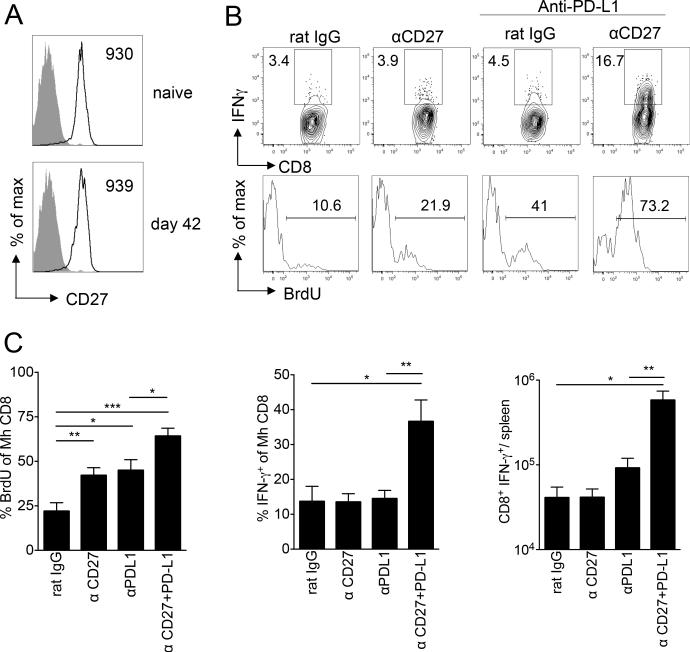
Exhaustion of chronically stimulated CD8(+) T cells is a significant obstacle to immune control of chronic infections or tumors. Although coinhibitory checkpoint blockade with anti-programmed death ligand 1 (PD-L1) Ab can restore functions to exhausted T cell populations, recovery is often incomplete and dependent upon the pool size of a quiescent T-bet(high) subset that expresses lower levels of PD-1. In a model in which unhelped, HY-specific CD8(+) T cells gradually lose function following transfer to male bone marrow transplantation recipients, we have explored the effect of shifting the balance away from coinhibition and toward costimulation by combining anti-PD-L1 with agonistic Abs to the TNFR superfamily members, OX40 and CD27. Several weeks following T cell transfer, both agonistic Abs, but especially anti-CD27, demonstrated synergy with anti-PD-L1 by enhancing CD8(+) T cell proliferation and effector cytokine generation. Anti-CD27 and anti-PD-L1 synergized by downregulating the expression of multiple quiescence-related genes concomitant with a reduced frequency of T-bet(high) cells within the exhausted population. However, in the presence of persistent Ag, the CD8(+) T cell response was not sustained and the overall size of the effector cytokine-producing pool eventually contracted to levels below that of controls. Thus, CD27-mediated costimulation can synergize with coinhibitory checkpoint blockade to switch off molecular programs for quiescence in exhausted T cell populations, but at the expense of losing precursor cells required to maintain a response.


Figures

Figure 1
Figure 1. The effect of CD4+ T cell help upon the development of CD8+ T cell exhaustion
1 × 10⁶ CD45.1+ Mh transgenic CD8+ T cells were transferred with or without 2.5 × 10⁶ female B6 CD4+ T cells, 1 week after lethal irradiation of B6 male mice and reconstitution with female B6 BM. A) Graph shows mean percentage ± SEM of Mh CD8+ T cells that produced IFN-γ in response to UTY peptide on day 8 and 42 after T cell transfer. Data are pooled from 2 independent experiments (helped n=5-6/group; unhelped n=3-12/group). B) Graph showing mean percentage ± SEM of PD-1high Mh CD8+ T cells (n=5/group). C) Irradiated male recipients were given 200ug anti-PD-L1 blocking antibody on day 36 and 39 following T cell transfer (n=5 +CD4, n=6 no CD4) or isotype control (n=6 +CD4, n=7 no CD4) before analysis on day 42. From left to right, graphs show mean percentage ± SEM of Mh CD8+ T cells that had incorporated BrdU, mean percentage ± SEM of Mh CD8+ T cells that produced IFN-γ and mean ± SEM absolute numbers of Mh CD8+IFN-γ+ cells/spleen. Data are pooled from 2 independent experiments. Statistical comparisons performed using two-tailed, unpaired student t-test: *p<0.05, **p<0.01, ***p<0.001.
Figure 2
Figure 2. Effect of agonistic anti-OX40 and/or blocking anti-PD-L1 antibody upon helped or unhelped donor Mh CD8+ T cell effector functions
A) Representative histograms showing OX40 expression upon gated Mh CD8+ T cells (open histograms) in recipient spleens on day 42 following transfer to irradiated B6 male mice (mean % OX40+ was 10.5 ±2.2 in helped vs. 11.4 ±2.4 in unhelped; naïve 1.5 ±0.5). Filled histograms show OX40 staining of endogenous cells in same host. B) Male BMT recipients were given anti-OX40 i.p. day 35 following Mh CD8+ T cell transfer (with or without CD4+ T cells, n=4/group) or 200ug anti-PD-L1 blocking antibody i.p. day 36 and 39 (n=5 in CD4+, n=6 in no CD4+) or both antibodies (n=6 in + CD4+, and n=7 in no CD4+). Control mice received the same number of i.p. injections with the relevant isotype control (n=6-7). Top two rows- representative contour plots show IFN-γ production by helped Mh CD8+ T cells following exposure to UTY peptide, with gates set according to irrelevant peptide and representative histograms showing the percentage of Mh CD8+ T cells incorporating BrdU. Bottom two rows –representative contour plots for IFN-γ production and BrdU incorporation following transfer of unhelped Mh CD8+ T cells. C) From left to right, graphs show mean percentage ± SEM of Mh CD8+ T cells that had incorporated BrdU, mean percentage ± SEM of Mh CD8+ T cells that produced IFN-γ and mean ± SEM absolute numbers of Mh CD8+IFN-γ+ cells/spleen. Data are pooled from 2 independent experiments. Statistical comparisons performed using two-tailed, unpaired student t-test: *p<0.05, **p<0.01, ***p<0.001.
Figure 3
Figure 3. Effect of agonistic anti-CD27 and/or blocking anti-PD-L1 antibody upon unhelped Mh CD8+ T cell effector functions
A) From experiments of similar design to those in Figure 2, representative histograms showing CD27 expression upon naïve input Mh CD8+ T cells or on day 42 (open histograms) following transfer to irradiated male mice and isolation from recipient spleens. Filled histograms show isotype control staining. Numbers indicate mean fluorescence intensity. B) BMT recipients were given anti-CD27 on day 35 following unhelped Mh CD8+ T cell transfer (n=8) or 200ug anti-PD-L1 blocking antibody on day 36 and 39 (n=9) or both antibodies (n=9). Control mice received the same number of i.p. injections with the relevant isotype control (n=7). Top-representative contour plots show IFN-γ production by Mh CD8+ T cells following exposure overnight to UTY peptide, with gates set according to irrelevant peptide. Bottom- representative histograms showing the percentage of Mh CD8+ T cells incorporating BrdU. C) Graphs show mean percentage ± SEM of Mh CD8+ T cells that had incorporated BrdU, mean percentage ± SEM of Mh CD8+ T cells that produced IFN-γ and mean ± SEM absolute numbers of Mh CD8+IFN-γ+ cells/spleen. Data are pooled from 2 independent experiments. Statistical comparisons performed using two-tailed, unpaired student t-test: *p<0.05, **p<0.01, ***p<0.001.
Figure 4
Figure 4. Comparison of agonistic anti-CD27 and anti-OX40 in combination with anti-PD-L1 antibody upon unhelped Mh CD8+ T cell effector functions
Experimental design as set out in Figures 2 and 3. Graphs show mean percentage ± SEM of Mh CD8+ T cells that had incorporated BrdU, mean percentage ± SEM of Mh CD8+ T cells that expressed surface CD107a or intracellular IFN-γ upon restimulation, and mean ± SEM absolute numbers of Mh CD8+IFN-γ+ cells/spleen. Data are pooled from 2 independent experiments (n=7-9/group). Statistical comparisons performed using two-tailed, unpaired student t-test: *p<0.05, **p<0.01, ***p<0.001.
Figure 5
Figure 5. Effect of anti-OX40 and anti-CD27 alone or in combination with anti-PD-L1 upon expression of genes linked to tolerance or effector function
Experimental design as set out in Figures 2-4. On day 42, unhelped Mh CD8+ T cells were flow sorted to high purity from recipient spleens and mRNA extracted (n=3 mice in controls, n=4 mice/group in antibody treatment groups). A) Heat map showing quantitative RT-PCR for 84 genes tested (see Materials and Methods). Color code for each group shown within Figure. B) Venn diagram showing pattern for >2.0 fold reduction in anergy/quiescence genes according to treatment group. C) Graphs showing fold change expression compared to controls for a panel of 10 anergy-quiescence-related genes. The p values were calculated based on an unpaired t-test of the replicate 2 (-delta Ct) values for each gene in the control group and treatment groups: *p<0.05, **p<0.01, ***p<0.001.
Figure 6
Figure 6. Effect of agonistic anti-CD27 with or without anti-PD-L1 antibody upon unhelped Mh CD8+ T cell T-box factor expression and long term effector functions
Experimental design as set out in Figure 3 except that anti-CD27 was given on day 59 and anti-PD-L1 on day 59 and day 62, with isotype control antibodies given on the same days (n=4/group). A) Frequency of blood Mh CD8+ T cells (as % of live gate) following indicated antibody treatment. Statistical comparisons are for combined treatment group versus isotype control. B-C) Analyses performed on day 65 in peripheral blood. B) Left- Mean ± SEM absolute number of blood Mh CD8+ T cells. Middle- Mean percentage ± SEM of blood Mh CD8+ T cells that produced IFN-γ. Right- Mean ± SEM absolute number of Mh CD8+IFN-γ+ cells/ml blood. C) Graphs showing mean % of blood Mh CD8+ T cells that were T-bethighEomeslow (left) or T-betlowEomeshigh (right) on day 65 following transfer. D-F) Analyses performed on day 120 in spleen. D) Mean percentage ± SEM of Mh CD8+ T cells that produced IFN-γ (left) and mean ± SEM absolute numbers of Mh CD8+IFN-γ+ cells/spleen (right) at day 120 following transfer. E) Graphs showing mean % of spleen Mh CD8+ T cells that were T-bethighEomeslow (left) or T-betlowEomeshigh (right) on day 120 following transfer. F) Scatter plot showing correlation between frequency of IFN-γ+ Mh CD8+ T cells (x-axis) versus frequency of cells that were Eomes+ (y-axis). Data are representative of two independent experiments with similar design. Statistical comparisons performed using two-tailed, unpaired student t-test: *p<0.05, **p<0.01, ***p<0.001.

References

    1. Wherry EJ. T cell exhaustion. Nature immunology. 2011;12:492–499. - PubMed
    1. Blackburn SD, Shin H, Haining WN, Zou T, Workman CJ, Polley A, Betts MR, Freeman GJ, Vignali DA, Wherry EJ. Coregulation of CD8+ T cell exhaustion by multiple inhibitory receptors during chronic viral infection. Nat Immunol. 2009;10:29–37. - PMC - PubMed
    1. Brahmer JR, Tykodi SS, Chow LQ, Hwu WJ, Topalian SL, Hwu P, Drake CG, Camacho LH, Kauh J, Odunsi K, Pitot HC, Hamid O, Bhatia S, Martins R, Eaton K, Chen S, Salay TM, Alaparthy S, Grosso JF, Korman AJ, Parker SM, Agrawal S, Goldberg SM, Pardoll DM, Gupta A, Wigginton JM. Safety and activity of anti-PD-L1 antibody in patients with advanced cancer. The New England journal of medicine. 2012;366:2455–2465. - PMC - PubMed
    1. Doering TA, Crawford A, Angelosanto JM, Paley MA, Ziegler CG, Wherry EJ. Network analysis reveals centrally connected genes and pathways involved in CD8+ T cell exhaustion versus memory. Immunity. 2012;37:1130–1144. - PMC - PubMed
    1. Paley MA, Kroy DC, Odorizzi PM, Johnnidis JB, Dolfi DV, Barnett BE, Bikoff EK, Robertson EJ, Lauer GM, Reiner SL, Wherry EJ. Progenitor and terminal subsets of CD8+ T cells cooperate to contain chronic viral infection. Science. 2012;338:1220–1225. - PMC - PubMed

Publication types

MeSH terms