Comment
. 2015 Mar;37(3):267-77.
doi: 10.1002/bies.201400129. Epub 2014 Nov 18.

Plentiful PtdIns5P from scanty PtdIns(3,5)P2 or from ample PtdIns? PIKfyve-dependent models: Evidence and speculation (response to: DOI 10.1002/bies.201300012)

Affiliations
Comment

Plentiful PtdIns5P from scanty PtdIns(3,5)P2 or from ample PtdIns? PIKfyve-dependent models: Evidence and speculation (response to: DOI 10.1002/bies.201300012)

Assia Shisheva et al. Bioessays. 2015 Mar.

Abstract

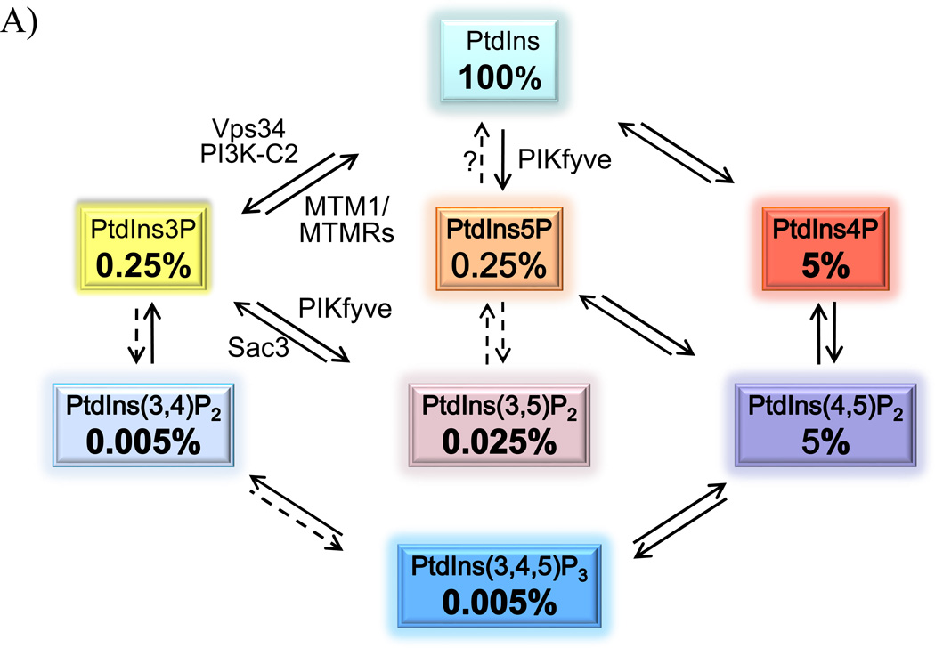
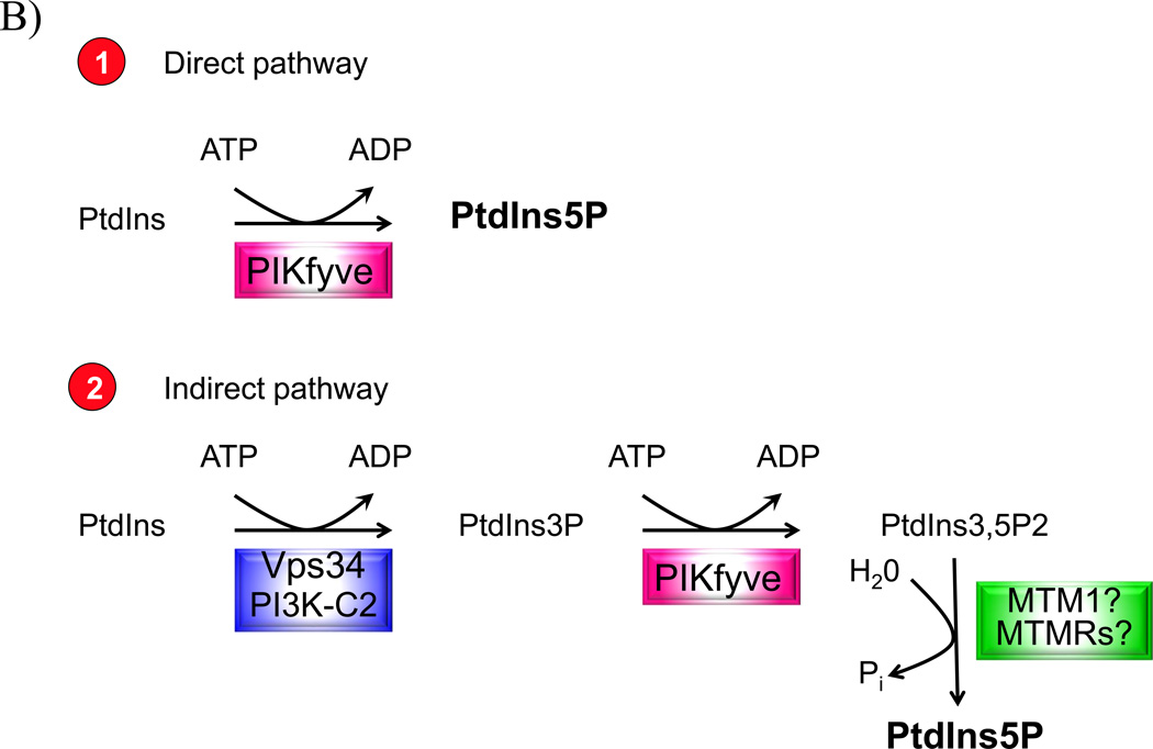
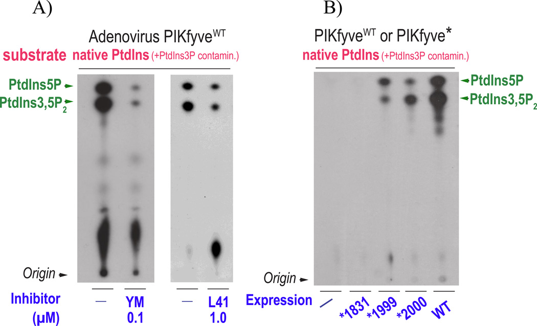
Recently, we have presented data supporting the notion that PIKfyve not only produces the majority of constitutive phosphatidylinositol 5-phosphate (PtdIns5P) in mammalian cells but that it does so through direct synthesis from PtdIns. Another group, albeit obtaining similar data, suggests an alternative pathway whereby the low-abundance PtdIns(3,5)P2 undergoes hydrolysis by unidentified 3-phosphatases, thereby serving as a precursor for most of PtdIns5P. Here, we review the experimental evidence supporting constitutive synthesis of PtdIns5P from PtdIns by PIKfyve. We further emphasize that the experiments presented in support of the alternative pathway are also compatible with a direct mechanism for PIKfyve-catalyzed synthesis of PtdIns5P. While agreeing with the authors that constitutive PtdIns5P could theoretically be produced from PtdIns(3,5)P2 by 3-dephosphorylation, we argue that until direct evidence for such an alternative pathway is obtained, we should adhere to the existing experimental evidence and quantitative considerations, which favor direct PIKfyve-catalyzed synthesis for most constitutive PtdIns5P.

Keywords: 3-phosphatases; PIKfyve; PtdIns(3,5)P2; PtdIns(3,5)P2-PtdIns3P conversion; PtdIns3P; direct PtdIns5P synthesis; phosphoinositides.


Conflict of interest statement

Conflict of Interest: The authors have no conflict of interest.

Figures

Figure 1
Figure 1
A: The seven PIs and their relative abundance in resting mammalian cells. The approximate relative abundance is derived from our work [23, 87] and that of others [88,89] in various resting mammalian cells. Indicated are experimentally confirmed cellular pathways involved in PI production (solid arrows). In vitro established conversions are indicated by dashed arrows. Only the enzymes relevant for this discussion are denoted. B: The two pathways for PIKfyve-catalyzed synthesis of constitutive PtdIns5P in resting mammalian cells. The direct pathway (1), for which evidence is detailed in this paper, consumes one mol ATP and uses PtdIns substrate that is in huge excess. The indirect pathway (2), proposed by the authors [32, 36], consumes 2 PIKfymol ATP and incorporates a third reaction of 3-dephosphorylation of the PtdIns(3,5)P2 substrate that in quiescent cells constitutes ~1/10 of the product PtdIns5P. The enzyme(s) involved in the 3-dephosphorylation reaction of PtdIns(3,5)P2 is(are) still unidentified.
Figure 1
Figure 1
A: The seven PIs and their relative abundance in resting mammalian cells. The approximate relative abundance is derived from our work [23, 87] and that of others [88,89] in various resting mammalian cells. Indicated are experimentally confirmed cellular pathways involved in PI production (solid arrows). In vitro established conversions are indicated by dashed arrows. Only the enzymes relevant for this discussion are denoted. B: The two pathways for PIKfyve-catalyzed synthesis of constitutive PtdIns5P in resting mammalian cells. The direct pathway (1), for which evidence is detailed in this paper, consumes one mol ATP and uses PtdIns substrate that is in huge excess. The indirect pathway (2), proposed by the authors [32, 36], consumes 2 PIKfymol ATP and incorporates a third reaction of 3-dephosphorylation of the PtdIns(3,5)P2 substrate that in quiescent cells constitutes ~1/10 of the product PtdIns5P. The enzyme(s) involved in the 3-dephosphorylation reaction of PtdIns(3,5)P2 is(are) still unidentified.
Figure 2
Figure 2
Schematic model of PIKfyve association with ArPIKfyve and Sac3 in the PAS complex. The PtdIns3P - PtdIns(3,5)P2 cycling on endosomal membrane microdomains is achieved through a stable protein complex comprising PI 5-kinase PIKfyve, PI 5-phosphatase Sac3 and dimerized scaffolding regulator ArPIKfyve [40]. The complex attaches to RabGTP-early endosome platforms, enriched in PtdIns3P [90], presumed to be generated mainly by Vps34, with a possible contribution by the other two PI3K classes [11, 83, 84]. Indicated are the conserved domains in PIKfyve: FYVE, DEP, Cpn60_TCP1, CH homology/Spectrin repeats and lipid kinase homology. The two antagonistic enzymes act in concert to regulate PtdIns3P-PtdIns(3,5)P2 cycling. Whereas ArPIKfyve and Sac3 are clearly responsible for PIKfyve-dependent synthesis of PtdIns5P from PtdIns, whether this occurs in parallel with PtdIns(3,5)P2 synthesis on endosomal membranes is unknown. Interaction mapping details are from [49, 50].
Figure 3
Figure 3
In vitro production of PtdIns(3,5)P2 and PtdIns5P is equally inhibited by PIKfyve inhibitors but differentially affected by PIKfyve mutagenesis. A: HEK293 cells transduced with adenoviral vector encoding HA-PIKfyveWT were lysed in RIPA buffer (containing two detergents, 1% NP-40 and 0.5% Na deoxycholate) and immunoprecipitated with PIKfyve antibodies. PIKfyve protein expressed from adenoviral vector is >30-fold higher than the endogenous, making specific co-immunoprecipitation of potentially PIKfyve-associated myotubularins unlikely. Immunoprecipitates adsorbed onto Protein A Sepharose beads, extensively washed (>10 times) with RIPA buffer and high-salt buffers, were preincubated for 15 min with a DMSO solvent (−) or with DMSO-dissolved inhibitors at concentrations 0.1 µM of YM201636 or 1 µM of L41 (a novel PIKfyve inhibitor, [51]). The in vitro lipid kinase assay was carried out for 15 min at 30° C in the presence of 12.5 µCi of [γ-32P]ATP (50 µM), 100 µM freshly sonicated PtdIns substrate (natural form soybean, Avanti Polar Lipids, containing small amounts of natural PtdIns3P as a contaminant) and an assay buffer [25 mM Hepes, pH 7.5, 120 mM NaCl, 2.5 mM MnCl2, 2.5 mM MgCl2, 5 mM β–glycerophosphate (phosphatase inhibitor) and 1mM dithiothreitol [20]. This ionic composition prevents any generation of PtdIns3P during the assay as we described elsewhere [46, 52]. Lipids were resolved by TLC using an acidic solvent system [65:35 (v/v) 1-propanol:2 M acetic acid]. Indicated are PtdIns(3,5)P2 and PtdIns5P products confirmed by HPLC analyses of the radioactive spots. Apparent is similar reduction of the two products consistent with their direct production by and inhibition of PIKfyve during the in vitro reaction. Data are reported in Refs. [34, 51, 52]. B: Parallel dishes of COS cells, transiently transfected with wild-type (WT) and point mutants (*) of HA-PIKfyve in pCMV5 expression vector, or with an empty vector (−), were lysed in RIPA buffer, followed by immunoprecipitation with HA antibodies and lipid separation by TLC, as described in A. Whereas production of both lipids is perturbed by mutagenesis within the predicted PIKfyve substrate-binding activation loop, apparent is greater reduction of PtdIns5P compared to PtdIns(3,5)P2 with PIKfyveK2000E and, vice versa, greater reduction of PtdIns(3,5)P2 compared to PtdIns5P, with PIKfyveK1999E. Note that under identical immunoprecipitation conditions, synthesis of the two lipids is selectively affected, nullifying hydrolysis by hypothetical 3-phosphatases underlying the observed differences. These data support direct synthesis of both PtdIns(3,5)P2 and PtdIns5P by PIKfyve. No production of either lipid is seen by the PIKfyveK1831E mutant that harbors mutation in the predicted ATP-binding lysine [17]. Modified from [20].
Figure 4
Figure 4
Differential effect between PIKfyveK1999 and PIKfyveK2000 on cell morphology. A,C: COS cells transiently transfected with pEGFP-PIKfyveK2000E and pEGFP-PIKfyveK1999E were observed by fluorescence microscopy based on GFP fluorescence signals. PIKfyveK1999E mutant, exhibiting greatly perturbed PtdIns(3,5)P2 synthesis (shown in Fig. 3B) and binding to PtdIns3P-enriched liposomes vs. wild-type, examined by in vitro lipid kinase activity and liposome-binding assays [54], respectively, provokes the characteristic aberrant phenotype in the form of cytoplasmic vacuolation. The PIKfyveK2000E mutant, with preferentially impaired PtdIns5P synthesis vs. PtdIns(3,5)P2 (shown in Fig. 3B) and intact binding to PtdIns3P-enriched liposomes [54], does not trigger an aberrant phenotype. B,D: phase-contrast images of A and C, respectively. Modified from [20].

Comment on

References

    1. Di Paolo G, De Camilli P. Phosphoinositides in cell regulation and membrane dynamics. Nature. 2006;443:651–657. - PubMed
    1. Shisheva A. PIKfyve and its lipid products in health and in sickness. Curr Top Microbiol Immunol. 2012;362:127–162. - PubMed
    1. Hakim S, Bertucci MC, Conduit SE, Vuong DL, et al. Inositol polyphosphate phosphatases in human disease. Curr Top Microbiol Immunol. 2012;362:247–314. - PubMed
    1. Balla T. Phosphoinositides: tiny lipids with giant impact on cell regulation. Physiol Rev. 2013;93:1019–1137. - PMC - PubMed
    1. Viaud J, Lagarrigue F, Ramel D, Allart S, et al. Phosphatidylinositol 5-phosphate regulates invasion through binding and activation of Tiam1. Nat Commun. 2014;5:4080. - PubMed

MeSH terms

Substances