

2,3,7,8-Tetrachlorodibenzo-p-dioxin (TCDD) inhibits human ovarian cancer cell proliferation
- PMID: 25404385
- PMCID: PMC5367904
- DOI: 10.1007/s13402-014-0206-4
2,3,7,8-Tetrachlorodibenzo-p-dioxin (TCDD) inhibits human ovarian cancer cell proliferation
Abstract
Purpose: The aryl hydrocarbon receptor (AhR), a ligand-activated transcription factor, mediates a broad spectrum of biological processes, including ovarian growth and ovulation. Recently, we found that an endogenous AhR ligand (ITE) can inhibit ovarian cancer proliferation and migration via the AhR. Here, we tested whether 2,3,7,8-tetrachlorodibenzo-p-dioxin (TCDD, an exogenous AhR ligand) may exert similar anti-ovarian cancer activities using human ovarian cancer and non-cancerous human ovarian surface epithelial cells.
Methods: Two human ovarian cancer cell lines (SKOV-3 and OVCAR-3) and one human ovarian surface epithelial cell line (IOSE-385) were used. Cell proliferation and migration activities were determined using crystal violet and FluoroBlok insert system assays, respectively. AhR protein expression was assessed by Western blotting. Expression of cytochrome P450, family 1, member A1 (CYP1A1) and member B1 (CYP1B1) mRNA was assessed by qPCR. Small interfering RNAs (siRNAs) were used to knock down AhR expression.
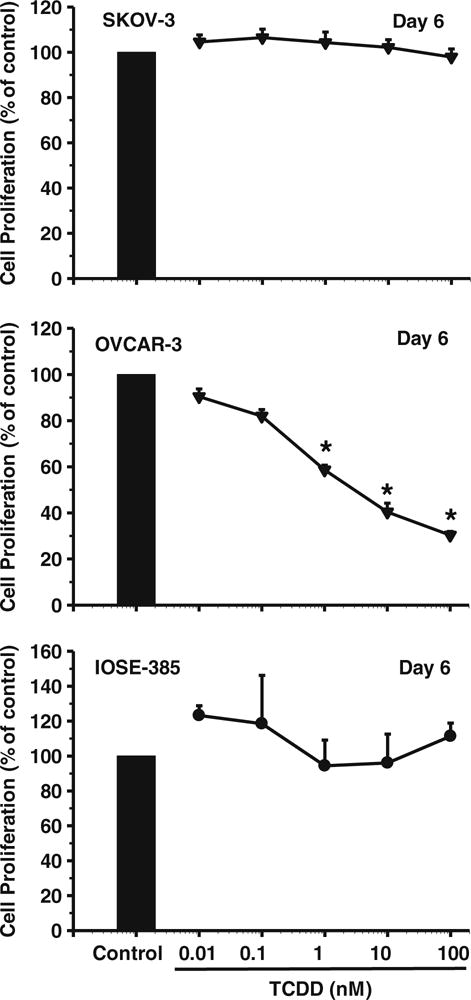
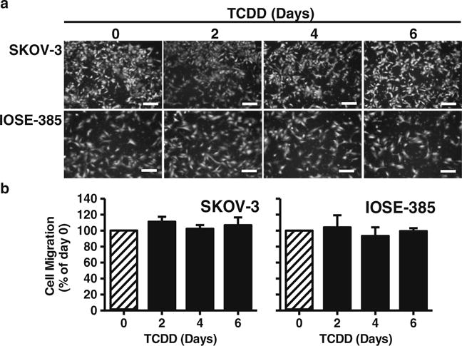
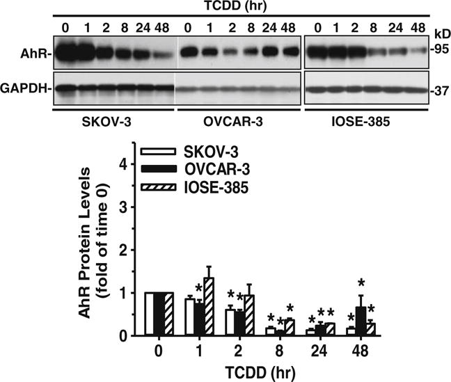
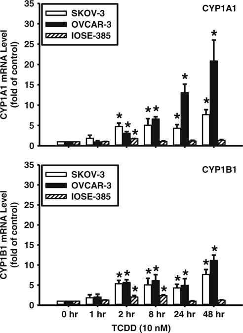
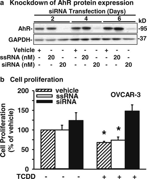
Results: We found that TCDD dose-dependently suppressed OVCAR-3 cell proliferation, with a maximum effect (~70% reduction) at 100 nM. However, TCDD did not affect SKOV-3 and IOSE-385 cell proliferation and migration. The estimated IC50 of TCDD for inhibiting OVCAR-3 cell proliferation was 4.6 nM. At 10 nM, TCDD time-dependently decreased AhR protein levels, while it significantly increased CYP1A1 and CYP1B1 mRNA levels in SKOV-3, OVCAR-3 and IOSE-385 cells, indicating activation of AhR signaling. siRNA-mediated AhR knockdown readily blocked TCDD-mediated suppression of OVCAR-3 cell proliferation.
Conclusion: Our data indicate that TCDD can suppress human ovarian cancer cell proliferation via the AhR signaling pathway and that TCDD exhibits an anti-proliferative activity in at least a subset of human ovarian cancer cells.
Conflict of interest statement
Figures
Similar articles
-
Activation of the aryl hydrocarbon receptor pathway enhances cancer cell invasion by upregulating the MMP expression and is associated with poor prognosis in upper urinary tract urothelial cancer.Carcinogenesis. 2010 Feb;31(2):287-95. doi: 10.1093/carcin/bgp222. Epub 2009 Sep 15. Carcinogenesis. 2010. PMID: 19755661
-
Aryl hydrocarbon receptor pathway activation enhances gastric cancer cell invasiveness likely through a c-Jun-dependent induction of matrix metalloproteinase-9.BMC Cell Biol. 2009 Apr 16;10:27. doi: 10.1186/1471-2121-10-27. BMC Cell Biol. 2009. PMID: 19371443 Free PMC article.
-
An endogenous aryl hydrocarbon receptor ligand inhibits proliferation and migration of human ovarian cancer cells.Cancer Lett. 2013 Oct 28;340(1):63-71. doi: 10.1016/j.canlet.2013.06.026. Epub 2013 Jul 9. Cancer Lett. 2013. PMID: 23851185 Free PMC article.
-
Characterization of the aryl hydrocarbon receptor and aryl hydrocarbon responsiveness in human ovarian carcinoma cell lines.Cancer Res. 1993 Apr 15;53(8):1802-7. Cancer Res. 1993. PMID: 8385571
-
The role of AHR-inducible cytochrome P450s in metabolism of polyunsaturated fatty acids.Drug Metab Rev. 2016 Aug;48(3):342-50. doi: 10.1080/03602532.2016.1197240. Epub 2016 Jun 30. Drug Metab Rev. 2016. PMID: 27358009 Free PMC article. Review.
Cited by
-
A bioassay to measure energy metabolism in mouse colonic crypts, organoids, and sorted stem cells.Am J Physiol Gastrointest Liver Physiol. 2015 Jul 1;309(1):G1-9. doi: 10.1152/ajpgi.00052.2015. Epub 2015 May 14. Am J Physiol Gastrointest Liver Physiol. 2015. PMID: 25977509 Free PMC article.
-
AhR and Cancer: From Gene Profiling to Targeted Therapy.Int J Mol Sci. 2021 Jan 13;22(2):752. doi: 10.3390/ijms22020752. Int J Mol Sci. 2021. PMID: 33451095 Free PMC article. Review.
-
INHIBITORY EFFECT OF LYCOPENE AGAINST THE GROWTH OF HUMAN GASTRIC CANCER CELLS.Afr J Tradit Complement Altern Med. 2016 Jul 3;13(4):184-190. doi: 10.21010/ajtcam.v13i4.24. eCollection 2016. Afr J Tradit Complement Altern Med. 2016. PMID: 28852735 Free PMC article.
-
Proper modulation of AHR signaling is necessary for establishing neural connectivity and oligodendrocyte precursor cell development in the embryonic zebrafish brain.Front Mol Neurosci. 2022 Nov 29;15:1032302. doi: 10.3389/fnmol.2022.1032302. eCollection 2022. Front Mol Neurosci. 2022. PMID: 36523606 Free PMC article.
-
Polymorphisms within the Boule Gene Detected by Tetra-Primer Amplification Refractory Mutation System PCR (T-ARMS-PCR) are Significantly Associated with Goat Litter Size.Animals (Basel). 2019 Nov 1;9(11):910. doi: 10.3390/ani9110910. Animals (Basel). 2019. PMID: 31683986 Free PMC article.
References
-
- Yap TA, Carden CP, Kaye SB. Beyond chemotherapy: targeted therapies in ovarian cancer. Nat Rev Cancer. 2009;9:167–181. - PubMed
-
- Di J, Duiveman-de Boer T, Zusterzeel PL, Fig CG, Massuger LF, Torensma R. The stem cell markers Oct4A, Nanog and c-Myc are expressed in ascites cells and tumor tissue of ovarian cancer patients. Cell Oncol. 2013;36:363–374. - PubMed
-
- Krijgsman O, Israeli D, van Essen HF, Eijk PP, Berens ML, Mellink CH, Nieuwint AW, Weiss MM, Steenbergen RD, Meijer GA, Ylstra B. Detection limits of DNA copy number alterations in heterogeneous cell populations. Cell Oncol. 2013;36:27–36. - PubMed
-
- Safe S, McDougal A. Mechanism of action and development of selective aryl hydrocarbon receptor modulators for treatment of hormone-dependent cancers (review) Int J Oncol. 2002;20:1123–1128. - PubMed
Publication types
MeSH terms
Substances
Grants and funding
LinkOut - more resources
Full Text Sources
Other Literature Sources
Medical
