. 2015 Feb;197(3):542-52.
doi: 10.1128/JB.02136-14. Epub 2014 Nov 17.

Escherichia coli SecG is required for residual export mediated by mutant signal sequences and for SecY-SecE complex stability

Affiliations

Escherichia coli SecG is required for residual export mediated by mutant signal sequences and for SecY-SecE complex stability

Dominique Belin et al. J Bacteriol. 2015 Feb.

Abstract

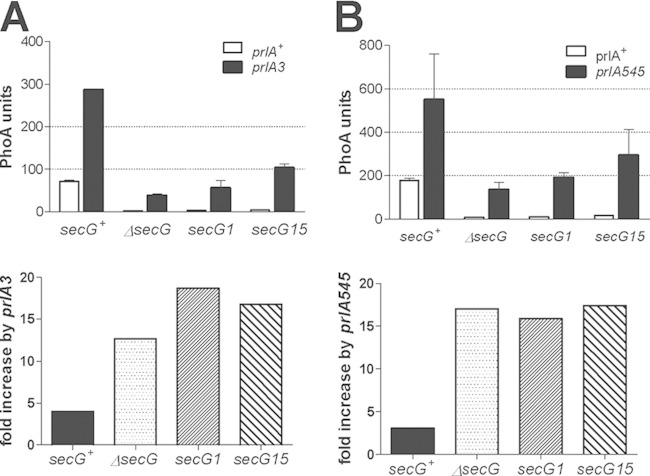
Protein export to the bacterial periplasm is achieved by SecYEG, an inner membrane heterotrimer. SecY and SecE are encoded by essential genes, while SecG is not essential for growth under standard laboratory conditions. Using a quantitative and sensitive export assay, we show that SecG plays a critical role for the residual export mediated by mutant signal sequences; the magnitude of this effect is not proportional to the strength of the export defect. In contrast, export mediated by wild-type signal sequences is only barely retarded in the absence of SecG. When probed with mutant signal sequences, secG loss of function mutations display a phenotype opposite to that of prlA mutations in secY. The analysis of secG and prlA single and double mutant strains shows that the increased export conferred by several prlA alleles is enhanced in the absence of SecG. Several combinations of prlA alleles with a secG deletion cannot be easily constructed. This synthetic phenotype is conditional, indicating that cells can adapt to the presence of both alleles. The biochemical basis of this phenomenon is linked to the stability of the SecYE dimer in solubilized membranes. With prlA alleles that can be normally introduced in a secG deletion strain, SecG has only a limited effect on the stability of the SecYE dimer. With the other prlA alleles, the SecYE dimer can often be detected only in the presence of SecG. A possible role for the maintenance of SecG during evolution is proposed.


Figures

FIG 1
FIG 1
Effects of secG mutations on PhoA export. The indicated wild-type and mutant signal sequences were fused to the mature portion of PhoA and expressed from the arabinose PBAD promoter. The kinetics of PhoA export after induction with 0.2% arabinose was measured in isogenic strains carrying the indicated secG alleles (DB617 to DB620). Each curve represents the average results from three independent cultures; error bars indicate the standard deviations (SD).
FIG 2
FIG 2
The additional export defect in ΔsecG strains does not correlate with the intrinsic defect of individual signal sequence mutations. PhoA export was measured in cultures induced for 60 min. For each signal sequence, the activities in the secG+ and ΔsecG strains (DB617 and DB618) are connected by a line. With the three wild-type signal sequences (*), as well as with the RbsB16 pseudorevertant (S16H, Ψ), the horizontal lines reflect an equivalent export in the presence and in the absence of SecG.
FIG 3
FIG 3
Kinetics of export measured by pulse-chase and immunoprecipitation. (A) Kinetics of MalE and PhoA export in secG+ and ΔsecG strains. The proteins, expressed from the chromosomal genes, contained either the wild-type alleles or the indicated signal sequence mutations. Cells were pulse-labeled at 37°C for 30 s and chased for 3 and 10 min. Lysates were immunoprecipitated with anti-MalE or anti-PhoA antibodies. The percentages of mature proteins were calculated from the intensities of the mature and precursor bands. The values represent the averages from two independent cultures; the error bars indicate ranges. (B) Hybrid proteins containing the indicated signal sequences fused to the mature portion of OmpA were expressed from the PBAD promoter in strain DB686 (secG+) or DB685 (ΔsecG), in which the chromosomal ompA gene is deleted. After 5 min of induction with 0.2% arabinose, the cultures were pulse-labeled for 20 s and chased for 10 min. Lysates were immunoprecipitated with anti-OmpA antibodies. The values represent the percentages of mature OmpA and are the averages from three independent cultures; error bars indicate the SD.
FIG 4
FIG 4
Combined effects of secG and prlA mutations on PhoA export. The MalE14 mutant signal sequence was fused to the mature portion of PhoA and expressed from the arabinose PBAD promoter (pDB613). The level of PhoA export 60 min after induction with 0.2% arabinose was measured in isogenic strains derived from DB687 (ΔsecG). (A) prlA3 mutant strain; (B) prlA545 mutant strain. The indicated secG alleles were expressed from pACYC184-derived plasmids (27). The prlA alleles were introduced by cotransduction with the gspA::Tn10 marker. Each value represents the average of results from two to four independent cultures assayed twice; error bars indicate the SD. In the bottom panels, the ratios of mean PhoA export in the prlA mutant and prlA+ strains are indicated.
FIG 5
FIG 5
Transduction frequencies of the ΔsecG allele in prlA strains. Representative members of two sets of prlA alleles (12, 23) were analyzed in each panel. All strains are derivatives of DB504 that contain the gspA::Tn10 marker and the indicated prlA alleles. Transductions were carried out with P1 lysates grown on strain DB638, to introduce the secG null allele, and on strain CAG12131, to determine the transduction efficiency of each culture. The numbers of Knr secG null transductants were normalized to those of Knr leuO transductants. Different lysates grown on CAG12131 were used in each panel. Each value represents the average of results from three independent cultures; error bars indicate the range.
FIG 6
FIG 6
Enhanced effects of some prlA mutations on PhoA export in the absence of secG. The prlA alleles were introduced in DB504 by cotransduction with the gspA::Tn10 marker, and pDB613 was introduced by transformation. KnR secG null and control Knr leuO transductants were isolated, and PhoA export was assayed 60 min after induction with 0.2% arabinose. Each value represents the average result for three independent transductants assayed twice; error bars indicate the SD. In the bottom panels, the ratios of mean PhoA export in the prlA mutant and prlA+ strains are indicated.
FIG 7
FIG 7
Stability of the different SecYE dimers in solubilized membranes. DB559 cells carrying derivatives of pBAD-HisEYG were induced and labeled with [35S]methionine as described previously (27). Isolated membranes were solubilized by dodecylmaltoside in the presence of E. coli polar lipids. His-tagged SecE was purified on Ni2+-agarose. The complexes were either kept all the time at 4°C or washed once at 37°C for 5 min. (A) Left panel, comparison of SecY+, PrlA517 (G81E), and PrlA545 (A71V). Right panel, cultures labeled as described for the preparation of membranes or chased for an additional 5 min were lysed and immunoprecipitated with an anti-SecY antibody; the amounts of accumulated SecY were normalized to that detected at the end of the labeling period with the SecY+-SecG+ encoding plasmid. (B) Comparison of SecY+, PrlA7 (L407R), and PrlA207 (I278S). While experiments performed at 4°C were highly reproducible, binding of SecY+ at 37°C in the absence of SecG was more variable, as can be seen by comparison with results shown in panel A. (C) Autoradiogram of part of the experiments quantified in panel B.
FIG 8
FIG 8
Localization of the prlA alleles on the crystal structure of the M. jannaschii SecYEG complex. Top, lateral view from the membrane plane; bottom, from the cytoplasm. Each subunit is color coded and shown as a ribbon. The side chains of residues modified by prlA mutations are indicated by their volume. Residues that interfere with the introduction of a ΔsecG allele by P1 transduction are shown in red. Residues that do not affect the introduction of a ΔsecG allele are shown in green. The structure (PDB ID 1RHZ [3]) was drawn with DeepViewSwiss-Pdbviewer v4.0.4.

Similar articles

Cited by

References

    1. Veenendaal AK, van der Does C, Driessen AJ. 2004. The protein-conducting channel SecYEG. Biochim Biophys Acta 1694:81–95. doi:10.1016/j.bbamcr.2004.02.009. - DOI - PubMed
    1. Rusch SL, Kendall DA. 2007. Oligomeric states of the SecA and SecYEG core components of the bacterial Sec translocon. Biochim Biophys Acta 1768:5–12. doi:10.1016/j.bbamem.2006.08.013. - DOI - PMC - PubMed
    1. Van den Berg B, Clemons WM Jr, Collinson I, Modis Y, Hartmann E, Harrison SC, Rapoport TA. 2004. X-ray structure of a protein-conducting channel. Nature 427:36–44. doi:10.1038/nature02218. - DOI - PubMed
    1. Clemons WM Jr, Menetret JF, Akey CW, Rapoport TA. 2004. Structural insight into the protein translocation channel. Curr Opin Struct Biol 14:390–396. doi:10.1016/j.sbi.2004.07.006. - DOI - PubMed
    1. Pohlschroder M, Prinz WA, Hartmann E, Beckwith J. 1997. Protein translocation in the three domains of life: variations on a theme. Cell 91:563–566. doi:10.1016/S0092-8674(00)80443-2. - DOI - PubMed

Publication types

MeSH terms

Associated data

LinkOut - more resources