. 2015 Feb;197(3):553-62.
doi: 10.1128/JB.02413-14. Epub 2014 Nov 17.

Utilization of host iron sources by Corynebacterium diphtheriae: multiple hemoglobin-binding proteins are essential for the use of iron from the hemoglobin-haptoglobin complex

Affiliations

Utilization of host iron sources by Corynebacterium diphtheriae: multiple hemoglobin-binding proteins are essential for the use of iron from the hemoglobin-haptoglobin complex

Courtni E Allen et al. J Bacteriol. 2015 Feb.

Abstract

The use of hemin iron by Corynebacterium diphtheriae requires the DtxR- and iron-regulated ABC hemin transporter HmuTUV and the secreted Hb-binding protein HtaA. We recently described two surface anchored proteins, ChtA and ChtC, which also bind hemin and Hb. ChtA and ChtC share structural similarities to HtaA; however, a function for ChtA and ChtC was not determined. In this study, we identified additional host iron sources that are utilized by C. diphtheriae. We show that several C. diphtheriae strains use the hemoglobin-haptoglobin (Hb-Hp) complex as an iron source. We report that an htaA deletion mutant of C. diphtheriae strain 1737 is unable to use the Hb-Hp complex as an iron source, and we further demonstrate that a chtA-chtC double mutant is also unable to use Hb-Hp iron. Single-deletion mutants of chtA or chtC use Hb-Hp iron in a manner similar to that of the wild type. These findings suggest that both HtaA and either ChtA or ChtC are essential for the use of Hb-Hp iron. Enzyme-linked immunosorbent assay (ELISA) studies show that HtaA binds the Hb-Hp complex, and the substitution of a conserved tyrosine (Y361) for alanine in HtaA results in significantly reduced binding. C. diphtheriae was also able to use human serum albumin (HSA) and myoglobin (Mb) but not hemopexin as iron sources. These studies identify a biological function for the ChtA and ChtC proteins and demonstrate that the use of the Hb-Hp complex as an iron source by C. diphtheriae requires multiple iron-regulated surface components.


Figures

FIG 1
FIG 1
(A) Native gel containing Hb, Hp, and Hb-Hp complex (at a 1:1 molar ratio). (B) Growth of various C. diphtheriae strains in low-iron mPGT media containing Hb, Hb-Hp, or Hp as the sole iron source. All strains exhibited similar levels of growth with Hp; only the Hp result for 1737 is shown (Hp was used at 35 μg/ml). Growth with Hp is significantly reduced relative to growth with Hb-Hp for all strains. Values represent the means from three independent experiments (±SD).
FIG 2
FIG 2
(A) Genetic map of the hmu gene cluster. Arrows indicate direction of transcription. CR domains for HtaA and HtaB are shown. Below the map are shown the regions deleted in the various nonpolar deletion mutants constructed from strain 1737. (B) Growth of strain 1737-wt and various nonpolar deletion mutants in low-iron media containing either Hb or Hb-Hp. The asterisk indicates that growth with Hb-Hp is significantly different from that of the wild type (P < 0.0001). (C) Growth of strain 1737htaAΔ carrying various pKN-2.6 plasmids. Growth conditions are the same as described for panel B. The asterisk indicates that growth with Hb-Hp is significantly different from that with the pKN2.6-vector (P < 0.0001). Values shown for panels B and C represent the means from three independent experiments (±SD).
FIG 3
FIG 3
(A) HtaA and derivatives were assessed for the ability to bind Hb-Hp by ELISA. GST was included as a negative control. Experiments were repeated at least three times, with similar results. Results of a representative experiment are shown. (B) Binding of HtaA and various derivatives to Hb-Hp at a 200 nM concentration is shown. Binding of HtaA-Y361A and CR2-Y361A to Hb-Hp was significantly different from binding with HtaA-wt and CR2, respectively (**, P < 0.0001). Binding of CR1 to Hb-Hp was significantly different from that with HtaA-wt (***, P < 0.05). *, HtaA does not bind Hp (HtaA-Hp). Values represent the means from three independent experiments (±SD).
FIG 4
FIG 4
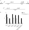
(A) Genetic map of the chtA-chtB and cirA-chtC regions. “P” (and arrow) indicates the direction of transcription from DtxR and iron-regulated promoters. “IS” indicates insertion sequences; “CR” indicates a conserved region containing hemin/Hb-binding domains. (B) Growth of 1737-wt and various nonpolar deletion mutants in low-iron media containing either Hb or Hb-Hp as the sole iron source. Values represent the means from three independent experiments (±SD). The asterisk indicates that growth with Hb-Hp is significantly different from that with the wild type (P < 0.0001).
FIG 5
FIG 5
(A) HtaA, ChtC, ChtA, and derivatives of ChtA were assessed for the ability to bind Hb by ELISA. Experiments were repeated at least three times, with similar results. Results of a representative experiment are shown. (B) The same proteins as in panel A were assessed for the ability to bind Hb-Hp by ELISA. Experiments were repeated at least three times, with similar results. Results of a representative experiment are shown. (C) Hb-Hp binding was assessed with proteins indicated in panel B at a 400 nM concentration. Values represent the means from three independent experiments (±SD). Binding of ChtA and ChtC to Hb-Hp was significantly different from that with HtaA (*, P < 0.001).
FIG 6
FIG 6
(A) Growth assay. C. diphtheriae 1737-wt and various nonpolar deletion mutants of 1737 were grown in low-iron medium containing heme-HSA, Mb, or heme-Hpx as the sole iron source. Values represent the means from three independent experiments (±SD). Asterisks indicate that growth is significantly different from that of the wild type (*, P < 0.0001; **, P < 0.0005). (B) An ELISA procedure was used to assess the binding of HtaA and associated proteins to heme-HSA. Experiments were repeated at least three times, with similar results. Results of a representative experiment are shown.
FIG 7
FIG 7
(A) Structural map of HtaA. CR domains and flanking cysteine residues (C) are indicated. TM, transmembrane region; SP, signal peptide. (B) Growth of strain 1737htaAΔ carrying various pKN2.6 plasmids. Plasmid pKNQ-cys carries the cloned htaA gene that contains Cys-Ala substitutions at all four cysteine residues. Strains were grown in low-iron medium containing either heme-HSA, Hb-Hp, or Mb as the sole iron source. Values represent the means from three independent experiments (±SD). The asterisk indicates that growth is significantly different from that with the pKN2.6-vector (P < 0.005). (C) Purified HtaA and derivatives were assessed for the ability to bind Mb by ELISA. Q-Cys is purified HtaA with Cys-Ala substitutions at all four cysteine residues. Experiments were repeated at least three times, with similar results. Results of a representative experiment are shown. (D) Mb binding was assessed with proteins indicated in panel C at a 50 nM concentration. Values represent the means from three independent experiments (±SD). Binding of HtaA, CR2, and CR1 to Mb was significantly different from that of Q-Cys (*, P < 0.005).
FIG 8
FIG 8
Model of Hb-Hp-iron utilization in C. diphtheriae. The Hb-Hp complex is composed of a Hp tetramer in which each dimer binds an αβ dimer of Hb. HtaA in association with ChtA or ChtC is proposed to bind Hb-Hp at the cell surface by an unknown mechanism (?). Following binding, hemin is either extracted or naturally released from Hb-Hp and subsequently transported into the bacterium by a cascade process involving various surface proteins, including HtaA, HtaB, and HmuT. An ABC transporter, composed of HmuU and HmuV, mobilizes heme into the cytosol, where iron is released by the action of the heme oxygenase HmuO.

Similar articles

Cited by

References

    1. Barksdale L. 1970. Corynebacterium diphtheriae and its relatives. Bacteriol Rev 34:378–422. - PMC - PubMed
    1. Collier RJ. 2001. Understanding the mode of action of diphtheria toxin: a perspective on progress during the 20th century. Toxicon 39:1793–1803. doi:10.1016/S0041-0101(01)00165-9. - DOI - PubMed
    1. Mandlik A, Swierczynski A, Das A, Ton-That H. 2007. Corynebacterium diphtheriae employs specific minor pilins to target human pharyngeal epithelial cells. Mol Microbiol 64:111–124. doi:10.1111/j.1365-2958.2007.05630.x. - DOI - PMC - PubMed
    1. Kunkle CA, Schmitt MP. 2005. Analysis of a DtxR-regulated iron transport and siderophore biosynthesis gene cluster in Corynebacterium diphtheriae. J Bacteriol 187:422–433. doi:10.1128/JB.187.2.422-433.2005. - DOI - PMC - PubMed
    1. Allen CE, Schmitt MP. 2009. HtaA is an iron-regulated hemin binding protein involved in the utilization of heme iron in Corynebacterium diphtheriae. J Bacteriol 191:2638–2648. doi:10.1128/JB.01784-08. - DOI - PMC - PubMed

Publication types