. 2015 Jan 2;290(1):350-8.
doi: 10.1074/jbc.M114.612721. Epub 2014 Nov 17.

Increased SHP-1 protein expression by high glucose levels reduces nephrin phosphorylation in podocytes

Affiliations

Increased SHP-1 protein expression by high glucose levels reduces nephrin phosphorylation in podocytes

Benoit Denhez et al. J Biol Chem. .

Abstract

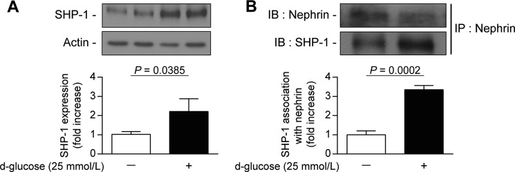
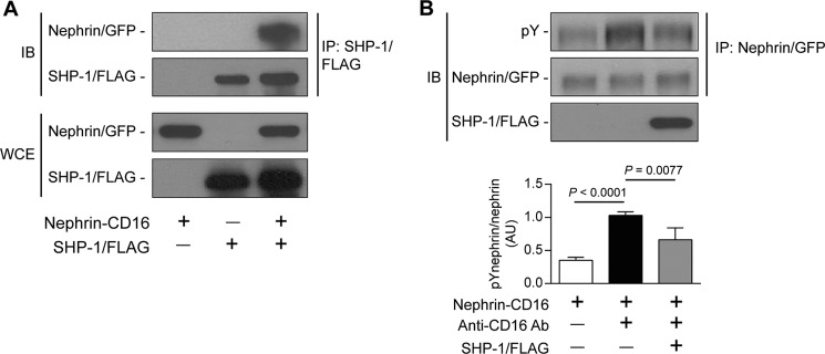
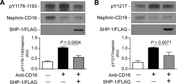
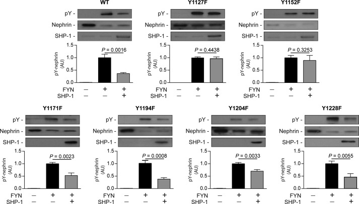
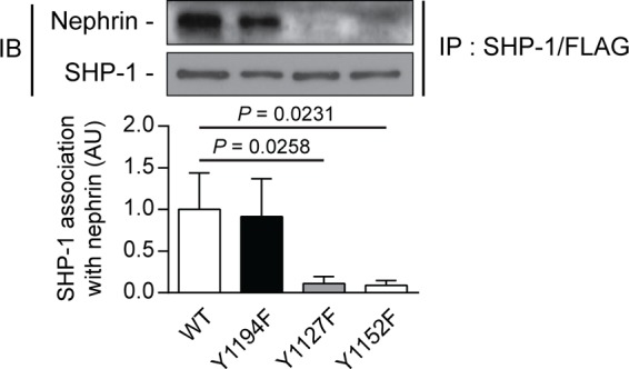
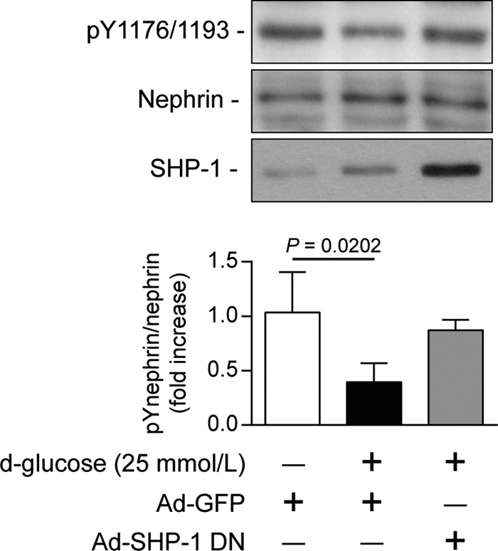
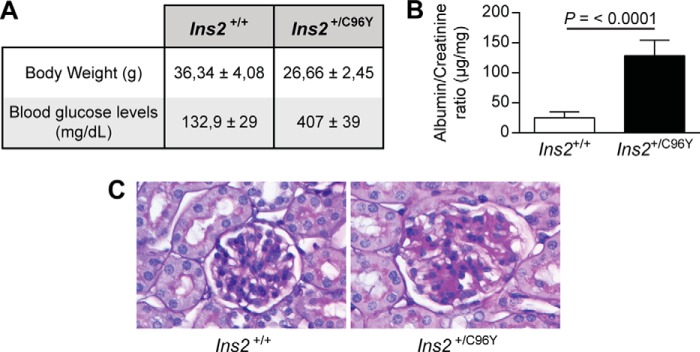
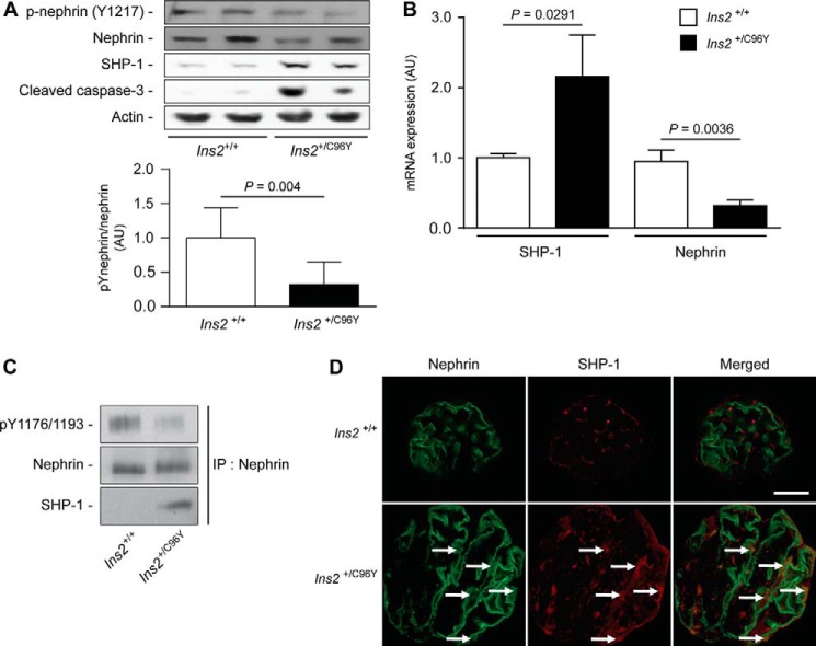
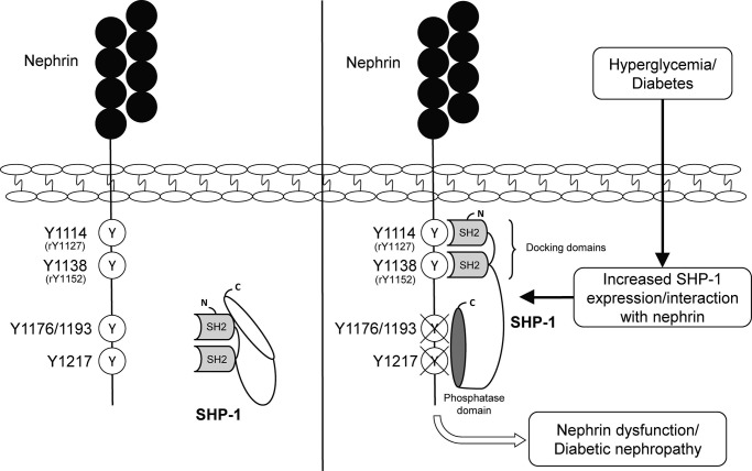
Nephrin, a critical podocyte membrane component that is reduced in diabetic nephropathy, has been shown to activate phosphotyrosine signaling pathways in human podocytes. Nephrin signaling is important to reduce cell death induced by apoptotic stimuli. We have shown previously that high glucose level exposure and diabetes increased the expression of SHP-1, causing podocyte apoptosis. SHP-1 possesses two Src homology 2 domains that serve as docking elements to dephosphorylate tyrosine residues of target proteins. However, it remains unknown whether SHP-1 interacts with nephrin and whether its elevated expression affects the nephrin phosphorylation state in diabetes. Here we show that human podocytes exposed to high glucose levels exhibited elevated expression of SHP-1, which was associated with nephrin. Coexpression of nephrin-CD16 and SHP-1 reduced nephrin tyrosine phosphorylation in transfected human embryonic kidney 293 cells. A single tyrosine-to-phenylalanine mutation revealed that rat nephrin Tyr(1127) and Tyr(1152) are required to allow SHP-1 interaction with nephrin. Overexpression of dominant negative SHP-1 in human podocytes prevented high glucose-induced reduction of nephrin phosphorylation. In vivo, immunoblot analysis demonstrated that nephrin expression and phosphorylation were decreased in glomeruli of type 1 diabetic Akita mice (Ins2(+/C96Y)) compared with control littermate mice (Ins2(+/+)), and this was associated with elevated SHP-1 and cleaved caspase-3 expression. Furthermore, immunofluorescence analysis indicated increased colocalization of SHP-1 with nephrin in diabetic mice compared with control littermates. In conclusion, our results demonstrate that high glucose exposure increases SHP-1 interaction with nephrin, causing decreased nephrin phosphorylation, which may, in turn, contribute to diabetic nephropathy.

Keywords: Diabetes; Nephrin; Nephrology; Phosphotyrosine; Podocyte; Protein-Tyrosine Phosphatase (Tyrosine Phosphatase); Src Homology 2 domain (SH2 domain).


Figures

FIGURE 1.
FIGURE 1.
Increased expression of SHP-1 by high glucose levels is associated with nephrin in podocytes. Shown are protein expression of SHP-1 (A) and coimmunoprecipitation with nephrin (B) following an immunoblot of SHP-1 and nephrin in podocytes exposed to high glucose levels (25 mmol/liters) for 96 h. Protein expression was detected by Western blot, and densitometric quantitation was measured. Results are shown as mean ± S.D. of three independent experiments. IB, immunoblot; IP, immunoprecipitation.
FIGURE 2.
FIGURE 2.
Interaction of SHP-1 and nephrin caused the reduction of nephrin phosphorylation. Shown are coimmunoprecipitation of SHP-1/FLAG with nephrin was performed in HEK cells transfected with nephrin/CD16/GFP and SHP-1/FLAG (A) and stimulated with anti-CD16 antibody (B). Protein expression of nephrin and SHP-1 and tyrosine phosphorylation of nephrin was detected by Western blot, and densitometric quantitation was measured. Results are shown as mean ± S.D. of three independent experiments. AU, arbitrary units; IB, immunoblot; IP, immunoprecipitation; WCE, whole cell extract.
FIGURE 3.
FIGURE 3.
SHP-1 reduced Tyr1176/1193 and Tyr1217 phosphorylation of nephrin. Shown is nephrin phosphorylation of Tyr1176/1193 (A) and Tyr1217 (B) in HEK cells transfected with nephrin/CD16/GFP and SHP-1/FLAG and then stimulated with anti-CD16 antibody. Protein expression was detected by Western blot, and densitometric quantitation was measured. Results are shown as mean ± S.D. of three to four independent experiments. AU, arbitrary units.
FIGURE 4.
FIGURE 4.
Nephrin mutation of Tyr1127 or Tyr1152 is required for SHP-1-induced nephrin dephosphorylation. Expression of SHP-1 and nephrin and phosphorylation of nephrin in HEK cells transfected with rat Fyn, nephrin, and SHP-1. Protein expression was detected by Western blot, and densitometric quantitation was measured. Results are shown as mean ± S.D. of three to four independent experiments. AU, arbitrary units.
FIGURE 5.
FIGURE 5.
Loss of nephrin Tyr1127 or Tyr1152 prevented SHP-1 binding with nephrin. Shown is coimmunoprecipitation of SHP-1 with either nephrin WT, Y1194F-, Y1127F-, and Y1152F-transfected HEK293T cells. Protein expression was detected by Western blot, and densitometric quantitation was measured. Results are shown as mean ± S.D. of four independent experiments. AU, arbitrary units; IB, immunoblot; IP, immunoprecipitation.
FIGURE 6.
FIGURE 6.
SHP-1 inhibition preserved nephrin phosphorylation during high glucose level exposure. Human podocytes were transduced with either Ad-GFP or Ad-dnSHP-1 and then incubated with normal (5.6 mmol/liter) or high glucose (25 mmol/liter) for 96 h. Protein expression of SHP-1 nephrin and Tyr(P)1176/1193 of nephrin were detected by Western blot, and densitometric quantitation was measured. Results are shown as mean ± S.D. of four independent experiments.
FIGURE 7.
FIGURE 7.
Blood glucose, renal function, and pathology. Shown are body weight and fasting blood glucose levels (A), urine albumin/creatinine ratio (B), and renal glomeruli stained with periodic acid Schiff for renal pathology (C) of Ins2+/+ and Ins2+/C96Y mice. Results are shown as mean ± S.D. of five animals.
FIGURE 8.
FIGURE 8.
Expression of SHP-1 and nephrin is increased markedly in glomeruli of diabetic mice. A, nephrin, Tyr(P)1217 of nephrin, SHP-1, cleaved caspase-3, and actin protein expression from the renal cortex of Ins2+/+ and Ins2+/C96Y mice. B, mRNA expression of SHP-1 and nephrin in glomeruli using laser capture microdissection. C, immunoprecipitation (IP) assays of nephrin, Tyr(P)1176/1193 of nephrin, and SHP-1 from the renal cortex of Ins2+/+ and Ins2+/C96Y mice. Protein expression was detected by Western blot, and densitometric quantitation was measured. D, immunofluorescence labeling of podocytes (green, nephrin) and SHP-1 (red) within glomeruli of Ins2+/+ and Ins2+/C96Y mice. Results are shown as mean ± S.D. of five animals. Scale bar = 20 μm. AU, arbitrary units.
FIGURE 9.
FIGURE 9.
Schematic of the SHP-1 interaction with nephrin tyrosine residues.

References

    1. Greka A., Mundel P. (2012) Cell biology and pathology of podocytes. Annu. Rev. Physiol. 74, 299–323 - PMC - PubMed
    1. Kerjaschki D. (2001) Caught flat-footed: podocyte damage and the molecular bases of focal glomerulosclerosis. J. Clin. Invest. 108, 1583–1587 - PMC - PubMed
    1. Kaplan J. M., Kim S. H., North K. N., Rennke H., Correia L. A., Tong H. Q., Mathis B. J., Rodríguez-Pérez J. C., Allen P. G., Beggs A. H., Pollak M. R. (2000) Mutations in ACTN4, encoding alpha-actinin-4, cause familial focal segmental glomerulosclerosis. Nat. Genet. 24, 251–256 - PubMed
    1. Collins A. J., Kasiske B., Herzog C., Chavers B., Foley R., Gilbertson D., Grimm R., Liu J., Louis T., Manning W., McBean M., Murray A., St Peter W., Xue J., Fan Q., Guo H., Li Q., Li S., Qiu Y., Li S., Roberts T., Skeans M., Snyder J., Solid C., Wang C., Weinhandl E., Zhang R., Arko C., Chen S.-C. C., Dalleska F., Daniels F., Dunning S., Ebben J., Frazier E., Hanzlik C., Johnson R., Sheets D., Wang X., Forrest B., Berrini D., Constantini E., Everson S., Eggers P., Agodoa L. (2006) Excerpts from the United States Renal Data System 2006 Annual Data Report. Am. J. Kidney diseases 49, S1–S296 - PubMed
    1. Sarnak M. J., Levey A. S., Schoolwerth A. C., Coresh J., Culleton B., Hamm L. L., McCullough P. A., Kasiske B. L., Kelepouris E., Klag M. J., Parfrey P., Pfeffer M., Raij L., Spinosa D. J., Wilson P. W., American Heart Association Councils on Kidney in Cardiovascular Disease, High Blood Pressure Research, Clinical Cardiology, and Epidemiology and Prevention. (2003) Kidney disease as a risk factor for development of cardiovascular disease: a statement from the American Heart Association Councils on Kidney in Cardiovascular Disease, High Blood Pressure Research, Clinical Cardiology, and Epidemiology and Prevention. Circulation 108, 2154–2169 - PubMed

Publication types

MeSH terms