Review
. 2014 Nov 3:8:123.
doi: 10.3389/fnana.2014.00123. eCollection 2014.

Structural plasticity: mechanisms and contribution to developmental psychiatric disorders

Affiliations
Review

Structural plasticity: mechanisms and contribution to developmental psychiatric disorders

Yann Bernardinelli et al. Front Neuroanat. .

Abstract

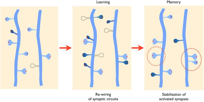
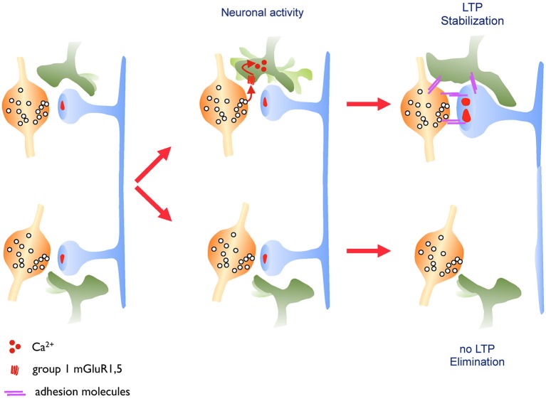
Synaptic plasticity mechanisms are usually discussed in terms of changes in synaptic strength. The capacity of excitatory synapses to rapidly modify the membrane expression of glutamate receptors in an activity-dependent manner plays a critical role in learning and memory processes by re-distributing activity within neuronal networks. Recent work has however also shown that functional plasticity properties are associated with a rewiring of synaptic connections and a selective stabilization of activated synapses. These structural aspects of plasticity have the potential to continuously modify the organization of synaptic networks and thereby introduce specificity in the wiring diagram of cortical circuits. Recent work has started to unravel some of the molecular mechanisms that underlie these properties of structural plasticity, highlighting an important role of signaling pathways that are also major candidates for contributing to developmental psychiatric disorders. We review here some of these recent advances and discuss the hypothesis that alterations of structural plasticity could represent a common mechanism contributing to the cognitive and functional defects observed in diseases such as intellectual disability, autism spectrum disorders and schizophrenia.

Keywords: astrocyte; dendritic spines; excitatory synapses; morphology; plasticity.


Figures

Figure 1
Figure 1
Activity-mediated structural plasticity. Left panel: synaptic networks are characterized by a continuous process of growth (dark blue spine) and elimination (dotted line) of dendritic spines that is developmentally regulated. Middle panel: during learning or learning related activity, baseline turnover is significantly enhanced leading to an increased formation and elimination of synaptic contacts, thus allowing remodeling and adaptation of connectivity. New spines also tend to grow in the proximity of activated synapses favoring the formation of spine clusters. Right panel: newly formed and activated synapses are preferentially stabilized (dotted circles) allowing to maintain important functional connections.
Figure 2
Figure 2
Astrocytic structural plasticity contributes to the maintenance of activated synapses. Left panel: excitatory synapses are surrounded by astrocytic processes that show a high level of motility. Middle panel: this astrocytic motility is regulated by the released neurotransmitter, which activates glutamate metabotropic receptors on the astrocytic process leading to an increased calcium flux and motility of the astrocytic process. Right panel: when driven by learning related paradigms (LTP, upper synapse), this enhanced motility leads to an increased and more stable coverage of the synapse by the astrocytic process resulting in a long-lasting stabilization of the synapse. This mechanism might involve, among other possibilities, a contribution of adhesion molecules.
Figure 3
Figure 3
Regulation of local spine growth by nitric oxide (NO). Left panel: neuronal NO synthase (nNOS), present in the postsynaptic density and tightly associated with PSD-95 and NMDA receptors, generates NO upon NMDA receptor activation and calcium influx. Middle panel: NO generated at activated synapses by LTP diffuses locally and triggers the activation of a cGMP-dependent cascade leading to the phosphorylation of the cytoskeleton regulatory protein VASP by cGMP-dependent kinase. This in turn promotes actin filament elongation and spine formation. Right panel: this mechanisms allows the formation of clusters of spine synapses around activated contacts, a phenomenon absent in nNOS deficient mice. The electron microscopy image illustrates clusters of dendritic spines (filled in rose) in the hippocampus of wild type mouse raised in an enriched environment.

References

    1. Allen K. M., Gleeson J. G., Bagrodia S., Partington M. W., MacMillan J. C., Cerione R. A., et al. (1998). PAK3 mutation in nonsyndromic X-linked mental retardation. Nat. Genet. 20, 25–30. 10.1038/1675 - DOI - PubMed
    1. Ba W., van der Raadt J., Nadif Kasri N. (2013). Rho GTPase signaling at the synapse: implications for intellectual disability. Exp. Cell Res. 319, 2368–2374. 10.1016/j.yexcr.2013.05.033 - DOI - PubMed
    1. Becker N., Wierenga C. J., Fonseca R., Bonhoeffer T., Nägerl U. V. (2008). LTD induction causes morphological changes of presynaptic boutons and reduces their contacts with spines. Neuron 60, 590–597. 10.1016/j.neuron.2008.09.018 - DOI - PubMed
    1. Bednarek E., Caroni P. (2011). β-Adducin is required for stable assembly of new synapses and improved memory upon environmental enrichment. Neuron 69, 1132–1146. 10.1016/j.neuron.2011.02.034 - DOI - PubMed
    1. Bernardinelli Y., Randall J., Janett E., Nikonenko I., Konig S., Jones E. V., et al. (2014). Activity-dependent structural plasticity of perisynaptic astrocytic domains promotes excitatory synapse stability. Curr. Biol. 24, 1679–1688. 10.1016/j.cub.2014.06.025 - DOI - PubMed

LinkOut - more resources