Review
. 2014 Nov 18;5(5):603-13.
doi: 10.5312/wjo.v5.i5.603.

Flap reconstruction of the knee: A review of current concepts and a proposed algorithm

Affiliations
Review

Flap reconstruction of the knee: A review of current concepts and a proposed algorithm

Andreas Gravvanis et al. World J Orthop. .

Abstract

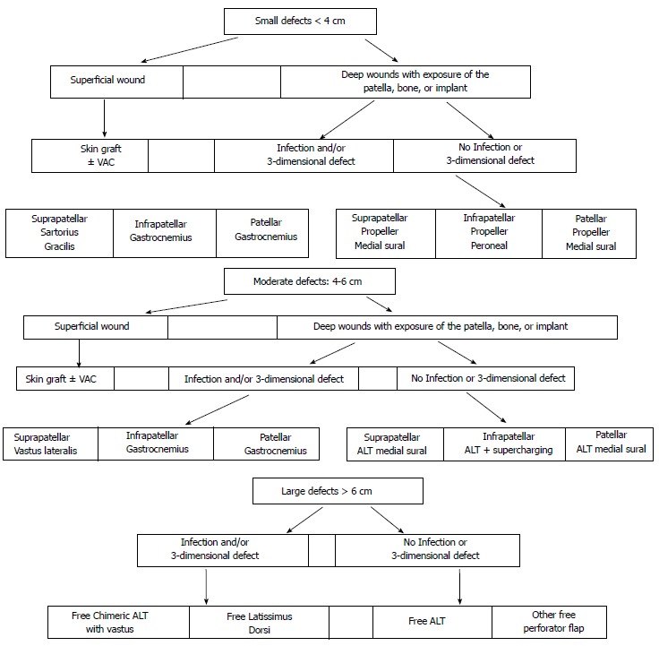
A literature search focusing on flap knee reconstruction revealed much controversy regarding the optimal management of around the knee defects. Muscle flaps are the preferred option, mainly in infected wounds. Perforator flaps have recently been introduced in knee coverage with significant advantages due to low donor morbidity and long pedicles with wide arc of rotation. In the case of free flap the choice of recipient vessels is the key point to the reconstruction. Taking the published experience into account, a reconstructive algorithm is proposed according to the size and location of the wound, the presence of infection and/or 3-dimensional defect.

Keywords: Free flap; Knee reconstruction; Local flap; Pedicled flap; Recipient vessels.


Figures

Figure 1
Figure 1
A 28-year-old man suffered a postburn unhealed wound around the right knee that was managed with pedicled gastrocnemius flap. A: The exposed patella was associated with flexion contracture; B: A medial gastrocnemius muscle flap was elevated as an island flap based on the medial sural artery. The muscle was freed from its origin and its motor nerve, as well, to allow greater arc of rotation; C: Four weeks postoperatively, gait without external supports was achieved. From the cosmetic point of view, although the muscle was denervated, a bulky pivot point was present that resulted in a cosmetic deformity.
Figure 2
Figure 2
A 24-year-old man suffered from skin and soft tissue defect over the right knee with exposure of the patella that was managed with a distally based anterolateral thigh flap. A: An anterolateral thigh flap was designed; B: The distally based flap was elevated, based on 2 perforators (arrowheads). Note the 20 cm length of the pedicle (perforator and descending branch of LCFA), as well as, the preservation of the proximal end of the LCFA (arrow) that was added to the flap; C: Satisfactory healing and contour achieved.
Figure 3
Figure 3
Patient with anterior compartment syndrome and vascular (femoral-popliteal) bypass, presented with skin and soft tissue defect over the lateral aspect of the knee. A: The descending branch of the lateral circumflex femoral vessels was dissected as recipients (arrow); B: The contralateral vastus lateralis muscle was dissected as a free flap and was anastomosed in end-to-end fashion to the distal end of the descending branch of the lateral circumflex femoral vessels; C: Satisfactory healing and contour achieved.
Figure 4
Figure 4
The use of superficial femoral vessels as recipient in free tissue transfer for around the knee defects. A: Femoral vessels skeletonised and dissected free from surrounding fat of the area; B: Penrose ”pull out” device in place facilitated the end to side anastomoses.
Figure 5
Figure 5
Infrapatellar exposure of a poorly placed plate in a 60-year-old man. A: Due to the questionable viability of the lateral gastrocnemius head, the medial head was dissected (arrow); B: The flap was medially rotated and covered the defect. The muscle was resurfaced with a split-thickness skin graft (arrow); C: Two months later, the plate was exposed in a more proximal position; D: Defect following thorough debridement; E: A free Anterolateral thigh flap based on a single perforator was raised; F: The free flap was anastomosed on end-to-end fashion to the anterior tibial vessels, and a satisfactory healing was achieved.
Figure 6
Figure 6
Knee reconstructive algorithm. VAC: Vacuum assisted closure; ALT: Alanine aminotransferase.

References

    1. Asko-Seljavaara S, Kilpi ML, Hytönen M, Sundell B. The burned hand. Early treatment and surgery of scars and contractions. Ann Chir Gynaecol. 1980;69:224–231. - PubMed
    1. Andres LA, Casey WJ, Clarke HD. Techniques in Soft Tissue Coverage Around the Knee. Techniques Knee Surg. 2009;8:119–125.
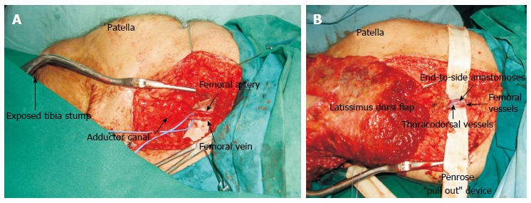
    1. Fang T, Zhang EW, Lineaweaver WC, Zhang F. Recipient vessels in the free flap reconstruction around the knee. Ann Plast Surg. 2013;71:429–433. - PubMed
    1. Panni AS, Vasso M, Cerciello S, Salgarello M. Wound complications in total knee arthroplasty. Which flap is to be used? With or without retention of prosthesis? Knee Surg Sports Traumatol Arthrosc. 2011;19:1060–1068. - PubMed
    1. Hill DS, O’Neill JK, Toms A, Watts AM. Pyoderma gangrenosum: a report of a rare complication after knee arthroplasty requiring muscle flap cover supplemented by negative pressure therapy and hyperbaric oxygen. J Plast Reconstr Aesthet Surg. 2011;64:1528–1532. - PubMed

LinkOut - more resources