. 2014 Oct;2(10):97.
doi: 10.3978/j.issn.2305-5839.2014.09.05.

Chemically defined serum-free and xeno-free media for multiple cell lineages

Affiliations

Chemically defined serum-free and xeno-free media for multiple cell lineages

Sümeyra Naz Usta et al. Ann Transl Med. 2014 Oct.

Abstract

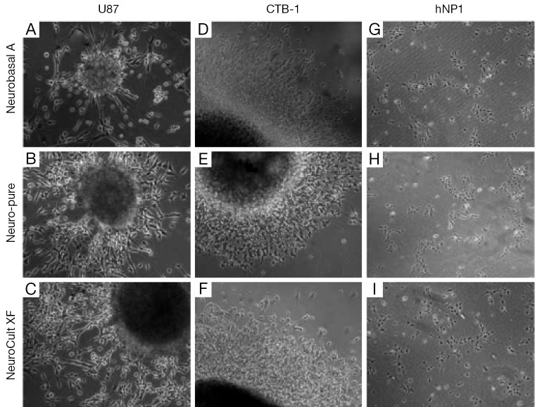
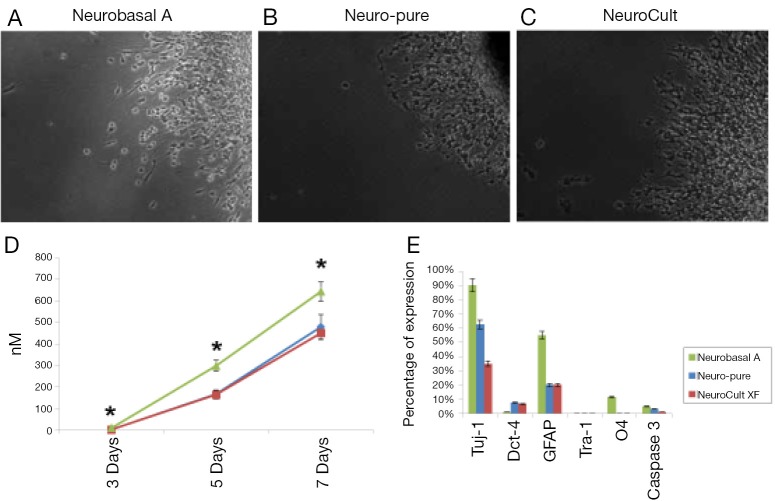
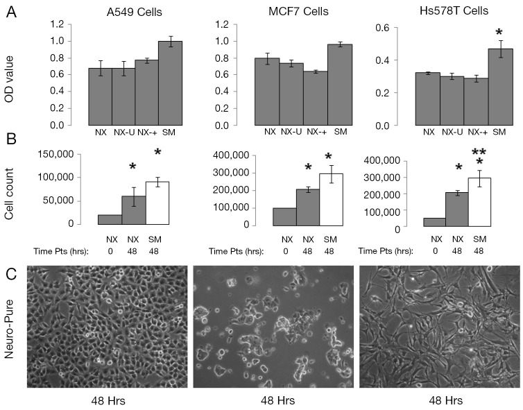
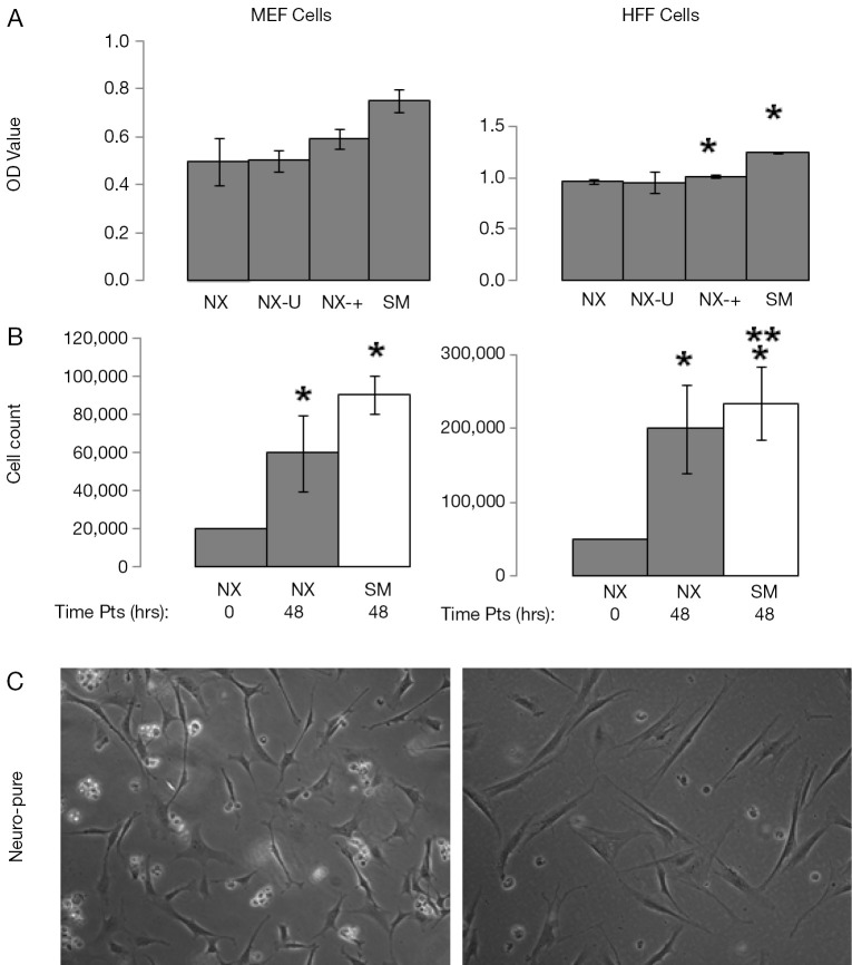
Cell culture is one of the most common methods used to recapitulate a human disease environment in a laboratory setting. Cell culture techniques are used to grow and maintain cells of various types including those derived from primary tissues, such as stem cells and cancer tumors. However, a major confounding factor with cell culture is the use of serum and animal (xeno) products in the media. The addition of animal products introduces batch and lot variations that lead to experimental variability, confounds studies with therapeutic outcomes for cultured cells, and represents a major cost associated with cell culture. Here we report a commercially available serum-free, albumin-free, and xeno free (XF) media (Neuro-Pure(TM)) that is more cost-effective than other commercial medias. Neuro-Pure was used to maintain and differentiate various cells of neuronal lineages, fibroblasts, as well as specific cancer cell lines; without the use of contaminants such serum, albumin, and animal products. Neuro-Pure allows for a controlled and reproducible cell culture environment that is applicable to translational medicine and general tissue culture.

Keywords: Serum-free; cell media; drug-development; stem cells; xeno free (XF).


Figures

Figure 1
Figure 1
Images (20×) of neurospheres generated and differentiated in Neurobasal A, Neuro-Pure and NeuroCult media. (A-C) U87; (D-F) CSC; and (G-I) hNP1 lines grown and differentiated (spontaneously) in Neurobasal A (Life Technology), Neuro-Pure (Jeevan Biosciences, Inc.), and NeuroCult-XF (Stem Cell Technologies). XF, xeno-free.
Figure 2
Figure 2
Quantitative analysis of spontaneously differentiated neurospheres derived from primary CSCs. CSCs differentiated in on lamin coated plates in (A) Neurobasal A; (B) Neuro-Pure; and (C) NeuroCult-XF media; (D) neurosphere area and neurite length were determined using NIS-Elements imaging software (Nikon). The area is the count of pixels detected in an object (computed by NIS-Elements imaging software). Neurite length was calculated as the distance between two points (computed by NIS-Elements imaging software). Measurements taken at day 3 (cells grown in Neuro-Pure ranged 0-9 µM and in Neurobasal ranged 5-17 µM) were statistically significant (P=0.0517); (E) quantitative analysis of specific cell type during differentiation was performed through immunocytochemistry. All antibodies (GFAP, O4, Tuj-1, Cleaved-Caspase-3, Oct-4, and Tra-1) were purchased from Cell Signaling Technology. Measurements from positively stained cells were taken from n=3 independent experiments with a (*P≤0.05) versus Neuro-Pure.
Figure 3
Figure 3
Growth characteristics of A549, MCF7, cancer cells grown in Neuro-Pure. (A) Cells were grown for 10 passages in Neuro-Pure under various condition; normal without additional factors (NX), unrefrigerated for 2 weeks (NX-U), with serum (NX-+) and in the recommend media with serum (SM). Cell were grown for 24 hrs and an MTT assay was conducted; (B) the same cells mentioned in (A), were grown for 10 passages under normal conditions at the initial plating density (0 hrs) and the density at 48 hours grown in Neuro-Pure (48 hrs) and at 48 hours in conventional serum medium as a control (Cont). Cells were counted manually using trypan blue and a hemocytometer; (C) images of cells taken at 20× to show morphology of cells are typical for their respective lineage. Cell counts were taken from n=3 independent experiments with a (*P≤0.05) versus Neuro-Pure [(A) 24 hrs and (B) 0 hrs], while (**P≤0.05) versus Neuro-Pure at the time point of 48 hrs (B).
Figure 4
Figure 4
Growth characteristics of MEF and HFF cells grown in Neuro-Pure. (A) Cells were grown in Neuro-Pure under various condition; normal without additional factors (NX), unrefrigerated for 2 weeks (NX-U), with serum (NX-+) and in the recommend media with serum (SM). Cell were grown for 24 hrs and an MTT assay was conducted; (B) the same cells mentioned in (A), were grown for 10 passages under normal conditions at the initial plating density (0 hrs) and the density at 48 hours grown in Neuro-Pure (48 hrs) and at 48 hours in conventional serum medium as a control (Cont). Cells were counted manually using trypan blue and a hemocytometer; (C) images of cells taken at 20× to show morphology of cells are typical for their respective lineage. Cell counts were taken from n=3 independent experiments with a (*P≤0.05) versus Neuro-Pure [(A) 24 hrs and (B) 0 hrs], while (**P≤0.05) versus Neuro-Pure at the time point of 48 hrs (B).

References

    1. Monterey MD, Szerlip NJ, Mathupala SP. Low-cost media formulation for culture of brain tumor spheroids (neurospheres). Biotechniques 2013;55:83-8. - PMC - PubMed
    1. Broedel SE, Jr, Papciak SM. The Case for Serum-Free Media. BioProcess Int 2003;1:56-8.
    1. Yao S, Chen S, Clark J, et al. Long-term self-renewal and directed differentiation of human embryonic stem cells in chemically defined conditions. Proc Natl Acad Sci U S A 2006;103:6907-12. - PMC - PubMed
    1. Tucker-Burden C, Chappa P, Krishnamoorthy M, et al. Lectins identify glycan biomarkers on glioblastoma-derived cancer stem cells. Stem Cells Dev 2012;21:2374-86. - PMC - PubMed
    1. Krishnamoorthy M, Gerwe BA, Scharer CD, et al. Ethanol alters proliferation and differentiation of normal and chromosomally abnormal human embryonic stem cell-derived neurospheres. Birth Defects Res B Dev Reprod Toxicol 2013;98:283-95. - PubMed