Review
. 2014 Sep;2(9):89.
doi: 10.3978/j.issn.2305-5839.2014.09.09.

Role of galectins in re-epithelialization of wounds

Affiliations
Review

Role of galectins in re-epithelialization of wounds

Noorjahan Panjwani. Ann Transl Med. 2014 Sep.

Abstract

Re-epithelialization is a critical contributing process in wound healing in the human body. When this process is compromised, impaired or delayed, serious disorders of wound healing may result that are painful, difficult to treat, and affect a variety of human tissues. Recent studies have demonstrated that members of the galectin class of β-galactoside-binding proteins modulate re-epithelialization of wounds by novel carbohydrate-based recognition systems. Galectins constitute a family of widely distributed carbohydrate-binding proteins with the affinity for the β-galactoside-containing glycans found on many cell surface and extracellular matrix (ECM) glycoproteins. There are 15 members of the mammalian galectin family that so far have been identified. Studies of the role of galectins in wound healing have revealed that galectin-3 promotes re-epithelialization of corneal, intestinal and skin wounds; galectin-7 promotes re-epithelialization of corneal, skin, kidney and uterine wounds; and galectins-2 and -4 promote re-epithelialization of intestinal wounds. Promising prospects for developing novel therapeutic strategies for the treatment of problematic, slow- or non-healing wounds are implicit in the findings that galectins stimulate the re-epithelialization of wounds of the cornea, skin, intestinal tract and kidney. Molecular mechanisms by which galectins modulate the process of wound healing are beginning to emerge and are described in this review.

Keywords: Wound healing; carbohydrate-based recognition; chronic wounds; galectins; non-healing wounds; re-epithelialization.


Figures
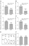
Figure 1
Figure 1
Corneal epithelial wound closure rate is significantly slower in gal3–/– mice. Corneas of gal3+/+ and gal3–/– mice with 2-mm transepithelial excimer laser ablations or alkali-burn wounds were allowed to partially heal in vivo for 16-18 h or in vitro for 20-22 h in serum-free media. At the end of the healing period, wound areas were quantified. Regardless of injury by excimer laser (A and B) or by alkali treatment (C and D) or whether corneas were allowed to heal in vivo (A and C) or in vitro (B and D), corneal epithelial wound closure rate expressed in mm2/h was significantly slower in gal3–/– mice compared with that in the gal3+/+ mice. A value of 1.0 was assigned to the healing rate of gal3+/+ corneas. The values for gal3–/– corneas are expressed as a change in healing rate with respect to gal3+/+ corneas. Wound closure rates expressed as 
mm2/h among different groups were: (A) excimer laser in vivo: gal3+/+, 0.076±0.003; gal3–/–, 0.060±0.004; (B) excimer laser in vitro: gal3+/+, 0.051±0.003; gal3–/–, 0.035±0.005; (C) alkali injury in vivo: gal3+/+, 0.182±0.003; gal3–/–, 0.150±0.008; and (D) alkali injury in vitro: gal3+/+, 0.106±0.005; gal3–/–, 0.081±0.004. Panel (E) shows outlines of remaining wound areas from one of the experiments (group: alkali injury, healing in vivo). There was no difference in wound closure rate between galectin-1+/+ and galectin-1–/– mice corneas (F). Mean ± SE of two or more experiments are shown. *, P<0.05 compared with the respective gal3+/+ group [Reprinted from (25)].
Figure 2
Figure 2
Exogenous Gal3 stimulates re-epithelialization of corneal wounds. Corneas with 2-mm alkali-burn wounds were allowed to heal in organ culture in serum-free media in the presence and absence of recombinant lectins and saccharides for 20-22 h. At the end of the healing period, wound areas were quantified and compared. (A) Galectin-3 stimulated corneal epithelial wound closure in a dose-dependent manner; (B) the stimulatory effect of exogenous galectin-3 on corneal epithelial wound closure is inhibited by β-lactose, a disaccharide that contains galactose but not by sucrose, which lacks galactose; (C) unlike galectin-3, galectin-1 did not accelerate corneal epithelial wound closure in gal3+/+ mice. A value of 1.0 was assigned to the healing rate of control corneas incubated in media alone. The value of corneas incubated in media containing galectins and saccharides is expressed as change in the healing rate with respect to the control corneas. Means ± SE of two or more experiments are shown. *, P<0.05 compared with the other three groups in panel (A); **, P<0.05 compared with gal3 (10 µg/mL) and gal3 + Suc groups in panel (B); Lac, β-lactose (0.1 M); Suc, sucrose (0.1 M) [Adapted from (25)].

References

    1. Singer AJ, Clark RA. Cutaneous wound healing. N Engl J Med 1999;341:738-46. - PubMed
    1. Raja Sivamani K, Garcia MS, et al. Wound re-epithelialization: Modulating keratinocyte migration in wound healing. Front Biosci 2007;12:2849-68. - PubMed
    1. Reinecke RD. eds. Ophthalmology annual. New York: Raven Press, 1989.
    1. Fonder MA, Lazarus GS, Cowan DA, et al. Treating the chronic wound: A practical approach to the care of nonhealing wounds and wound care dressings. J Am Acad Dermatol 2008;58:185-206. - PubMed
    1. Eaglstein WH, Falanga V. Chronic wounds. Surg Clin North Am 1997;77:689-700. - PubMed