

Editorial
doi: 10.1097/WNO.0000000000000192.
The promise of prediction: biomechanical analyses in neuro-ophthalmology
Affiliations
- PMID: 25405660
- PMCID: PMC8324323
- DOI: 10.1097/WNO.0000000000000192
Item in Clipboard
Editorial
The promise of prediction: biomechanical analyses in neuro-ophthalmology
J Neuroophthalmol.
2014 Dec.
No abstract available
Figures
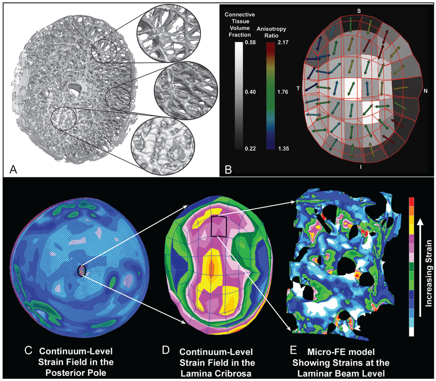
Regional differences in laminar microarchitecture in a normal eye and regional laminar density and anisotropy mapping. Characterization of the laminar microarchitecture (A) uses the element boundaries of a continuum finite mesh to partition the lamina cribrosa connective tissue into 45 subregions (B). The connective tissue volume fraction (CTVF) for each region is expressed as a percentage and mapped to a grayscale value in the background. The arrows indicate the pre-dominant orientation of the laminar beams in each region, with higher values (color-coded) indicating regions in which the beams are more highly oriented. Note that in the peripheral regions of the lamina, the beams are tethered radially into the scleral canal wall. Complexities in the posterior pole biomechanical response. C. The image shows the strain distribution in a macroscale model of the connective tissues of the posterior pole of the eye. Note that thickness variations in the sclera give rise to a nonuniform distribution of tensile strain within the scleral shell, and that the strains are lower in the sclera than in the more compliant lamina cribrosa. D. A detail is shown of the strain field within the macroscale representation of the lamina cribrosa derived from the individual mapping of laminar CTVF and beam orientation shown in (B). Although this portion of the model has been assigned regional material properties related to the amount and orientation of the laminar beams shown in (B), the continuum description represents a bulk homogenization of the specific microarchitecture in each element and only approximates the overall strain field. E. The distribution of mechanical strain at the microscale in the laminar beam microarchitecture demonstrates that strains concentrate focally in individual lamina cribrosa beams and around individual axonal pores. Adapted from Roberts et al (1) and Downs et al (2).
Comment on
-
Finite element modeling of optic chiasmal compression.J Neuroophthalmol. 2014 Dec;34(4):324-30. doi: 10.1097/WNO.0000000000000145. J Neuroophthalmol. 2014. PMID: 24978206
References
-
- Wang X, Neely AJ, McIlwain GG, Tahtali M, Lillicrap TT, Lueck CJ. Finite element modeling of optic chiasmal compression. J Neuroophthalmol. 2014. - PubMed
Publication types
MeSH terms
Grants and funding
LinkOut - more resources
Full Text Sources
Other Literature Sources
Medical
