. 2014 Nov 14;15(11):20982-96.
doi: 10.3390/ijms151120982.

Isolation and multiple differentiation potential assessment of human gingival mesenchymal stem cells

Affiliations

Isolation and multiple differentiation potential assessment of human gingival mesenchymal stem cells

Yuan Gao et al. Int J Mol Sci. .

Abstract

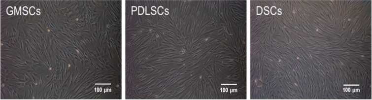
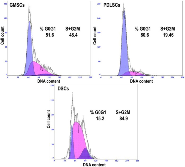
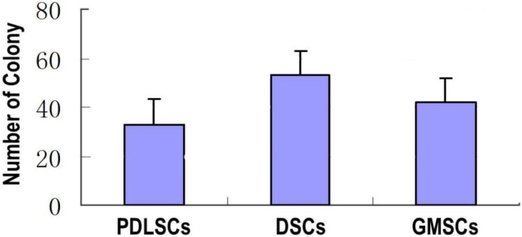
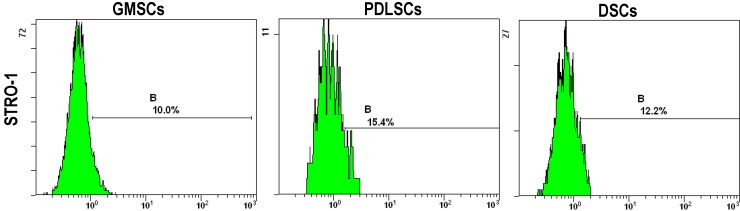
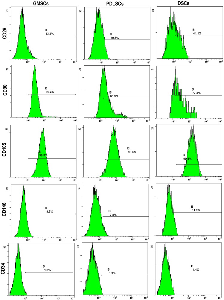
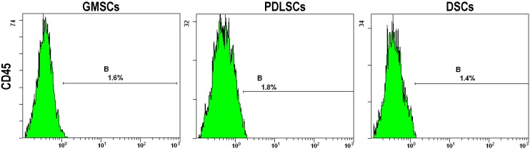
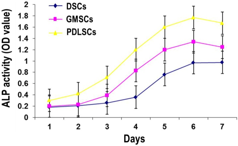
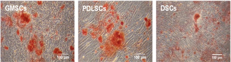
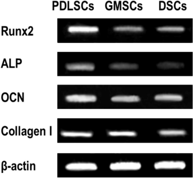
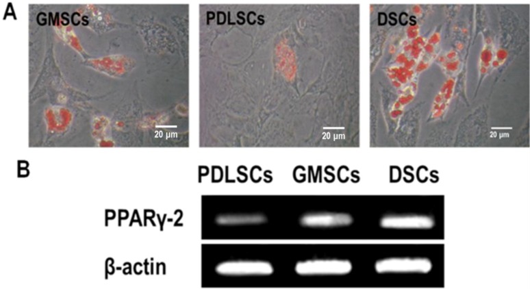
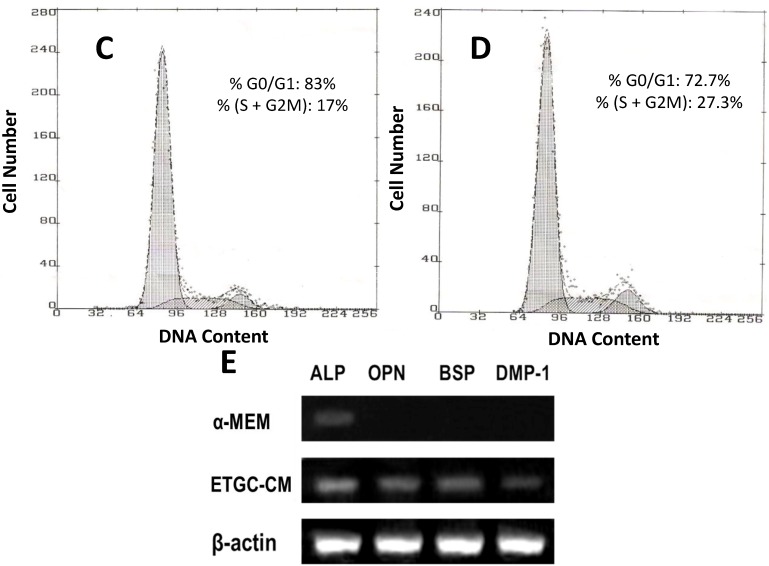
The aim of this study was to isolate human mesenchymal stem cells (MSCs) from the gingiva (GMSCs) and confirm their multiple differentiation potentials, including the odontogenic lineage. GMSCs, periodontal ligament stem cells (PDLSCs) and dermal stem cells (DSCs) cultures were analyzed for cell shape, cell cycle, colony-forming unit-fibroblast (CFU-F) and stem cell markers. Cells were then induced for osteogenic and adipogenic differentiation and analyzed for differentiation markers (alkaline phosphatase (ALP) activity, mineralization nodule formation and Runx2, ALP, osteocalcin (OCN) and collagen I expressions for the osteogenic differentiation, and lipid vacuole formation and PPARγ-2 expression for the adipogenic differentiation). Besides, the odontogenic differentiation potential of GMSCs induced with embryonic tooth germ cell-conditioned medium (ETGC-CM) was observed. GMSCs, PDLSCs and DSCs were all stromal origin. PDLSCs showed much higher osteogenic differentiation ability but lower adipogenic differentiation potential than DSCs. GMSCs showed the medial osteogenic and adipogenic differentiation potentials between those of PDLSCs and DSCs. GMSCs were capable of expressing the odontogenic genes after ETGC-CM induction. This study provides evidence that GMSCs can be used in tissue engineering/regeneration protocols as an approachable stem cell source.


Figures

Figure 1
Figure 1
Morphology of the primary mesenchymal stem cells (MSCs) established from human gingiva, periodontal ligament and dermis (P3).
Figure 2
Figure 2
Representative cell cycle distribution graphs of gingival MSCs (GMSCs), periodontal ligament stem cells (PDLSCs) and dermal stem cells (DSCs) (P3). The percentages of cells residing in the G0/G1 phase, S and G2/M phases are shown in the graphs.
Figure 3
Figure 3
The number of colony generated by GMSCs, PDLSCs and DSCs (P3). The single P3 cell suspensions were seeded in 10 cm culture dishes in α-MEM (10% fetal bovine serum (FBS)) at a density of 1 × 10³ cells/well and cultured for 14 days.
Figure 4
Figure 4
The expression of the stem cell surface markers including STRO-1, CD29, CD90, CD105 and CD146 and the leucocyte precursor markers CD45 and CD34 by GMSCs, PDLSCs and DSCs (P3). They were stained with antibodies for the markers and analyzed by flow cytometry. B indicates the expression levels of the surface markers.
Figure 4
Figure 4
The expression of the stem cell surface markers including STRO-1, CD29, CD90, CD105 and CD146 and the leucocyte precursor markers CD45 and CD34 by GMSCs, PDLSCs and DSCs (P3). They were stained with antibodies for the markers and analyzed by flow cytometry. B indicates the expression levels of the surface markers.
Figure 5
Figure 5
ALP activity of GMSCs, PDLSCs and DSCs (P3) after one to seven days of osteogenic induction.
Figure 6
Figure 6
Mineralized nodule formation by GMSCs, PDLSCs and DSCs (P3) after three weeks of osteogenic induction.
Figure 7
Figure 7
Expression of osteogenic genes including Runx2, ALP, OCN and collagen I by GMSCs, PDLSCs and DSCs (P3) after three weeks of osteogenic induction.
Figure 8
Figure 8
Adipogenic differentiation of GMSCs, PDLSCs and DSCs (P3) after four weeks of adipogenic induction. (A) Representative images of intracellular lipid vacuoles that appeared in all tested cells, as confirmed by Oil Red O staining; and (B) The expression of an adipogenic gene PPARγ detected by RT-PCR.
Figure 9
Figure 9
Odontogenic differentiation of GMSCs after two weeks of induction. (A) Representative images of embryonic Sprague-Dawley rats; (B) Representative images of tooth germ cells (TGCs). Representative cell cycle distribution graphs of GMSCs cultured with α-MEM (C) and ETGC-CM (D) for eight days; and (E) The expression of odontogenic genes detected by RT-PCR.
Figure 9
Figure 9
Odontogenic differentiation of GMSCs after two weeks of induction. (A) Representative images of embryonic Sprague-Dawley rats; (B) Representative images of tooth germ cells (TGCs). Representative cell cycle distribution graphs of GMSCs cultured with α-MEM (C) and ETGC-CM (D) for eight days; and (E) The expression of odontogenic genes detected by RT-PCR.

References

    1. Pittenger M.F., Mackay A.M., Beck S.C., Jaiswal R.K., Douglas R., Mosca J.D., Moorman M.A., Simonetti D.W., Craig S., Marshak D.R. Multilineage potential of adult human mesenchymal stem cells. Science. 1999;284:143–147. doi: 10.1126/science.284.5411.143. - DOI - PubMed
    1. Huang G.T., Gronthos S., Shi S. Mesenchymal stem cells derived from dental tissues vs. those from other sources: Their biology and role in regenerative medicine. J. Dent. Res. 2009;88:792–806. doi: 10.1177/0022034509340867. - DOI - PMC - PubMed
    1. Seo B.M., Miura M., Gronthos S., Bartold P.M., Batouli S., Brahim J., Young M., Robey P.G., Wang C.Y., Shi S. Investigation of multipotent postnatal stem cells from human periodontal ligament. Lancet. 2004;364:149–155. doi: 10.1016/S0140-6736(04)16627-0. - DOI - PubMed
    1. Graziano A., d’Aquino R., Laino G., Papaccio G. Dental pulp stem cells: A promising tool for bone regeneration. Stem Cell Rev. 2008;4:21–26. doi: 10.1007/s12015-008-9015-3. - DOI - PubMed
    1. Wang J., Wang X., Sun Z., Wang X., Yang H., Shi S., Wang S. Stem cells from human-exfoliated deciduous teeth can differentiate into dopaminergic neuron-like cells. Stem Cells Dev. 2010;19:1375–1383. doi: 10.1089/scd.2009.0258. - DOI - PMC - PubMed

LinkOut - more resources