. 2014 Nov 18;4(11):e483.
doi: 10.1038/tp.2014.124.

Hippocampal structural and functional changes associated with electroconvulsive therapy response

Affiliations

Hippocampal structural and functional changes associated with electroconvulsive therapy response

C C Abbott et al. Transl Psychiatry. .

Abstract

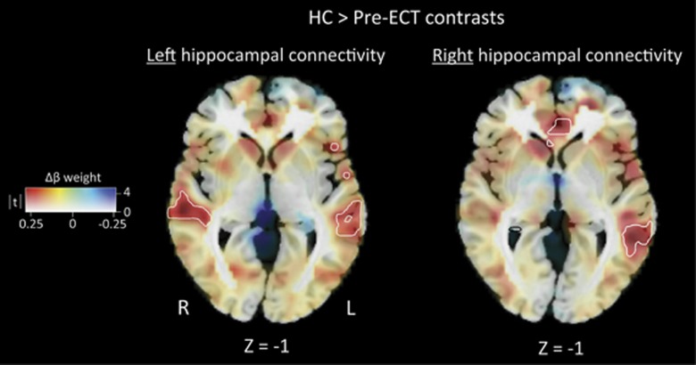
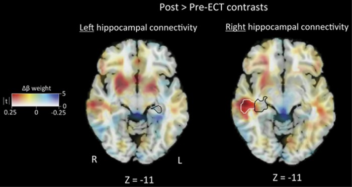
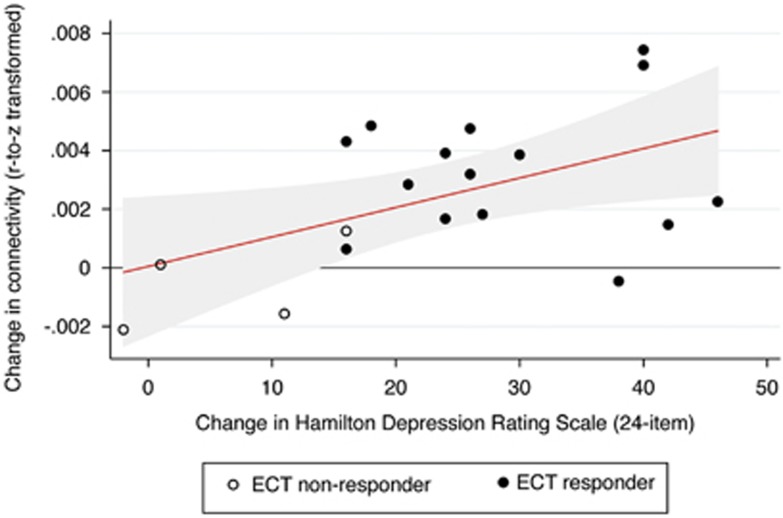
Previous animal models and structural imaging investigations have linked hippocampal neuroplasticity to electroconvulsive therapy (ECT) response, but the relationship between changes in hippocampal volume and temporal coherence in the context of ECT response is unknown. We hypothesized that ECT response would increase both hippocampal resting-state functional magnetic resonance imaging connectivity and hippocampal volumes. Patients with major depressive disorder (n=19) were scanned before and after the ECT series. Healthy, demographically matched comparisons (n=20) were scanned at one-time interval. Longitudinal changes in functional connectivity of hippocampal regions and volumes of hippocampal subfields were compared with reductions in ratings of depressive symptoms. Right hippocampal connectivity increased (normalized) after the ECT series and correlated with depressive symptom reduction. Similarly, the volumes of the right hippocampal cornu ammonis (CA2/3), dentate gyrus and subiculum regions increased, but the hippocampal subfields were unchanged relative to the comparison group. Connectivity changes were not evident in the left hippocampus, and volume changes were limited to the left CA2/3 subfields. The laterality of the right hippocampal functional connectivity and volume increases may be related to stimulus delivery method, which was predominately right unilateral in this investigation. The findings suggested that increased hippocampal functional connectivity and volumes may be biomarkers for ECT response.


Figures

Figure 1
Figure 1
The healthy comparison (HC) >pre-electroconvulsive therapy (ECT) contrast shows decreased left hippocampal connectivity in the left and right temporal lobe. Similarly, the HC >pre-ECT contrast shows decreased right hippocampal connectivity in the left medial temporal and anterior cingulate cortex. Differences in connectivity are mapped to color hue; t-statistic magnitude is mapped to transparency. White contour lines denote familywise error-corrected areas of significance with a cluster-defining threshold of P=0.005.
Figure 2
Figure 2
The post-electroconvulsive therapy (ECT) >pre-ECT contrast shows no significant differences with left hippocampal connectivity. The post-ECT >pre-ECT contrast shows increased right hippocampal connectivity among ECT responders in the left superior temporal lobe. The black contour lines denote the study-specific right hippocampal seed region.
Figure 3
Figure 3
The scatter plot and linear fit show the relationship between differences in connectivity and symptom change among all electroconvulsive therapy (ECT) patients (n=19, r=0.52, P=0.024). The red line represents the linear fit and the shaded region represents 95% confidence intervals.

References

    1. Lucassen PJ, Muller MB, Holsboer F, Bauer J, Holtrop A, Wouda J, et al. Hippocampal apoptosis in major depression is a minor event and absent from subareas at risk for glucocorticoid overexposure. Am J Pathol. 2001;158:453–468. - PMC - PubMed
    1. Gould E, Tanapat P, McEwen BS, Flugge G, Fuchs E. Proliferation of granule cell precursors in the dentate gyrus of adult monkeys is diminished by stress. Proc Natl Acad Sci USA. 1998;95:3168–3171. - PMC - PubMed
    1. Sahay A, Drew MR, Hen R. Dentate gyrus neurogenesis and depression. Prog Brain Res. 2007;163:697–722. - PubMed
    1. Steffens DC, Byrum CE, McQuoid DR, Greenberg DL, Payne ME, Blitchington TF, et al. Hippocampal volume in geriatric depression. Biol Psychiatry. 2000;48:301–309. - PubMed
    1. Sheline YI, Wang PW, Gado MH, Csernansky JG, Vannier MW. Hippocampal atrophy in recurrent major depression. Proc Nati Acad Sci USA. 1996;93:3908–3913. - PMC - PubMed

Publication types

LinkOut - more resources