Review
. 2016 Feb;22(1):83-97.
doi: 10.1177/1073858414559409. Epub 2014 Nov 18.

Cerebellar Transcranial Direct Current Stimulation (ctDCS): A Novel Approach to Understanding Cerebellar Function in Health and Disease

Affiliations
Review

Cerebellar Transcranial Direct Current Stimulation (ctDCS): A Novel Approach to Understanding Cerebellar Function in Health and Disease

Giuliana Grimaldi et al. Neuroscientist. 2016 Feb.

Abstract

The cerebellum is critical for both motor and cognitive control. Dysfunction of the cerebellum is a component of multiple neurological disorders. In recent years, interventions have been developed that aim to excite or inhibit the activity and function of the human cerebellum. Transcranial direct current stimulation of the cerebellum (ctDCS) promises to be a powerful tool for the modulation of cerebellar excitability. This technique has gained popularity in recent years as it can be used to investigate human cerebellar function, is easily delivered, is well tolerated, and has not shown serious adverse effects. Importantly, the ability of ctDCS to modify behavior makes it an interesting approach with a potential therapeutic role for neurological patients. Through both electrical and non-electrical effects (vascular, metabolic) ctDCS is thought to modify the activity of the cerebellum and alter the output from cerebellar nuclei. Physiological studies have shown a polarity-specific effect on the modulation of cerebellar-motor cortex connectivity, likely via cerebellar-thalamocortical pathways. Modeling studies that have assessed commonly used electrode montages have shown that the ctDCS-generated electric field reaches the human cerebellum with little diffusion to neighboring structures. The posterior and inferior parts of the cerebellum (i.e., lobules VI-VIII) seem particularly susceptible to modulation by ctDCS. Numerous studies have shown to date that ctDCS can modulate motor learning, and affect cognitive and emotional processes. Importantly, this intervention has a good safety profile; similar to when applied over cerebral areas. Thus, investigations have begun exploring ctDCS as a viable intervention for patients with neurological conditions.

Keywords: cerebellum; cognitive; ctDCS; direct current stimulation; emotion; language; learning; modeling; motor; plasticity; safety; transcranial; working memory.


Conflict of interest statement

Declaration of Conflicting Interests: The author(s) declared the following potential conflicts of interest with respect to the research, authorship, and/or publication of this article: GG received funds from the European Commission for the European grant “Tremor” (ICT-2007-224051). MM receives honoraria from Cambridge University Press and Springer Science+Business Media, and has received research grants from the Communauté Française, the European Commission, and the FNRS–Belgium. MM serves as editor-in-chief of The Cerebellum, Cerebellum and Ataxias, and as associate editor for the Journal of NeuroEngineering and Rehabilitation. RF and AP are shareholders of Newronika s.r.l, Milan, a spin-off company of the University of Milan and of the Fondazione IRCCS Ca’ Granda, Milan devoted to the development of neurotechnologies, including tDCS stimulators.

Figures

Figure 1.
Figure 1.
Cerebellar–primary motor cortex (M1) connectivity measure assessed by transcranial magnetic stimulation (TMS). The scheme represents the current interpretation of how the circuitry from the cerebellar cortex to M1 is engaged when assessed with TMS. A conditioning TMS pulse (CS, blue lightning) delivered over the cerebellum activates many Purkinje cells (PCs) leading to inhibition of the deep cerebellar nuclei (DCN). Since the DCN has a disynaptic excitatory connection to M1, inhibition of the DCN by PC stimulation leads to reduce excitation of M1. This inhibition is evidenced by the reduced amplitude of motor evoked potentials (MEPs) in response to test stimuli (TS, black lightning) to M1 when compared with unconditioned TS. The difference in MEP amplitudes (black–blue traces) represent the magnitude of inhibition the cerebellum (CB) is exerting over M1, a measured known as cerebellar inhibition or CBI. Please note that MEPs are recorded from a muscle typically from the hand (i.e., first dorsal interosseuos as shown in the figure) or leg using surface electromyography electrodes.
Figure 2.
Figure 2.
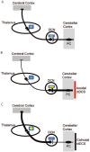
Illustration showing the current interpretation of how cerebellar transcranial direct current stimulation (ctDCS) affects the cerebellar–thalamocortical pathway. (A) Purkinje cell (PC; major neurons of the cerebellar cortex) exert physiologically an inhibition (−) over the deep cerebellar nuclei (DCN). These latter project on contralateral thalamic nuclei (+: excitatory thalamic pathway), which project themselves diffusely on the cerebral cortex. (B) Anodal ctDCS (red) is thought to increase the excitability of PC. In this model, the inhibition from the cerebellar cortex to DCN is augmented, hence reducing nucleothalamic facilitatory drive to cortical areas. (C) Cathodal ctDCS (black) decreases the activity of the cerebellar cortex. Thus, the DCN is disinhibited releasing the inhibition of the nucleothalamic drive.
Figure 3.
Figure 3.
The figure shows the electrical field strength and distribution over the cerebellum (A) when a unilateral hemispheric montage is used (B). Note the position of the active electrode (black square) targeting the lateral cerebellum (black dot represents the actual target). When using this montage, cerebellar transcranial direct current stimulation (ctDCS) delivers a fairly unilateral high electrical strength over the anterior and posterior cerebellar hemisphere. Modified from Rampersad and others (2014).
Figure 4.
Figure 4.
Results from Galea and others (2011). End-point error (degrees) are shown during baseline (Pre1, 2), adaptation (Adapt), and de-adaptation with no visual feedback (Post1, 2, 3) for the sham (black), CB (cerebellar anodal tDCS, red) and M1 (M1 anodal, blue) groups (mean ± SEM of 8 trial epochs). The shaded area represents blocks in which transcranial direct current stimulation (tDCS) was applied (Pre2, Adapt). Bar graphs insets indicate mean end-point error in degrees (±SEM) for Sham (black), CB (red), M1 (blue). For each block, separate one-way analyses of variance compared these values across groups. *P < 0.05. It is evident that anodal ctDCS led to faster acquisition (rapid error correction) whereas anodal M1 tDCS caused greater retention (longer presence of movements with the previously learned pattern).
Figure 5.
Figure 5.
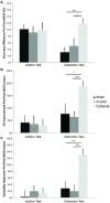
Changes in two working memory tasks (addition = PASAT and subtraction = PASST) expressed as percentage from session one (pre-stimulation) to session two (post-stimulation; n = 20). (A) This figure shows the gain in accuracy that participants’ experienced between stimulation sessions on the subtraction task, but not the addition task after cathodal stimulation only. (B) Improvement in response speed from pre-to post-stimulation. (C) Reduction in response latency variability. Participants performed calculations more quickly and paced them more consistently after cathodal, than after anodal or sham stimulation on the subtraction task only. Asterisks indicate significant differences (P < 0.05) as revealed with post hoc t tests (values are mean + 1 SEM). Modified from Pope and Miall (2012).
Figure 6.
Figure 6.
The figure shows the effect of cerebellar transcranial direct current stimulation (ctDCS) on negative emotional recognition (sadness and anger). Y-axis (arbitrary units, AU) represents trial to trial grand average of reaction times (RTs) across task stimulus presentation before and after ctDCS. X-axis represents answers, note that the X-axis graphically represents the time elapsing between the end of the task execution before stimulation and the beginning of the task execution after ctDCS. Note that anodal (left panel, solid line) and cathodal (right panel, dashed line) ctDCS both reduce baseline RTs for negative emotions. The trial-to-trial representation highlights the finding that anodal and cathodal curves differ from sham curves for negative emotions. Modified from Ferrucci and others (2013).

References

    1. Baddeley AD. 1986. Working memory. Oxford, England: Clarendon Press.
    1. Batsikadze G, Moliadze V, Paulus W, Kuo MF, Nitsche MA. 2013. Partially non-linear stimulation intensity-dependent effects of direct current stimulation on motor cortex excitability in humans. J Physiol 591(Pt 7):1987–2000. - PMC - PubMed
    1. Block HJ, Celnik P. 2012. Can cerebellar transcranial direct current stimulation become a valuable neurorehabilitation intervention? Expert Rev Neurother 12:1275–7. - PMC - PubMed
    1. Block H, Celnik P. 2013. Stimulating the cerebellum affects visuomotor adaptation but not intermanual transfer of learning. Cerebellum 12:781–93. - PMC - PubMed
    1. Boehringer A, Macher K, Dukart J, Villringer A, Pleger B. 2013. Cerebellar transcranial direct current stimulation modulates verbal working memory. Brain Stimul 6:649–53. - PubMed

Publication types

MeSH terms

LinkOut - more resources