. 2014 Dec 26;289(52):36070-88.
doi: 10.1074/jbc.M114.610535. Epub 2014 Nov 18.

Whirlin and PDZ domain-containing 7 (PDZD7) proteins are both required to form the quaternary protein complex associated with Usher syndrome type 2

Affiliations

Whirlin and PDZ domain-containing 7 (PDZD7) proteins are both required to form the quaternary protein complex associated with Usher syndrome type 2

Qian Chen et al. J Biol Chem. .

Abstract

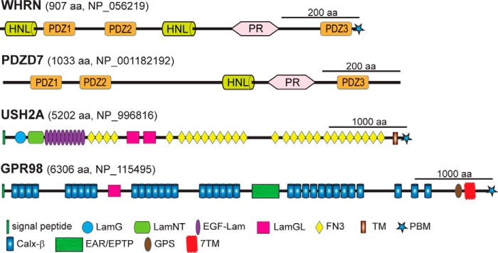
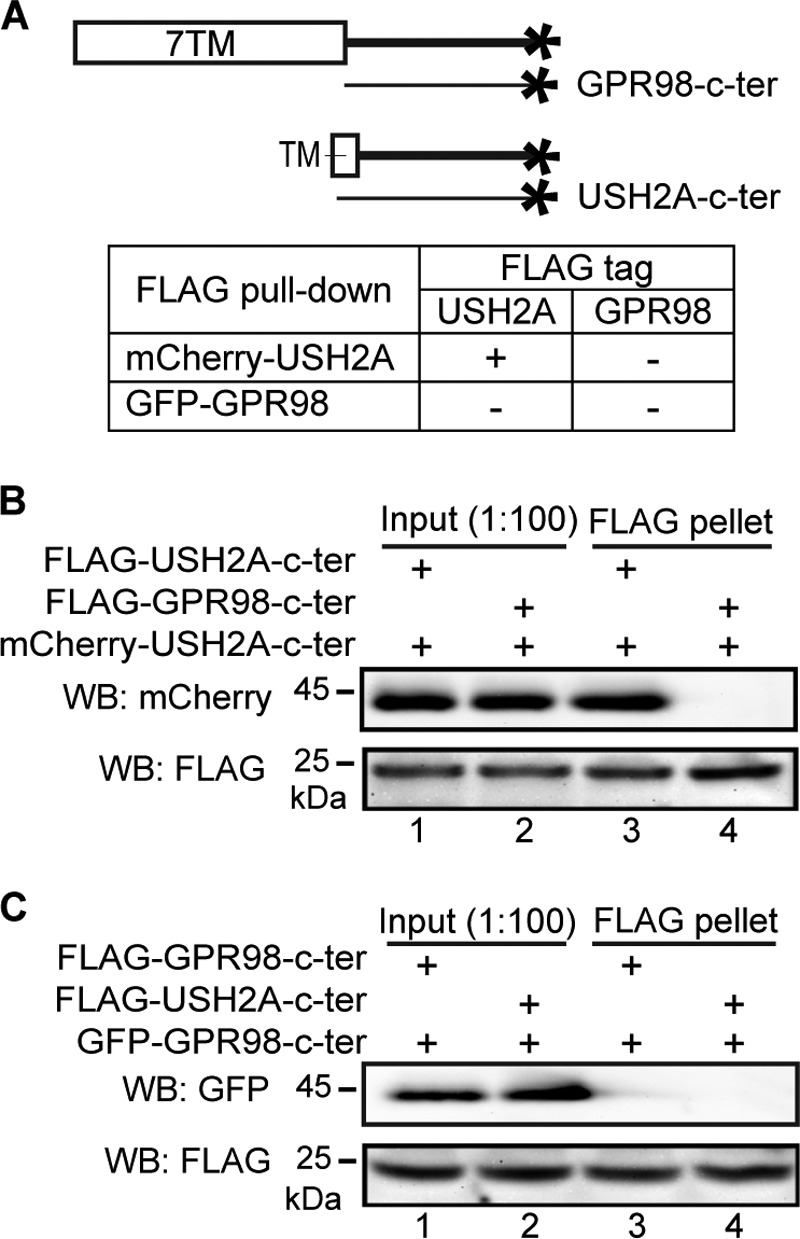
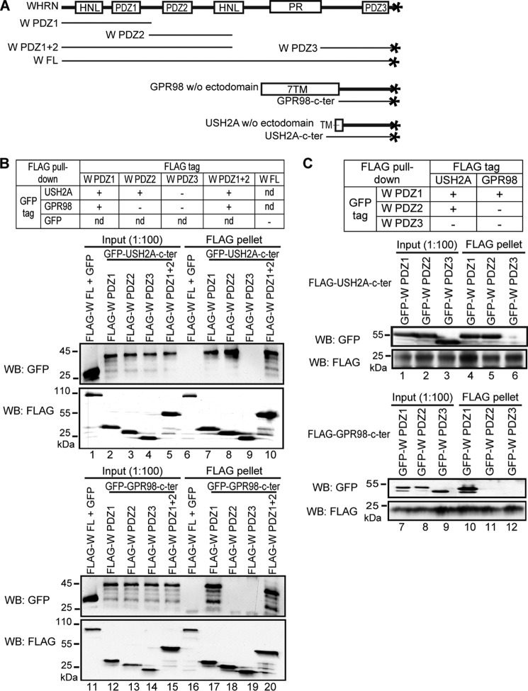
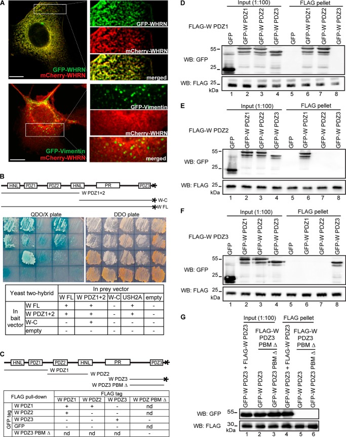
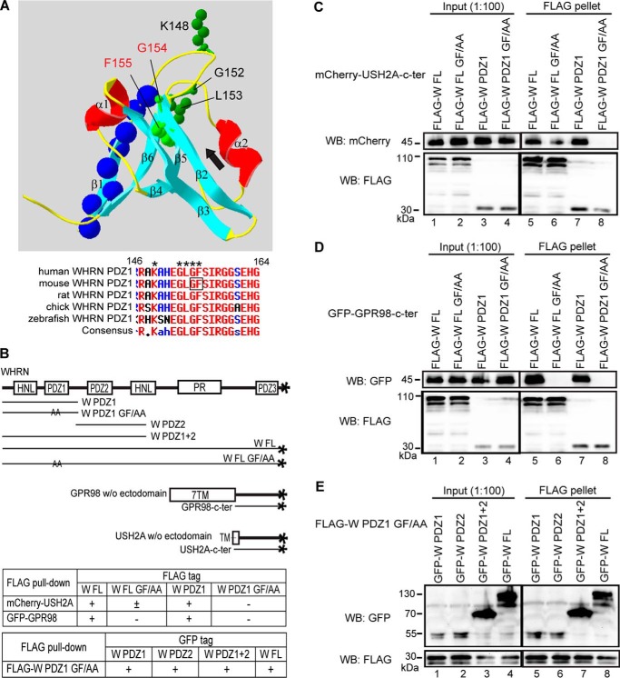
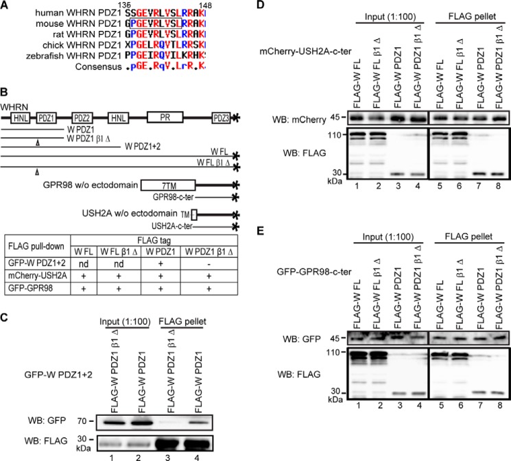
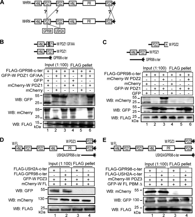
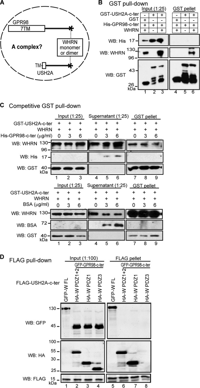
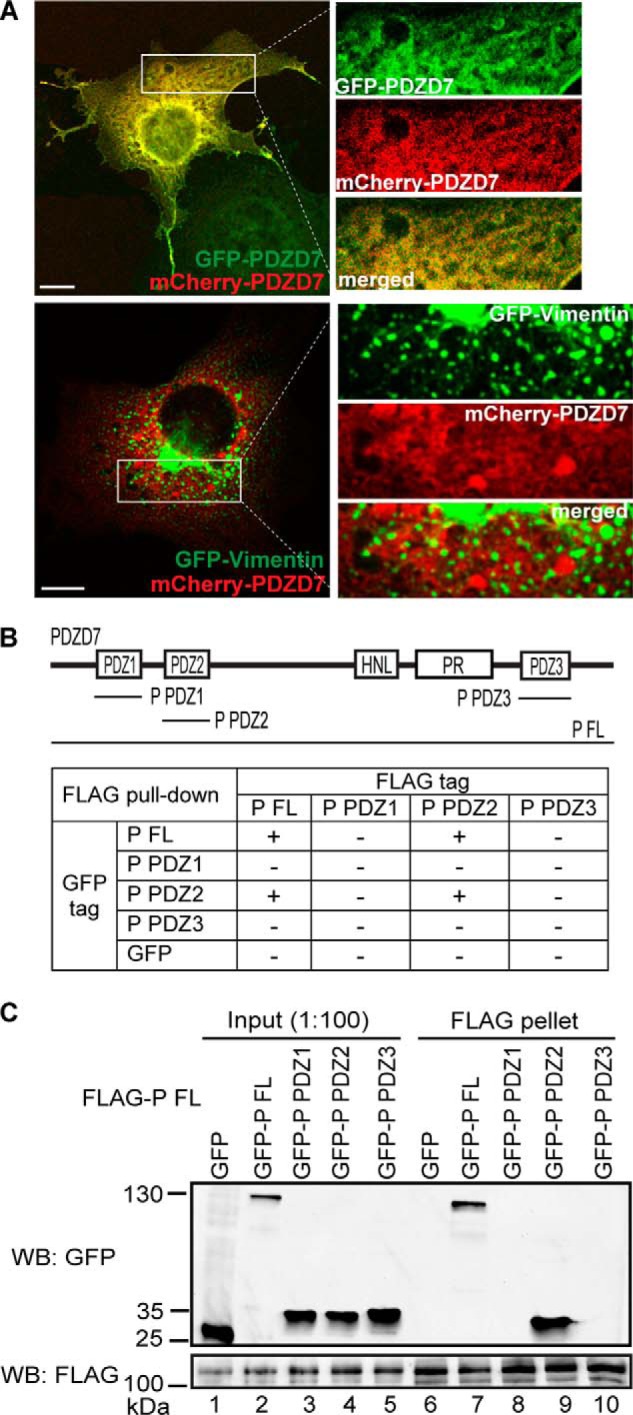
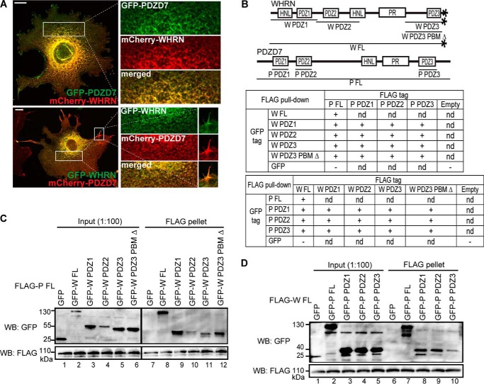
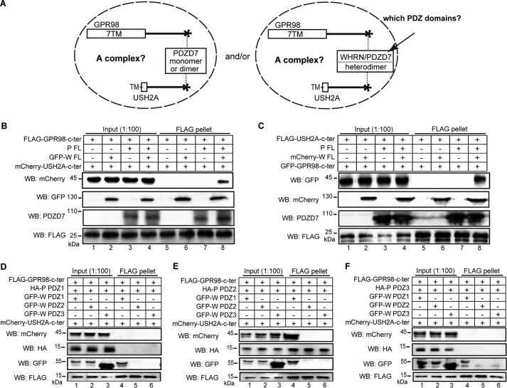
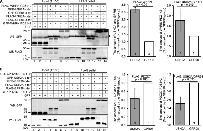
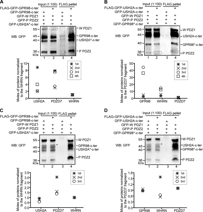
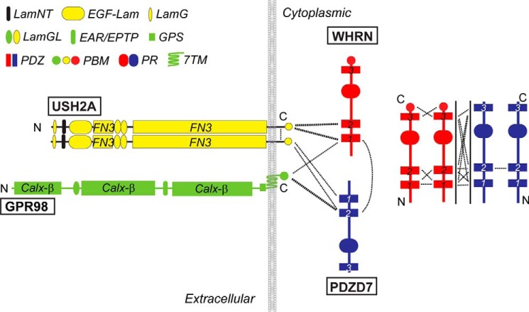
Usher syndrome (USH) is the leading genetic cause of combined hearing and vision loss. Among the three USH clinical types, type 2 (USH2) occurs most commonly. USH2A, GPR98, and WHRN are three known causative genes of USH2, whereas PDZD7 is a modifier gene found in USH2 patients. The proteins encoded by these four USH genes have been proposed to form a multiprotein complex, the USH2 complex, due to interactions found among some of these proteins in vitro, their colocalization in vivo, and mutual dependence of some of these proteins for their normal in vivo localizations. However, evidence showing the formation of the USH2 complex is missing, and details on how this complex is formed remain elusive. Here, we systematically investigated interactions among the intracellular regions of the four USH proteins using colocalization, yeast two-hybrid, and pull-down assays. We show that multiple domains of the four USH proteins interact among one another. Importantly, both WHRN and PDZD7 are required for the complex formation with USH2A and GPR98. In this USH2 quaternary complex, WHRN prefers to bind to USH2A, whereas PDZD7 prefers to bind to GPR98. Interaction between WHRN and PDZD7 is the bridge between USH2A and GPR98. Additionally, the USH2 quaternary complex has a variable stoichiometry. These findings suggest that a non-obligate, short term, and dynamic USH2 quaternary protein complex may exist in vivo. Our work provides valuable insight into the physiological role of the USH2 complex in vivo and informs possible reconstruction of the USH2 complex for future therapy.

Keywords: GPR98; Hair Cell; PDZ Domain; PDZD7; Photoreceptor; Protein Complex; Protein-Protein Interaction; USH2A; Usher Syndrome; WHRN.


Figures

FIGURE 1.
FIGURE 1.
Predicted functional domains of WHRN, PDZD7, USH2A, and GPR98. The longest alternatively spliced isoforms of WHRN, PDZD7, USH2A, and GPR98 are shown. The numbers of amino acids and GenBankTM accession numbers of human sequences are shown at the top of each protein. PR, proline-rich region; LamG, thrombospondin-type laminin G domain; LamNT, N-terminal globular laminin domain; EGF-Lam, laminin EGF-like domain; LamGL, laminin globular-like domain; FN3, fibronectin type III repeat; Calxβ, calx-β motif; EAR/EPTP, epilepsy-associated repeats/epitemptin; GPS, GPCR proteolytic site.
FIGURE 2.
FIGURE 2.
USH2A and GPR98 cytoplasmic fragments do not interact directly, whereas USH2A cytoplasmic fragment interacts with itself. A, diagram showing GPR98 and USH2A fragments used in this experiment and summary of the results. + and −, existence and absence of interactions, respectively. B, FLAG-USH2A (lane 3) but not FLAG-GPR98 (lane 4) cytoplasmic fragment was able to pull down mCherry-USH2A cytoplasmic fragment. C, neither FLAG-GPR98 (lane 3) nor FLAG-USH2A (lane 4) cytoplasmic fragment was able to pull down the GFP-GPR98 cytoplasmic fragment. The anti-FLAG blots in B and C demonstrate success of the FLAG pull-down assays. Lanes 1 and 2 (B and C) are input samples as controls. + (B and C), presence of protein fragments in the reaction.
FIGURE 3.
FIGURE 3.
WHRN PDZ domains interact differently with USH2A and GPR98. A, diagram showing WHRN (W), GPR98, and USH2A fragments used. B, top, summary of the results. Bottom, FLAG-W PDZ1 (lane 7), PDZ2 (lane 8), and PDZ1+2 (lane 10) but not PDZ3 (lane 9) fragments could pull down GFP-USH2A cytoplasmic fragment, whereas FLAG-W PDZ1 (lane 17) and PDZ1+2 (lane 20) but not PDZ2 (lane 18) or PDZ3 (lane 19) fragments could pull down GFP-GPR98 cytoplasmic fragment. FLAG-W FL protein and GFP were used as negative controls (lanes 6 and 16). C, top, summary of the results. Bottom, FLAG-USH2A cytoplasmic fragment could pull down GFP-WHRN PDZ1 (lane 4) and PDZ2 (lane 5) but not PDZ3 (lane 6) fragments, whereas FLAG-GPR98 cytoplasmic fragment could pull down GFP-WHRN PDZ1 (lane 10) but not PDZ2 (lane 11) or PDZ3 (lane 12) fragment. The anti-FLAG blots (B and C) demonstrate the success of the FLAG pull-down assays. Lanes 1–5, 11–15 (B), and lanes 1–3, 7–9 (C) are input samples as controls. +, existence of interactions; −, absence of interactions; nd, not determined.
FIGURE 4.
FIGURE 4.
WHRN forms homodimers through interactions among its multiple regions. A, GFP-WHRN and mCherry-WHRN colocalize in the cytoplasm when cotransfected in COS7 cells (yellow, top panels). As a negative control, GFP-vimentin and mCherry-WHRN show no colocalization in cotransfected COS7 cells (bottom panels). Signals in white boxes were enlarged and shown (right) in individual and merged channels. Scale bars, 10 μm. B, yeast two-hybrid analysis demonstrates that WHRN (W) FL and W PDZ1+2 fragments form homodimers, whereas the W-C fragment does not. The USH2A cytoplasmic fragment (USH2A) and empty vectors (empty) represent positive and negative controls, respectively. Sample arrangements in the images and table correspond. QDO/X, quadruple dropout medium SD/−Ade/−His/−Leu/−Trp with X-α-Gal to show the existence of interactions. DDO, double dropout medium SD/−Leu/−Trp to show the success of cotransformations. C, diagram of WHRN (W) fragments used in the FLAG pull-down assays (D–G) and summary of the results. D, FLAG-W PDZ1 fragment could pull down GFP-W PDZ1 (lane 6) and PDZ2 (lane 7) fragments but not GFP-W PDZ3 fragment (lane 8) or GFP (lane 5). E, FLAG-W PDZ2 fragment could pull down the GFP-W PDZ1 fragment (lane 6), but not other GFP-W fragments (lanes 7 and 8) or GFP (lane 5). F, FLAG-W PDZ3 fragment could pull down the GFP-W PDZ3 fragment (lane 8) but not other GFP-W fragments (lanes 6 and 7) or GFP (lane 5). G, FLAG-W PDZ3 PBM Δ fragment was unable to pull down the GFP-W PDZ3 fragment (lane 5) or the GFP-W PDZ3 PBM Δ fragment (lane 6). FLAG-W PDZ3 and GFP-W PDZ3 fragments were used as a positive control (lane 4). The anti-FLAG blots (D–G) demonstrate success of FLAG pull-down assays. +, existence of interactions; −, absence of interactions; nd, not determined.
FIGURE 5.
FIGURE 5.
The carboxylate-binding loop of WHRN PDZ1 domain binds to the USH2A/GPR98 PBM. A, three-dimensional structure of human WHRN PDZ1 domain (Protein Data Bank code IUEZ) (top) and sequence alignment of the WHRN PDZ1 carboxylate-binding loop across different species (bottom). From the N to C terminus, WHRN PDZ1 domain has the following β strands and α helixes, β1, β2, β3, α1, β4, β5, α2, and β6. The carboxylate-binding loop, Lys148-Xaa-Xaa-Xaa-Gly152-Leu153-Gly154-Phe155 (green side chains in top panel, asterisks in bottom panel), is located at the N-terminal end of the β2 strand. The PBM-binding groove (black arrow) lies between the β2 strand and α2 helix. The β1 strand is on the opposite side of the carboxylate-binding loop and PBM-binding groove. Residues deleted in the β1 Δ fragments (blue spheres) are labeled. The Gly154-Phe155 residues, replaced by two alanine residues in the GF/AA mutant fragments, are labeled using red type in the top panel and framed in the bottom panel. B, diagram of WHRN (W), GPR98, and USH2A fragments used in the FLAG pull-down assays (C–E) and summary of the results. +, existence of interactions; −, absence of interactions; ±, existence of weak interactions. C, FLAG-W FL GF/AA protein (lane 6) pulled down less mCherry-USH2A cytoplasmic fragment than wild-type FLAG-W FL protein (lane 5). Additionally, FLAG-W PDZ1 GF/AA fragment (lane 8) did not pull down the mCherry-USH2A cytoplasmic fragment, whereas wild-type FLAG-W PDZ1 fragment did (lane 7). D, the FLAG-W FL (lane 6) and PDZ1 (lane 8) GF/AA mutant fragments did not pull down the GFP-GPR98 cytoplasmic fragment, whereas the wild-type FLAG-W FL (lane 5) and PDZ1 (lane 7) fragments did. E, the FLAG-W PDZ1 GF/AA fragment was able to pull down GFP-tagged W PDZ1 (lane 5), PDZ2 (lane 6), PDZ1+2 (lane 7), and FL (lane 8) fragments. The anti-FLAG blots in C–E demonstrate success of the FLAG pull-down assays.
FIGURE 6.
FIGURE 6.
WHRN PDZ1 domain homo- and heterodimerizes through its β1 strand. A, sequence alignment of the WHRN PDZ1 β1 strand across different species. The deleted amino acids of the β1 stand are framed here and labeled in Fig. 5A. B, diagram of WHRN (W), GPR98, and USH2A fragments used in the FLAG pull-down assays (C–E) and summary of the results. +, existence of interactions; −, absence of interactions; nd, not determined. C, FLAG-W PDZ1 β1 Δ fragment could not pull down GFP-W PDZ1+2 fragment (lane 3), whereas the wild-type FLAG-W PDZ1 fragment could (lane 4). D and E, deletion of the WHRN PDZ1 β1 strand did not affect bindings of W FL (lanes 5 and 6) and W PDZ1 (lanes 7 and 8) fragments to USH2A (D) or GPR98 (E) cytoplasmic fragment. The anti-FLAG blots in C–E demonstrate success of the FLAG pull-down assays.
FIGURE 7.
FIGURE 7.
WHRN, while binding to GPR98 or USH2A, dimerizes only through its PDZ1 domain. A, diagram with the questions to be tested: whether one WHRN protein could bind to GPR98/USH2A and dimerize with another WHRN protein at the same time, and which WHRN regions mediated this dimerization if it occurred. B, FLAG-GPR98 cytoplasmic fragment could pull down GFP-WHRN (W) PDZ1 GF/AA fragment (lane 6) but not GFP (lane 4) in the presence of wild-type mCherry-W PDZ1 fragment. Additionally, the FLAG-GPR98 cytoplasmic fragment could not pull down the GFP-W PDZ1 GF/AA fragment in the presence of mCherry (lane 5). C, FLAG-GPR98 cytoplasmic fragment could not pull down the mCherry-W PDZ2 fragment in the presence of the GFP-W PDZ1 fragment (lane 6), although the FLAG-GPR98 cytoplasmic fragment could pull down the GFP-W PDZ1 fragment (lanes 5 and 6). mCherry and GFP proteins were negative controls (lane 4). Note that, in the anti-GFP blot, weak signals at 55 kDa in lanes 1 and 4 are artifacts caused by sample leaking from other lanes. D, FLAG-USH2A (lane 3) and FLAG-GPR98 (lane 4) cytoplasmic fragments could not pull down GFP-W PDZ3 fragment in the presence of mCherry-W FL protein, although these two FLAG-tagged proteins could pull down the mCherry-W FL protein. E, FLAG-USH2A (lane 4) and FLAG-GPR98 (lane 3) cytoplasmic fragments could not pull down the mCherry-W PDZ3 fragment in the presence of GFP-W FL PBM Δ protein, although these two FLAG-tagged proteins could pull down the GFP-W FL PBM Δ protein. B–E, diagrams of the protein fragments used are shown (top of each panel). The anti-FLAG blots demonstrate the success of the FLAG pull-down assays. + and −, presence and absence of protein fragments in the reaction, respectively.
FIGURE 8.
FIGURE 8.
USH2A, GPR98, and WHRN do not form a complex. A, diagram of the question to be tested: whether WHRN, USH2A, and GPR98 can form a complex. B, GST-USH2A cytoplasmic fragment could pull down WHRN but not the His-GPR98 cytoplasmic fragment when the three proteins were mixed (lane 6). Lanes 4 and 5, two different negative controls. C, increasing amounts of His-GPR98 cytoplasmic fragment (top panels) but not BSA protein (bottom panels) competitively removed WHRN from the GST pull-down pellet (lanes 7–9), where WHRN bound to the GST-USH2A cytoplasmic fragment, into the supernatant (lanes 4–6). D, FLAG-USH2A cytoplasmic fragment could not pull down GFP-GPR98 cytoplasmic fragment in the presence of either the HA-WHRN (W) PDZ1+2 (lane 6) or PDZ1 (lane 7) fragment. The HA-W PDZ3 fragment served as a negative control (lane 8). As a positive control, the FLAG-USH2A cytoplasmic fragment could pull down GFP-W FL protein (lane 5). The anti-GST blots in B and C and anti-FLAG blots in D demonstrate the success of the GST and FLAG pull-down assays, respectively. + and −, presence and absence of protein fragments in the reaction, respectively.
FIGURE 9.
FIGURE 9.
PDZD7 homodimerization is mediated by its PDZ2 domain. A, GFP-PDZD7 and mCherry-PDZD7 proteins colocalized with each other when cotransfected in COS7 cells (top panels). As a negative control, mCherry-PDZD7 and GFP-vimentin proteins did not show similar signal patterns in cotransfected COS7 cells (bottom panels). Signals in white boxes in the images on the left were enlarged and shown (right) in individual and merged channels. Scale bars, 10 μm. B, diagram of PDZD7 (P) fragments used to study PDZD7 homodimerization and summary of results from the FLAG pull-down assays. +, existence of interactions; −, absence of interactions. C, FLAG-P FL protein could pull down GFP-P FL (lane 7) and PDZ2 (lane 9) fragments but not GFP-P PDZ1 (lane 8), GFP-P PDZ3 (lane 10), or GFP (lane 6) fragment. The anti-FLAG blot demonstrates success of the FLAG pull-down assay.
FIGURE 10.
FIGURE 10.
Heterodimerization between WHRN and PDZD7 is mediated by their multiple PDZ domains. A, colocalization was observed between GFP-PDZD7 and mCherry-WHRN proteins (top panels) and between GFP-WHRN and mCherry-PDZD7 proteins (bottom panels) when they were cotransfected in COS7 cells. Signals in the cytoplasm (top and bottom images) and filopodia (bottom image), framed in white boxes in the left-hand images, were enlarged and shown on the right in individual and merged channels. Scale bars, 10 μm. B, diagram of WHRN (W) and PDZD7 (P) fragments used in this study and a summary of results from reciprocal FLAG pull-down assays. +, existence of interactions; −, absence of interactions; nd, not determined. C, FLAG-P FL protein could pull down GFP-W FL (lane 8), PDZ1 (lane 9), PDZ2 (lane 10), PDZ3 (lane 11), and PDZ3 PBM Δ (lane 12) fragments but not GFP (lane 7). D, FLAG-W FL protein could pull down GFP-P FL (lane 7), PDZ1 (lane 8), PDZ2 (lane 9), and PDZ3 (lane 10) fragments but not GFP (lane 6). The anti-FLAG blots (C and D) demonstrate the success of the FLAG pull-down assays.
FIGURE 11.
FIGURE 11.
USH2A, GPR98, WHRN, and PDZD7 proteins form a quaternary complex. A, diagram showing the questions to be tested: whether USH2A and GPR98 were able to form a complex with PDZD7 and/or with both PDZD7 and WHRN, and which PDZ domains of WHRN and PDZD7 contributed to the complex formation. B, FLAG-GPR98 cytoplasmic fragment could pull down mCherry-USH2A cytoplasmic fragment only in the presence of both WHRN (W) FL and PDZD7 (P) FL proteins (lane 8) but not in the presence of either W FL (lane 6) or P FL protein (lane 7) alone. Additionally, the FLAG-GPR98 cytoplasmic fragment could not pull down the mCherry-USH2A cytoplasmic fragment directly (lane 5). C, the FLAG-USH2A cytoplasmic fragment could pull down the GFP-GPR98 cytoplasmic fragment only in the presence of both W FL and P FL proteins (lane 8) and not in the presence of either W FL (lane 6) or P FL protein (lane 7) alone. FLAG-USH2A cytoplasmic fragment could not pull down the GFP-GPR98 cytoplasmic fragment directly (lane 5). D–F, FLAG-GPR98 cytoplasmic fragment could pull down the mCherry-USH2A cytoplasmic fragment only in the presence of W PDZ1 and P PDZ2 fragments (lane 4 in E) but not in the presence of any other combinations of W and P fragments (all other lanes in D–F). The anti-FLAG blots (B–F) demonstrate success of the FLAG pull-down assays. + and −, presence and absence of protein fragments in the reaction, respectively.
FIGURE 12.
FIGURE 12.
WHRN and PDZD7 proteins have different binding affinities to USH2A and GPR98. A, representative immunoblots showing the amount of GFP-USH2A (lane 11) and GFP-GPR98 (lane 12) cytoplasmic fragments pulled down by FLAG-WHRN PDZ1+2 fragment as well as the amount of GFP-WHRN PDZ1+2 fragment pulled down by FLAG-USH2A (lane 13) and FLAG-GPR98 (lane 14) cytoplasmic fragments. GFP served as a negative control (lanes 8–10). Bands labeled by an asterisk are nonspecific. Signal quantification from three independent experiments and p values from Student's t tests are shown (right). B, representative immunoblots showing the amount of GFP-USH2A (lane 11) and GFP-GPR98 (lane 12) cytoplasmic fragments pulled down by FLAG-PDZD7 PDZ1+2 fragment as well as the amount of GFP-PDZD7 PDZ1+2 fragment pulled down by FLAG-USH2A (lane 13) and FLAG-GPR98 (lane 14) cytoplasmic fragments. GFP is a negative control (lanes 8–10). Quantification of signals from three independent experiments and p values from Student's t tests are shown (right). Signals in lanes 11–14 (FLAG blots) and lanes 4–7 (GFP blots) were used to normalize the signals in lanes 11–14 (GFP blots). + and −, presence and absence of protein fragments in the reaction, respectively. Error bars, S.E.
FIGURE 13.
FIGURE 13.
The USH2 quaternary protein complex has a variable stoichiometry. A and C, representative anti-GFP immunoblots showing GFP-tagged WHRN (W) PDZ1 fragment, USH2A cytoplasmic fragment, and PDZD7 (P) PDZ2 fragment in the pellet pulled down by FLAG-GFP-GPR98 fragment from quadruply transfected cell lysate (lane 4 in A) or the mixed cell lysates individually transfected with each of the four fragments (lane 4 in C). Graphs (bottom) show the calculated moles of USH2A, WHRN PDZ1, and PDZD7 PDZ2 fragments in the FLAG pull-down pellet, normalized by the moles of WHRN PDZ1 fragment in the same pellet, from four (A) or three (C) independent trials. Data from the same experiment (i.e. same FLAG pull-down pellet) are labeled by the same symbols. B and D, representative anti-GFP immunoblots showing GFP-tagged W PDZ1 fragment, GPR98 cytoplasmic fragment, and P PDZ2 fragment in the pellet pulled down by FLAG-GFP-USH2A fragment from their quadruply transfected cell lysate (lane 4 in B) or the mixed cell lysates individually transfected with the four fragments (lane 4 in D). Graphs (bottom) show the calculated moles of GPR98, WHRN PDZ1, and PDZD7 PDZ2 fragments in the FLAG pull-down pellet, normalized by the moles of PDZD7 PDZ2 fragment in the same pellet, from four (B) or three (D) independent trials. Data from the same experiment (i.e. the same FLAG pull-down pellet) are labeled by the same symbols. Substitutions of FLAG-GFP-GPR98 and FLAG-GFP-USH2A fragments with GFP-GPR98 and GFP-USH2A fragments, respectively, are negative controls (lane 3, all panels). Bands circled by dashed lines are the W PDZ1 fragment (A) and P PDZ2 fragment (B). Bands (arrows in C and D) are nonspecific. +, presence of protein fragments in the reaction.
FIGURE 14.
FIGURE 14.
Model for USH2 protein complex formation and in vitro interactions among complex components. Dashed lines indicate the existence of interaction between protein domains they connect. The thickness of dashed lines corresponds to interaction strength. LamNT, N-terminal globular laminin domain; EGF-Lam, laminin EGF-like domain; LamG, thrombospondin-type laminin G domain; LamGL, laminin globular-like domain; EAR/EPTP, epilepsy-associated repeats/epitemptin; GPS, GPCR proteolytic site; PR, proline-rich region; Calxβ, calx-β motif; FN3, fibronectin type III repeat.

References

    1. Ebermann I., Phillips J. B., Liebau M. C., Koenekoop R. K., Schermer B., Lopez I., Schäfer E., Roux A. F., Dafinger C., Bernd A., Zrenner E., Claustres M., Blanco B., Nürnberg G., Nürnberg P., Ruland R., Westerfield M., Benzing T., Bolz H. J. (2010) PDZD7 is a modifier of retinal disease and a contributor to digenic Usher syndrome. J. Clin. Invest. 120, 1812–1823 - PMC - PubMed
    1. Ebermann I., Scholl H. P., Charbel Issa P., Becirovic E., Lamprecht J., Jurklies B., Millán J. M., Aller E., Mitter D., Bolz H. (2007) A novel gene for Usher syndrome type 2: mutations in the long isoform of whirlin are associated with retinitis pigmentosa and sensorineural hearing loss. Hum. Genet. 121, 203–211 - PubMed
    1. Eudy J. D., Weston M. D., Yao S., Hoover D. M., Rehm H. L., Ma-Edmonds M., Yan D., Ahmad I., Cheng J. J., Ayuso C., Cremers C., Davenport S., Moller C., Talmadge C. B., Beisel K. W., Tamayo M., Morton C. C., Swaroop A., Kimberling W. J., Sumegi J. (1998) Mutation of a gene encoding a protein with extracellular matrix motifs in Usher syndrome type IIa. Science 280, 1753–1757 - PubMed
    1. Weston M. D., Luijendijk M. W., Humphrey K. D., Möller C., Kimberling W. J. (2004) Mutations in the VLGR1 gene implicate G-protein signaling in the pathogenesis of Usher syndrome type II. Am. J. Hum. Genet. 74, 357–366 - PMC - PubMed
    1. Schneider E., Märker T., Daser A., Frey-Mahn G., Beyer V., Farcas R., Schneider-Ratzke B., Kohlschmidt N., Grossmann B., Bauss K., Napiontek U., Keilmann A., Bartsch O., Zechner U., Wolfrum U., Haaf T. (2009) Homozygous disruption of PDZD7 by reciprocal translocation in a consanguineous family: a new member of the Usher syndrome protein interactome causing congenital hearing impairment. Hum. Mol. Genet. 18, 655–666 - PubMed

Publication types

MeSH terms