

Review
doi: 10.1039/c4dt02846e.
A nuclear chocolate box: the periodic table of nuclear medicine
Affiliations
- PMID: 25406520
- PMCID: PMC6205633
- DOI: 10.1039/c4dt02846e
Item in Clipboard
Review
A nuclear chocolate box: the periodic table of nuclear medicine
Dalton Trans.
.
Abstract
Radioisotopes of elements from all parts of the periodic table find both clinical and research applications in radionuclide molecular imaging and therapy (nuclear medicine). This article provides an overview of these applications in relation to both the radiological properties of the radionuclides and the chemical properties of the elements, indicating past successes, current applications and future opportunities and challenges for inorganic chemistry.
Figures
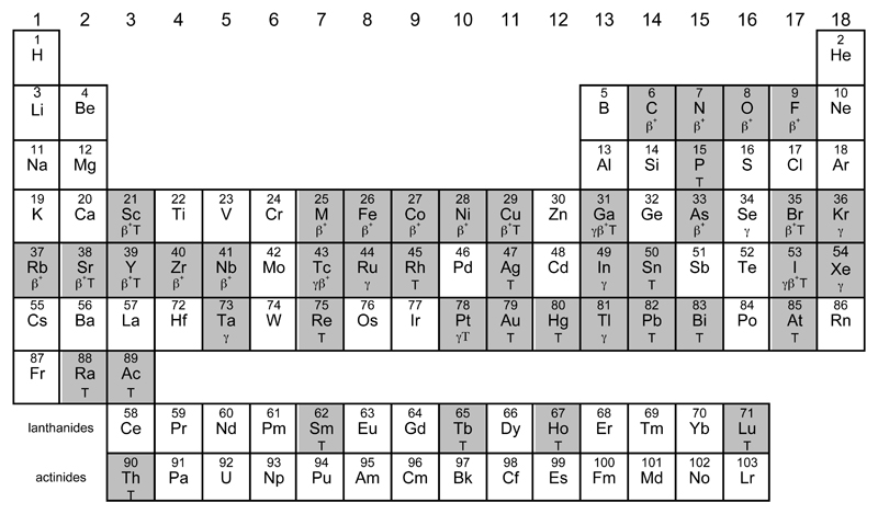
Periodic table highlighting (shaded) elements with radionuclides with uses or identified potential uses in molecular imaging or targeted radionuclide therapy. Main uses are identified by symbols: γ (gamma camera imaging/SPET), β+ (positron emission tomography) and T (therapy, using beta-, alpha, or Auger-emitting radioisotopes). Some unshaded elements also have indirect importance e.g. as parent radionuclides for directly-used radionuclides, or as cyclotron target materials, or chemical binding sites for biomolecule labelling, hence the “usefulness” is somewhat arbitrary.
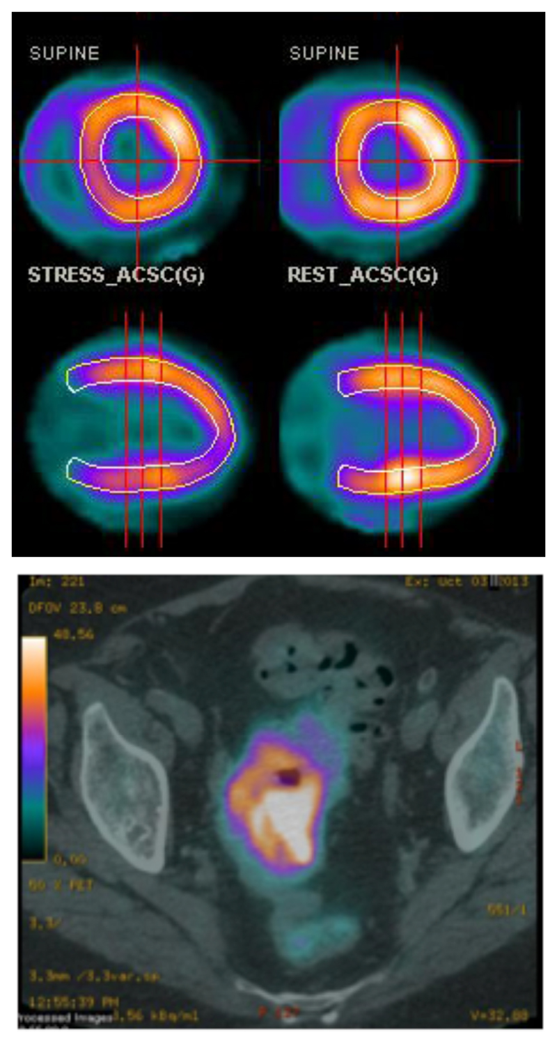
Top: Example of myocardial perfusion PET imaging using 82Rb+ (transverse (top) and sagittal (bottom) sections); bottom: PET-CT scan (transverse section, pelvic region) of a patient with colorectal cancer after injection of 82Rb+. (Images courtesy of Prof A. M. Groves, University College London Hospitals)
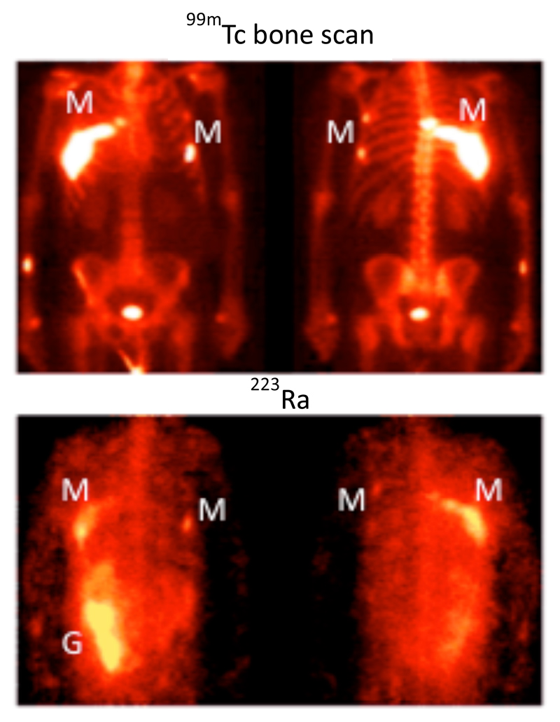
Biodistribution of 223Ra in a patient with metastatic prostate cancer after i.v. administration of 223RaCl2. The anterior (left) and posterior (right) views show specific uptake in the sites of bone metastases indicated by the 99mTc bone scan (M), and activity in the gut (G) indicating radioactivity that has undergone hepatobiliary excretion. The gamma camera image was possible by virtue of a low abundance gamma emission from 223Ra accompanying the alpha decay. (Adapted with permission from ref. 5).
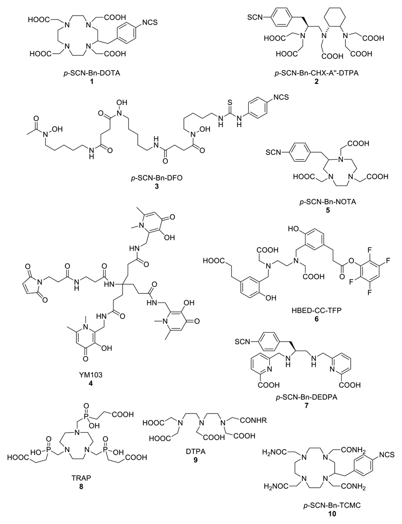
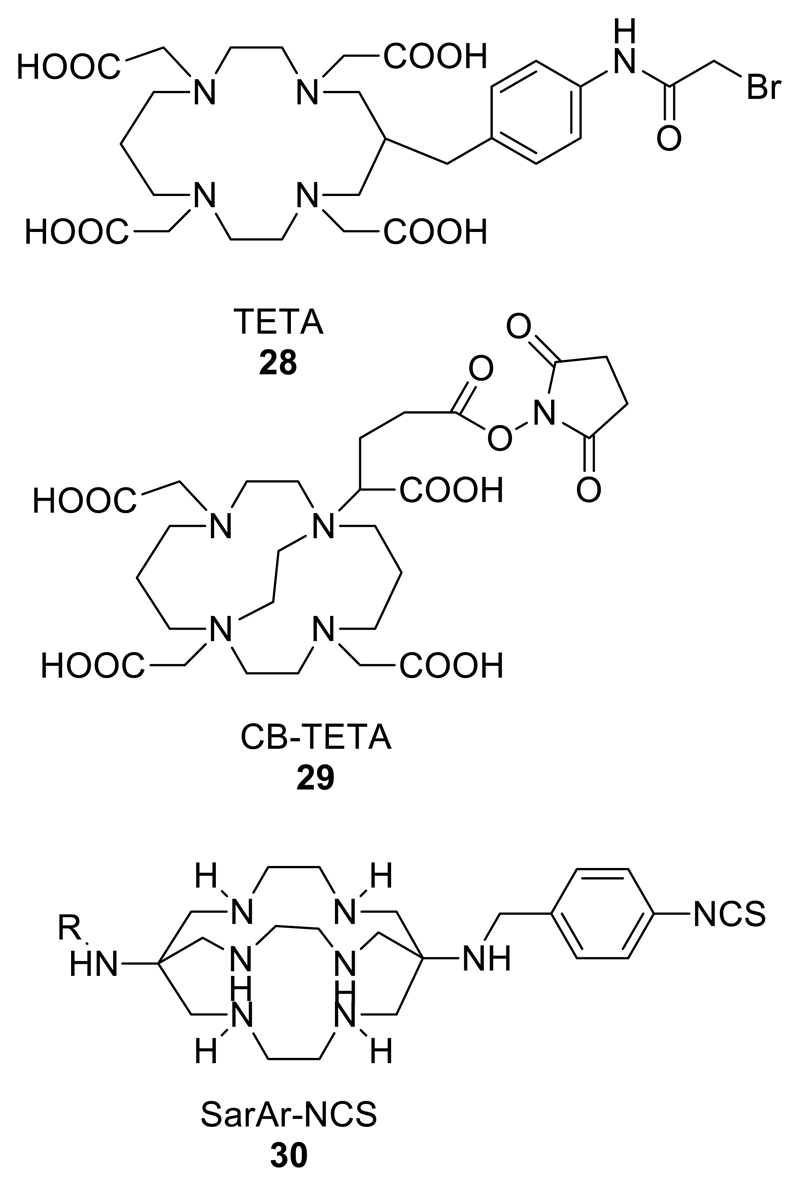
Structures of bifunctional chelators used for radioactive di-, tri- and tetravalent metal ions
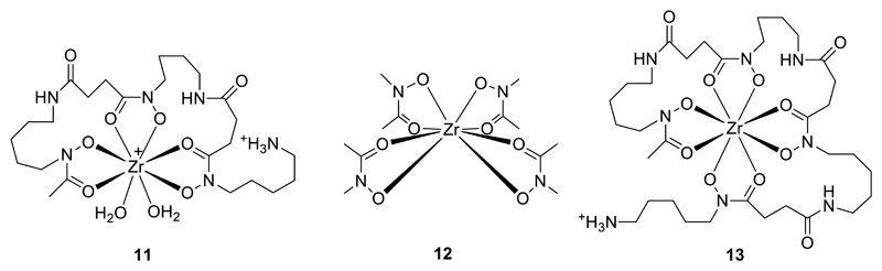
Left: Modelled coordination of Zr4+ by DFO (11); Centre: Schematic structure of ZrL4 (HL = N-methylacetohydroxamic acid) (12), serving as a template for the design of new octadentate chelators for Zr+ (note that disorder in the C and N orientations of the hydroxamate rings precludes determination of their preferred relative orientation and suggests that any such preferences are weak); Right: Schematic structure of Zr4+ complex 13 of an octadentate DFO homologue.
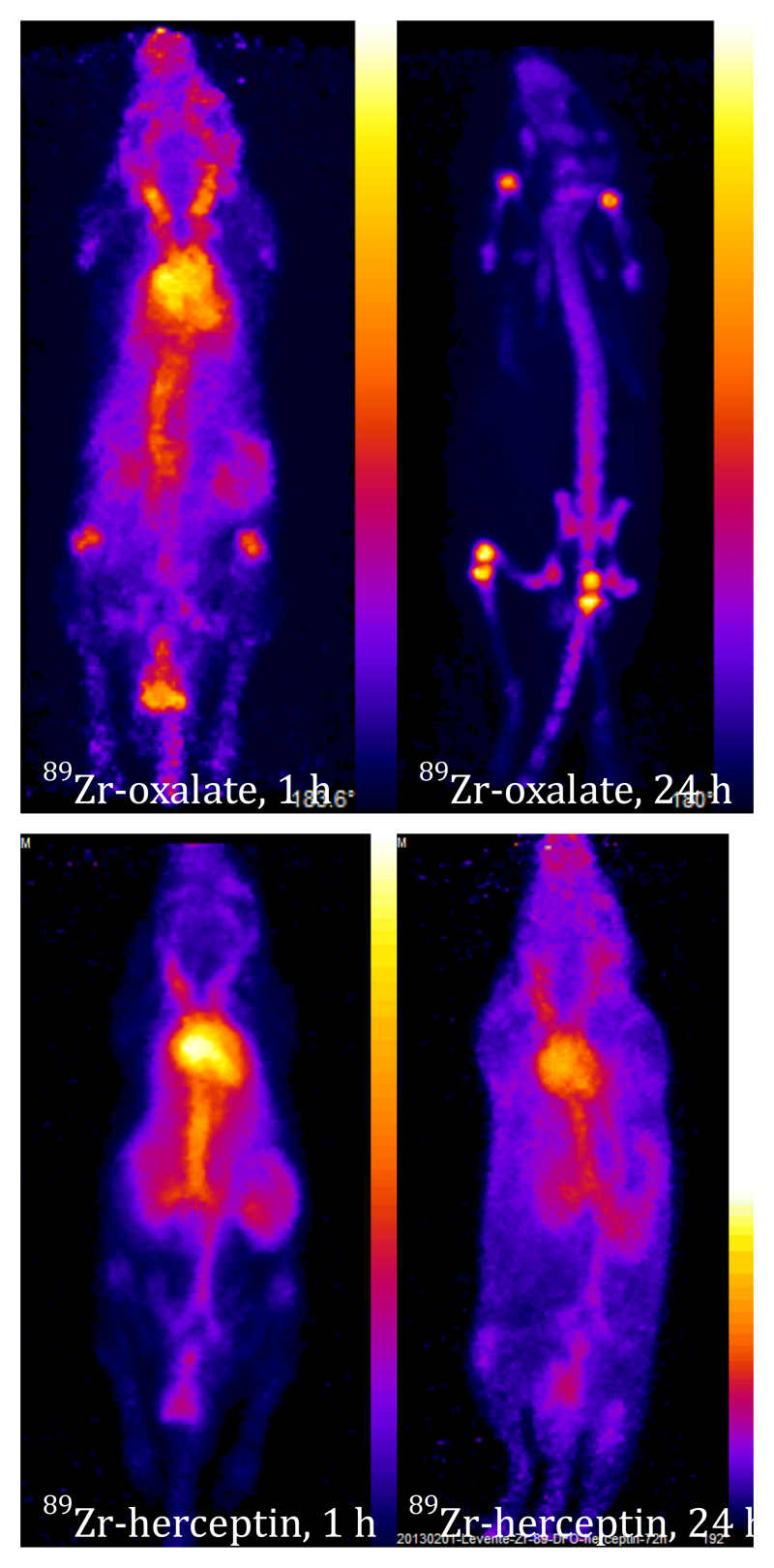
PET images showing biodistribution of 89Zr-oxalate complex (top) and 89Zr-labelled trastuzumab-DFO conjugate (bottom) at 1 h (left) and 4 h (right) post-injection in mice. Oxalate complex is gradually cleared from blood to bone and to some extent excreted through kidney, while the labelled antibody remains largely in the blood pool until at least 24 h, with only slight uptake in bone by 24 h.
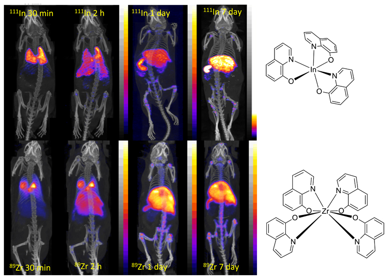
PET/CT scans comparing cell tracking with 111In(oxinate)3 (top) and 89Zr(oxinate)4 (bottom) labelled myeloma cells in mice. Cells initially accumulate in lungs, followed by migration to spleen, liver and bone marrow. Less uptake in kidney is observed for 89Zr, suggesting greater stability and cell survival.
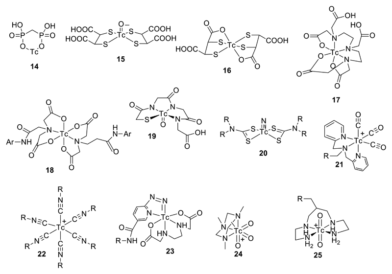
A selection of established 99mTc radiopharmaceuticals and core structures (if known): TcMDP (14), Tc(V)DMSA (15), TcDMSA (16), TcDTPA (17), TcHIDA (18) TcMAG3 (19), Tc(V)-nitridobis(dithiocarbamate) (20), Tc(I)-tricarbonyl (21), Tc(I)-hexakis(isonitrile) (22), Tc(V)-hynic (23), Tc(VII)-trioxo (24), and Tc(V)-dioxo (25). Structures of 15, 19, 20, 21, 22, 24 and 25 are known and well defined; the others are not clearly defined.
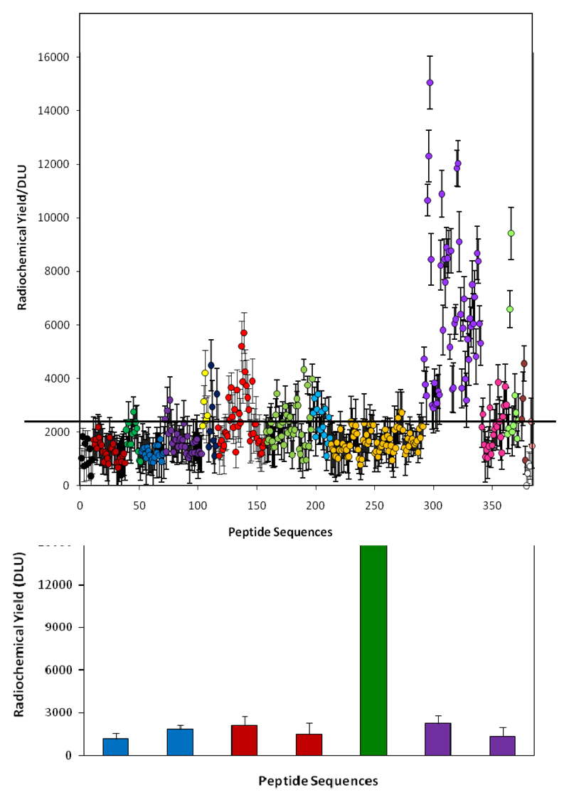
Optimisation of His-tag sequences for 99mTc tricarbonyl labelling from high throughput screening. Top: relative labelling efficiency of a range of peptides. Purple symbols represent His-tags with adjacent arginine or lysine residues (i.e. positively charged sequences). Other colours represent uncharged and negatively charged sequences. Bottom: Comparison of labelling efficiency of optimal binding sequence selected from the screening (green) with established his tags sequences.–
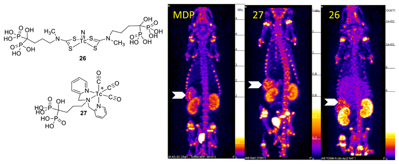
SPET images (maximum intensity projection) of vascular calcification in a mouse model 30 min after injection with [99mTc]-MDP, [99mTc]-27 and [99mTc]-26. Arrows indicate uptake in calcified mesenteric arteries. Uptake in bone and clearance via kidneys and bladder is also evident.
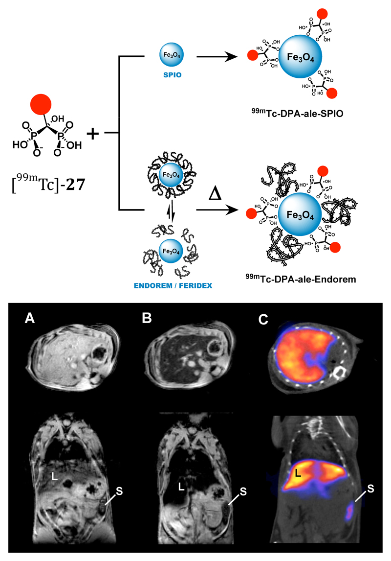
Dual-modality (MRI/SPET) contrast agent combining iron oxide nanoparticles with 99mTc radiolabel linked to the inorganic iron oxide surface via bisphosphonate groups. Top: assembly of composite particle; bottom: Mouse images, transverse sections through liver. A: MR image pre-injection of contrast agent; B: MR image post-injection of contrast agent showing darkening of liver due to accumulation of iron oxide nanoparticles; C: SPET/CT image showing co-localisation of 99mTc with iron oxide contrast in liver (L) and spleen (S).
Selection of chelators for radioisotopes of Cu2+; others of significance include 1 and 5 from Fig. 4.
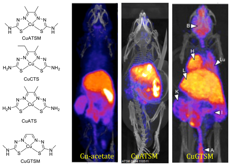
Structures of copper bis(thiosemicarbazone) complexes and PET imaging with 64Cu and its bis(thiosemicarbazone) complexes in mice 30 min post injection. Cu-acetate is rapidly taken up in liver and excreted into the gut; CuATSM (hypoxia-selective tracer) behaves in a superficially similar way; CuGTSM (non-hypoxia-selective tracer) likewise shows liver and gut activity but also shows high uptake in normal brain and myocardium.
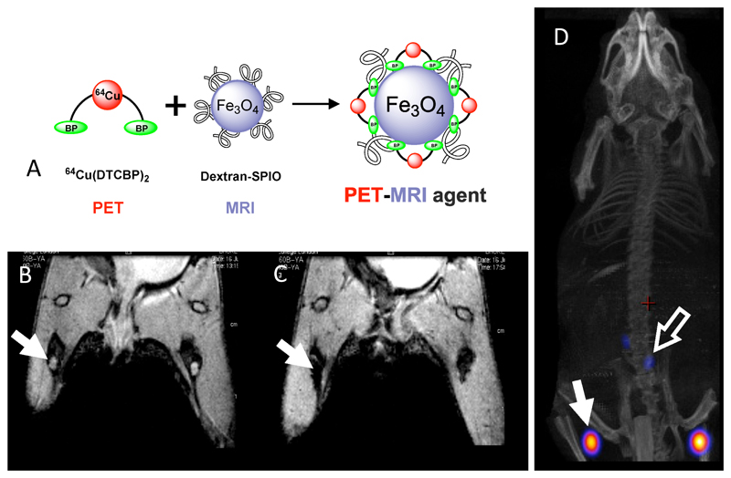
Dual modality (PET/MR) imaging of sentinel lymph nodes in mice. A: assembly of 64Cu-labelled iron oxide/dextran nanoparticles using a dithiocarbamate-bisphosphonate ligand (as used in technetium complex 26, see Fig. 10). B: MR scan (coronal section) of hind limbs and lower abdomen, pre-injection of contrast agent. Lymph nodes are visible as bright areas (arrow). C: MR scan post-injection of contrast agent, showing darkening of lymph nodes due to uptake of MR contrast. D: PET/CT scan showing radioactivity co-localised with magnetic particles (arrows). The radioactivity remains bound to the magnetic particles in vivo.
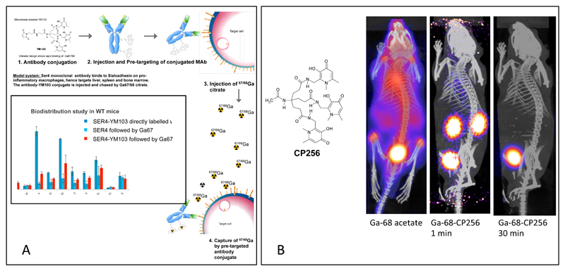
A. Concept of pre-targeting by in vivo transchelation of 68Ga, which is feasible with YM103 (4 in Fig. 4) because of its ability to transchelate Ga3+ from transferrin in plasma and in vivo. Inset: Uptake of 68Ga in most organs (including spleen which is the location of the macrophages expressing sialoadhesin, the SER4 antibody target) when given as a chase after administration of antibody-YM103 conjugate, is intermediate between that of the pre-labelled antibody and the distribution of 68Ga given as a chase after administration of unconjugated antibody. The exception is bone, which is the target of unchelated 68Ga (see part B, left image). This suggests that 68Ga is able to find and bind to the pre-targeted antibody-YM103 conjugate in vivo. B: Demonstration that the tris(hydroxypyridinone) chelator CP256 (non-derivatised form of YM103) can bind to 68Ga in vivo and excrete it rapidly via kidney. Left: Biodistribution of 68Ga administered in unchelated form; centre and right: biodistribution of 68Ga-CP256 complex 1 min and 30 min post-injection respectively, showing rapid renal excretion in progress and completed. Similar images are obtained if the 68Ga is administered first, followed by the CP256 ligand.
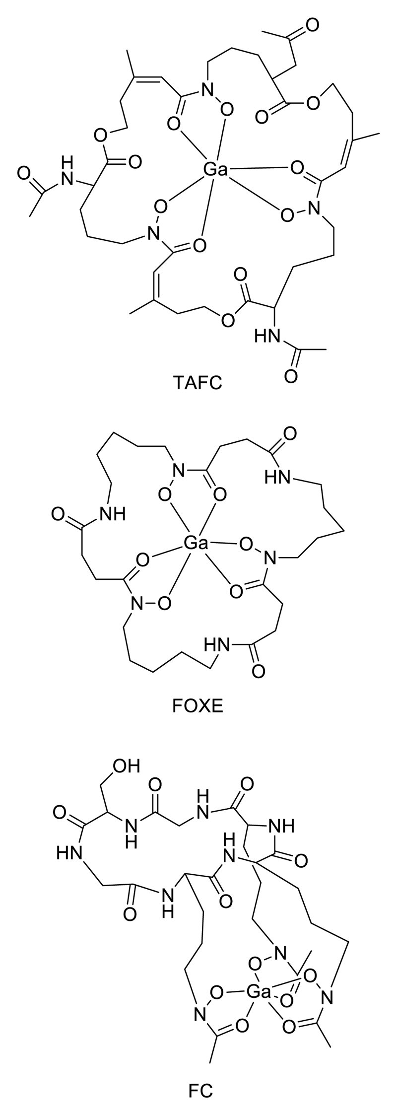
Siderophores labelled with 68Ga for targeting microorganisms: TAFC = triacetylfusarinine; FOXE = ferrixoamine E; FC = desferriferricrocin.
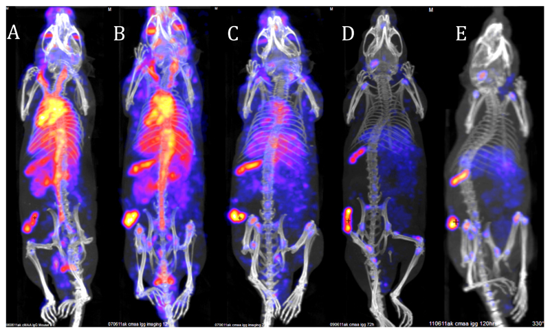
Typical tumour SPET/CT imaging with whole IgG monoclonal antibody in mouse bearing an experimental melanoma on the flank, illustrating the need for long half-life radionuclide (in this case 111In, half-life 2.8 days). CSPG4 IgG antibody, which binds to melanoma associated antigen, was labelled with 111In using the bifunctional chelator CHX-A”-DTPA. Images show predominantly blood pool at 4 h (A) and 24 h (B) with increasing tumour- and spleen-to-blood ratio 48 h and optimal images are not seen until 48-120 h.
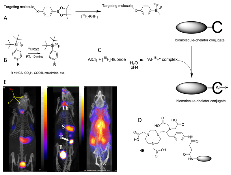
A. Nucleophilic radiolabelling of boronic ester prosthetic group; B: Isotopic exchange labelling of fluorosilane prosthetic group; C: Use of coordinatively unsaturated aluminium complex as a binding site for fluoride ions in biomolecule labelling; D: currently favoured ligand for chelation of Al3+ and labelling with [18F]-fluoride; E: PET-CT imaging of sodium/iodide symporter (NIS) activity mouse using NIS substrate [18F}-BF4-, 30 min post-injection. Left: normal mouse showing thyroid and stomach uptake and excretion into bladder; Centre: mouse with implanted NIS-expressing breast tumour showing tumour (T) uptake as well as thyroid (Th) and stomach (S) uptake; right: normal mouse pre-injected with perchlorate to block NIS activity, showing radioactivity largely confined to blood pool.
References
Publication types
MeSH terms
Substances
Grants and funding
LinkOut - more resources
Full Text Sources
Other Literature Sources
Miscellaneous