

Significance of main pulmonary artery dilation on imaging studies
- PMID: 25406836
- PMCID: PMC4298979
- DOI: 10.1513/AnnalsATS.201406-253PP
Significance of main pulmonary artery dilation on imaging studies
Abstract
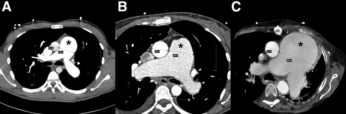
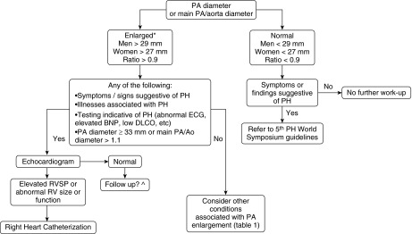
Proper and early identification of patients who harbor serious occult illness is the first step in developing a disease-management strategy. Identification of illnesses through the use of noninvasive techniques provides assurance of patient safety and is ideal. PA dilation is easily measured noninvasively and is due to a variety of conditions, including pulmonary hypertension (PH). The clinician should be able to thoroughly assess the significance of PA dilation in each individual patient. This involves knowledge of the ability of PA dilation to accurately predict PH, understand the wide differential diagnosis of causes of PA dilation, and reverse its life-threatening complications. We found that although PA dilation is suggestive of PH, data remain inconclusive regarding its ability to accurately predict PH. At this point, data are insufficient to place PA dilation into a PH risk-score equation. Here we review the causes and complications of PA dilation, define normal and abnormal PA measurements, and summarize the data linking its association to PH, while suggesting an algorithm designed to assist clinicians in patient work-up after recognizing PA dilation.
Keywords: pulmonary arterial hypertension; pulmonary artery diameter; pulmonary artery enlargement.
Figures
References
-
- Lee J, Kirschner J, Pawa S, Wiener DE, Newman DH, Shah K. Computed tomography use in the adult emergency department of an academic urban hospital from 2001 to 2007. Ann Emerg Med. 2010;56:591–596. - PubMed
-
- Smith-Bindman R, Miglioretti DL, Johnson E, Lee C, Feigelson HS, Flynn M, Greenlee RT, Kruger RL, Hornbrook MC, Roblin D, et al. Use of diagnostic imaging studies and associated radiation exposure for patients enrolled in large integrated health care systems, 1996-2010. JAMA. 2012;307:2400–2409. - PMC - PubMed
-
- Haimovici JB, Trotman-Dickenson B, Halpern EF, Dec GW, Ginns LC, Shepard JA, McLoud TC Massachusetts General Hospital Lung Transplantation Program. Relationship between pulmonary artery diameter at computed tomography and pulmonary artery pressures at right-sided heart catheterization. Acad Radiol. 1997;4:327–334. - PubMed
-
- Kuriyama K, Gamsu G, Stern RG, Cann CE, Herfkens RJ, Brundage BH. CT-determined pulmonary artery diameters in predicting pulmonary hypertension. Invest Radiol. 1984;19:16–22. - PubMed
Publication types
MeSH terms
Grants and funding
LinkOut - more resources
Full Text Sources
Other Literature Sources
Medical
