. 2015 Feb 26;58(4):1596-629.
doi: 10.1021/jm501234a. Epub 2014 Dec 2.

Selective inhibitors of protein methyltransferases

Affiliations

Selective inhibitors of protein methyltransferases

H Ümit Kaniskan et al. J Med Chem. .

Abstract

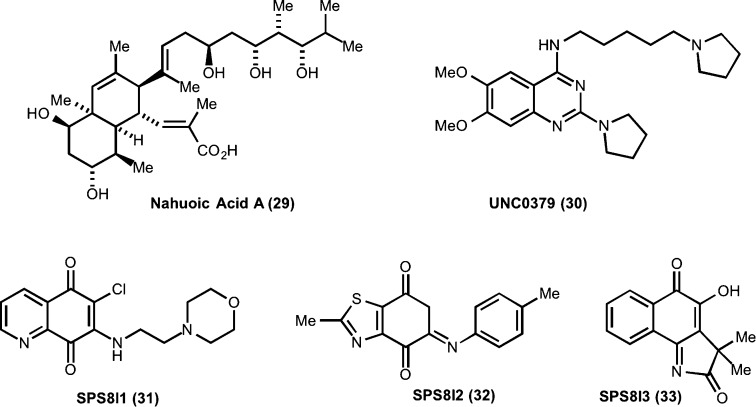
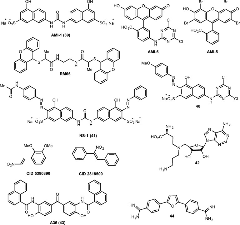
Mounting evidence suggests that protein methyltransferases (PMTs), which catalyze methylation of histone and nonhistone proteins, play a crucial role in diverse biological processes and human diseases. In particular, PMTs have been recognized as major players in regulating gene expression and chromatin state. PMTs are divided into two categories: protein lysine methyltransferases (PKMTs) and protein arginine methyltransferases (PRMTs). There has been a steadily growing interest in these enzymes as potential therapeutic targets and therefore discovery of PMT inhibitors has also been pursued increasingly over the past decade. Here, we present a perspective on selective, small-molecule inhibitors of PMTs with an emphasis on their discovery, characterization, and applicability as chemical tools for deciphering the target PMTs' physiological functions and involvement in human diseases. We highlight the current state of PMT inhibitors and discuss future directions and opportunities for PMT inhibitor discovery.


Figures

Figure 1
Figure 1
(A) Methylation of lysine (K) or arginine (R) residues of proteins by protein methyltransferases (PMTs). (B) Location of known methylation sites of histone 3 (H3) and histone 4 (H4). SAM, S-5′-adenosyl-l-methionine; SAH, S-5′-adenosyl-l-homocysteine.
Scheme 1
Scheme 1. Methylation States of Lysine and Arginine Residues
Figure 2
Figure 2
Phylogenetic tree of PMTs and known small-molecule inhibitors of PMTs.
Figure 3
Figure 3
Structures of (+)-chaetocin and related ETP compounds.
Figure 4
Figure 4
Structures of inhibitors 6 and 7.
Figure 5
Figure 5
X-ray co-crystal structure of the GLP–7 complex (PDB code: 3FPD). Protein residues, gray colored stick models; inhibitor, green colored stick model; and hydrogen bonds, red dashed line.
Figure 6
Figure 6
X-ray co-crystal structure of G9a–8 complex (PDB code: 3K5K). Inhibitor is shown as green colored stick model, and a fragment of the histone peptide (magenta) is transposed into this crystal structure to illustrate the lysine binding channel.
Figure 7
Figure 7
Structures of G9a and GLP inhibitors 8 and 9.
Figure 8
Figure 8
Structures of G9a and GLP inhibitors 1012 and their analogues.
Figure 9
Figure 9
Overlay of the X-ray co-crystal structure of G9a–8 complex (PDB code: 3K5K) with the one of the G9a–11 complex (PDB code: 3RJW). Inhibitor 8, green colored stick model; inhibitor 11, red colored stick model. A fragment of the histone peptide (magenta) was transposed into the crystal structures to illustrate the lysine binding channel.
Figure 10
Figure 10
Structure of the biotinylated G9a inhibitor 13.
Figure 11
Figure 11
Structures of compounds 1416.
Figure 12
Figure 12
X-ray co-crystal structure of the G9a–16 complex (PDB code: 4NVQ).
Figure 13
Figure 13
Structures of EZH2 inhibitors 17 and 18.
Figure 14
Figure 14
Structures of EZH2 inhibitors 19 and 20 and their analogues.
Figure 15
Figure 15
Structures of EZH2 and EZH1 inhibitor 21 and 21-based tool compounds.
Figure 16
Figure 16
Structures of EZH2 and EZH1 inhibitors 22 and 23.
Figure 17
Figure 17
Structures of inhibitors of H3K4 and H3K36 methyltransferases.
Figure 18
Figure 18
X-ray co-crystal structure of the SMYD2–25 complex (PDB code: 3S7B).
Figure 19
Figure 19
Structures of inhibitors 27 and 28.
Figure 20
Figure 20
Structures of SETD8 inhibitors.
Figure 21
Figure 21
Structures of DOT1L inhibitors.
Figure 22
Figure 22
Co-crystal structure of DOT1L–34 complex (PDB code: 4ER3) (left). Overlay of DOT1L–34 and DOT1L–37 complexes (PDB code: 4ER6) (right).
Figure 23
Figure 23
Structures of small-molecule inhibitors of PRMT1/PRMTs.
Figure 24
Figure 24
Structures of PRMT3 inhibitors.
Figure 25
Figure 25
Co-crystal structure of the dimeric PRMT3–45 complex (PDB code: 3SMQ) (left). Allosteric binding pocket and key interactions of 45 with PRMT3 (right).
Figure 26
Figure 26
Structures of selected inhibitors of CARM1 (PRMT4).

References

    1. Bernstein B. E.; Meissner A.; Lander E. S. The mammalian epigenome. Cell 2007, 128, 669–681. - PubMed
    1. Jones P. A.; Baylin S. B. The epigenomics of cancer. Cell 2007, 128, 683–692. - PMC - PubMed
    1. Berger S. L.; Kouzarides T.; Shiekhattar R.; Shilatifard A. An operational definition of epigenetics. Genes Dev. 2009, 23, 781–783. - PMC - PubMed
    1. Luger K.; Mader A. W.; Richmond R. K.; Sargent D. F.; Richmond T. J. Crystal structure of the nucleosome core particle at 2.8 Å resolution. Nature 1997, 389, 251–260. - PubMed
    1. Khorasanizadeh S. The nucleosome: from genomic organization to genomic regulation. Cell 2004, 116, 259–272. - PubMed

Publication types

MeSH terms