. 2015 Feb;77(2):363-9.
doi: 10.1038/pr.2014.183. Epub 2014 Nov 19.

Adrenergic receptor genotype influences heart failure severity and β-blocker response in children with dilated cardiomyopathy

Affiliations
Free PMC article

Adrenergic receptor genotype influences heart failure severity and β-blocker response in children with dilated cardiomyopathy

Sushma Reddy et al. Pediatr Res. 2015 Feb.
Free PMC article

Abstract

Background: Adrenergic receptor (ADR) genotypes are associated with heart failure (HF) and β-blocker response in adults. We assessed the influence of ADR genotypes in children with dilated cardiomyopathy (DCM).

Methods: Ninety-one children with advanced DCM and 44 with stable DCM were genotyped for three ADR genotypes associated with HF risk in adults: α2cdel322-325, β1Arg389, and β2Arg16. Data were analyzed by genotype and β-blocker use. Mean age at enrollment was 8.5 y.

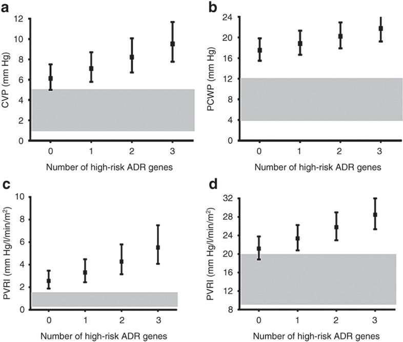
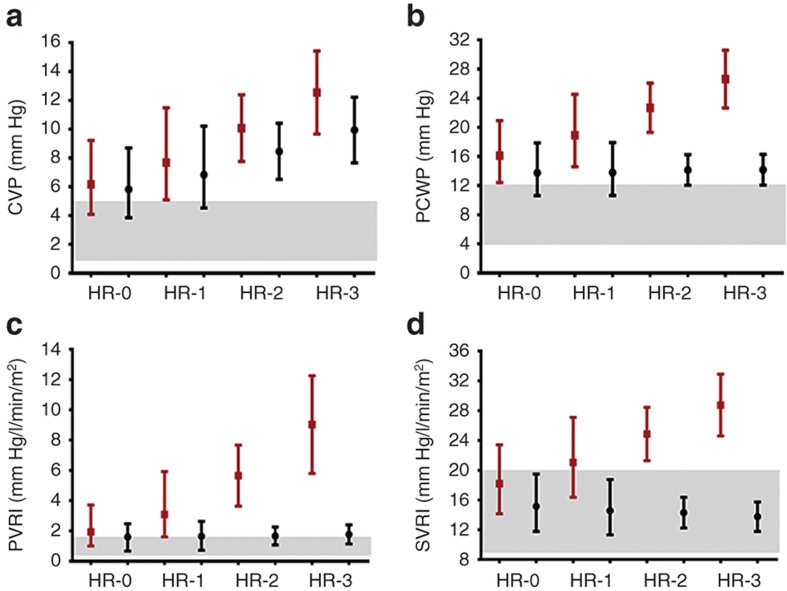
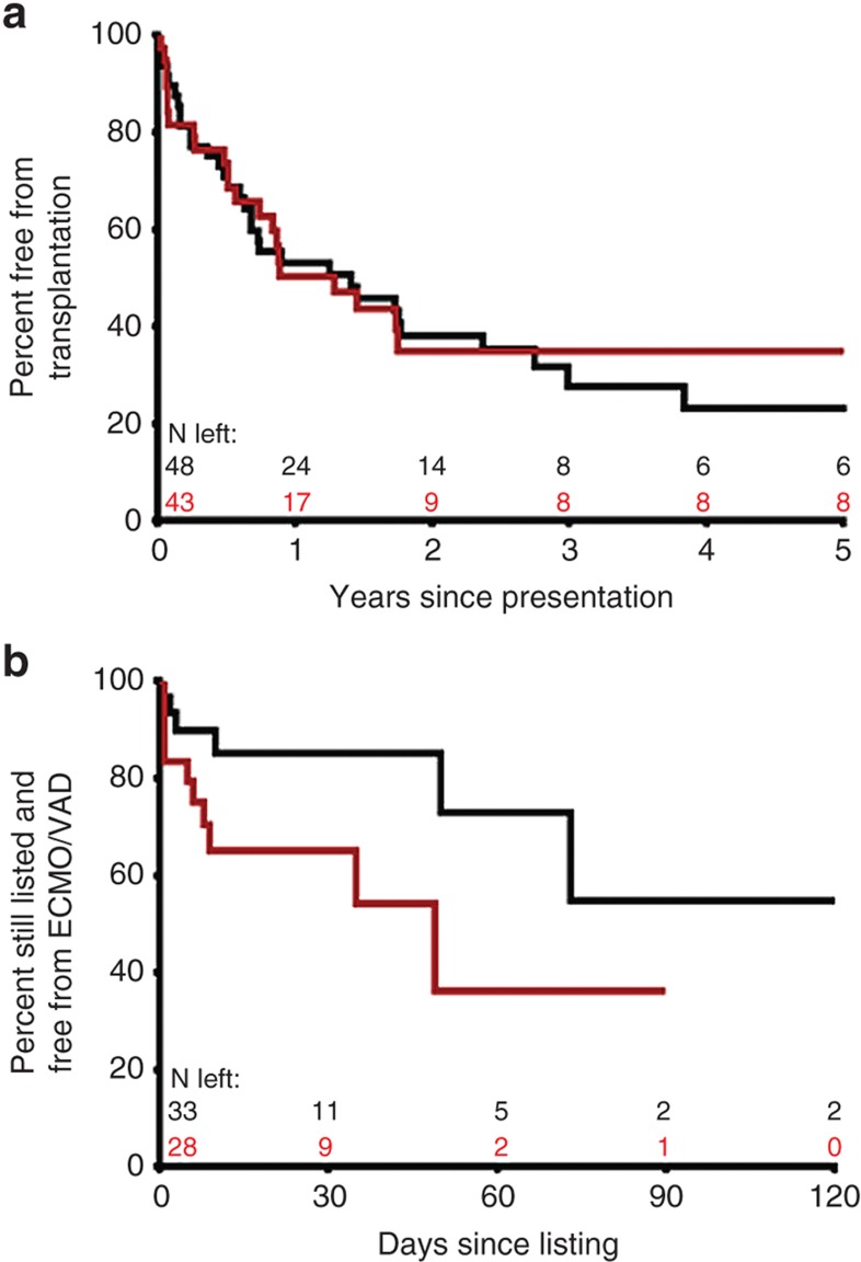
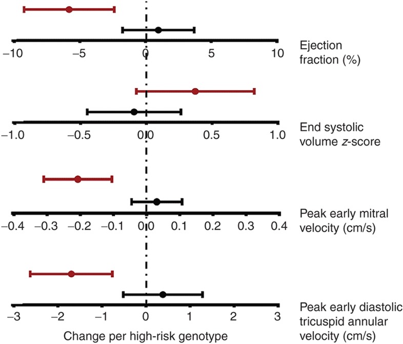
Results: One-year event-free survival was 51% in advanced and 80% in stable DCM. High-risk genotypes were associated with higher left ventricular (LV) filling pressures, higher systemic and pulmonary vascular resistance, greater decline in LV ejection fraction (P < 0.05), and a higher frequency of mechanical circulatory support while awaiting transplant (P = 0.05). While β-blockers did not reduce HF severity in the overall cohort, in the subset with multiple high-risk genotypes, those receiving β-blockers showed better preservation of cardiac function and hemodynamics compared with those not receiving β-blockers (interaction P < 0.05).

Conclusion: Our study identifies genetic risk markers that may help in the identification of patients at risk for developing decompensated HF and who may benefit from early institution of β-blocker therapy before progression to decompensated HF.


Figures

Figure 1
Figure 1
Cumulative effect of multiple adrenergic risk genotypes on cardiac hemodynamics (advanced HF cohort). The squares represent the point estimates from regression models, and the solid bars represent the 95% confidence interval around that estimate (n = 57). A higher number of adrenergic high-risk genotypes were associated with (a) higher CVP (P = 0.02), (b) higher PCWP (P = 0.06), (c) higher PVRI (P = 0.004), and (d) higher SVRI (P = 0.005). The gray zones represent normal hemodynamic ranges, i.e., CVP, 1–5 mm Hg; PCWP, 4–12 mm Hg; PVRI, 0.25–1.6 mm Hg/l/min/m2; and SVRI, 9–20 mm Hg/l/min/m2. ADR, adrenergic receptor; CVP, central venous pressure; PCWP, pulmonary capillary wedge pressure; PVRI, indexed pulmonary vascular resistance; SVRI, indexed systemic vascular resistance.
Figure 2
Figure 2
Interaction between adrenergic genotype and hemodynamic response to β-blockers (advanced HF cohort). The markers represent the point estimates (red: not on β-blockers, n = 27; black: on β-blockers, n = 28) from regression models, and the solid bars represent the 95% confidence interval around that estimate. Patients not receiving β-blockers showed (a) higher CVP (P = 0.02), (b) higher PCWP (P = 0.004), (c) higher PVRI (P = 0.0003), and (d) higher SVRI (P = 0.009) for every additional high-risk genotype. This increase in filling pressures and resistances was either blunted (CVP) or absent (PCWP, PVRI, and SVRI) in patients with high-risk genotypes receiving β-blockers. There was a significant interaction between ADR genotype and β-blocker therapy for PCWP (interaction P = 0.05), PVRI (interaction P = 0.03), and SVRI (interaction P = 0.02). Red, no β-blocker, black; β-blocker. The gray zones represent normal hemodynamic ranges, i.e., CVP, 1–5 mm Hg; PCWP, 4–12 mm Hg; PVRI, 0.25–1.6 mm Hg/l/min/m2; and SVRI, 9–20 mm Hg/l/min/m2. ADR, adrenergic receptor; CVP, central venous pressure; HR, high-risk genotype numbers; PCWP, pulmonary capillary wedge pressure; PVRI, indexed pulmonary vascular resistance; SVRI, indexed systemic vascular resistance.
Figure 3
Figure 3
Adrenergic genotype and event-free survival (advanced HF cohort). (a) Time to transplantation during follow-up was not different between patients with <2 (black, n = 48) vs. ≥2 (red, n = 43) ADR high-risk genotypes (hazard ratio: 0.88; 95% confidence interval: 0.52–1.46). (b) Patients listed for transplantation with ≥2 ADR high-risk genotypes (n = 28) showed lower freedom from mechanical circulatory support (VAD/ECMO) as a bridge to transplantation compared with those with <2 risk genotypes (n = 33) despite similar waiting times (hazard ratio: 2.57; 95% confidence interval: 1.05–7.23). Number of patients remaining at each time point are shown in black for patients with <2 high-risk genotypes and in red for patients with ≥2 high-risk genotypes. ADR, adrenergic receptor; ECMO, extracorporeal membrane oxygenation; VAD, ventricular assist device.
Figure 4
Figure 4
Adrenergic genotype and change in ventricular systolic and diastolic dysfunction (stable HF cohort). Regression analyses were performed to assess association of ADR genotypes with change in ventricular systolic and diastolic function on serial echocardiograms in 35 stable HF patients. Graphs show parameter estimate of change in echocardiographic measurements for each additional ADR risk genotype stratified by β-blocker use. Red, on no β-blockers; black, on β-blockers. There was a greater decline in LV ejection fraction for every additional ADR risk genotype, but this association was not seen in patients receiving β-blockers (interaction P = 0.003 vs. those not receiving β-blockers). There was a greater increase in LV end-systolic volume z-score for every additional ADR risk genotype, but this did not reach statistical significance between those receiving vs. those not receiving β-blockers (interaction P = 0.12). There was a greater decline in peak mitral E wave velocity for every additional ADR risk genotype, but this association was not seen in patients receiving β-blockers (interaction P < 0.001 vs. those not receiving β-blockers). There was a greater decline in early diastolic tricuspid annular velocity with each additional ADR risk genotype, but this association was not seen in patients receiving β-blockers (interaction P = 0.001 vs. those not receiving β-blockers) (n = 7 for 0, n = 22 for 1, n = 22 for 2, and n = 4 for 3 risk genotypes).

References

    1. Majewski H. Modulation of noradrenaline release through activation of presynaptic beta-adrenoreceptors. J Auton Pharmacol. 1983;3:47–60. - PubMed
    1. Brede M, Wiesmann F, Jahns R.et al.Feedback inhibition of catecholamine release by two different alpha2-adrenoceptor subtypes prevents progression of heart failure Circulation 20021062491–6. - PubMed
    1. Hein L, Kobilka BK. Adrenergic receptor signal transduction and regulation. Neuropharmacology. 1995;34:357–66. - PubMed
    1. Kaye DM, Smirk B, Finch S, Williams C, Esler MD. Interaction between cardiac sympathetic drive and heart rate in heart failure: modulation by adrenergic receptor genotype. J Am Coll Cardiol. 2004;44:2008–15. - PubMed
    1. Small KM, Wagoner LE, Levin AM, Kardia SL, Liggett SB. Synergistic polymorphisms of beta1- and alpha2C-adrenergic receptors and the risk of congestive heart failure. N Engl J Med. 2002;347:1135–42. - PubMed

Publication types

MeSH terms