. 2014 Nov 18;15(1):982.
doi: 10.1186/1471-2164-15-982.

Retinal transcriptome profiling at transcription start sites: a cap analysis of gene expression early after axonal injury

Affiliations

Retinal transcriptome profiling at transcription start sites: a cap analysis of gene expression early after axonal injury

Masayuki Yasuda et al. BMC Genomics. .

Abstract

Background: Glaucoma is characterized by progressive loss of the visual field and death of retinal ganglion cells (RGCs), a process that is mediated, in part, by axonal injury. However, the molecular pathomechanisms linking RGC death and axonal injury remain largely unknown. Here, we examined these mechanisms with a cap analysis of gene expression (CAGE), which allows the comprehensive quantification of transcription initiation across the entire genome. We aimed to identify changes in gene expression patterns and to predict the resulting alterations in the protein network in the early phases of axonal injury in mice.

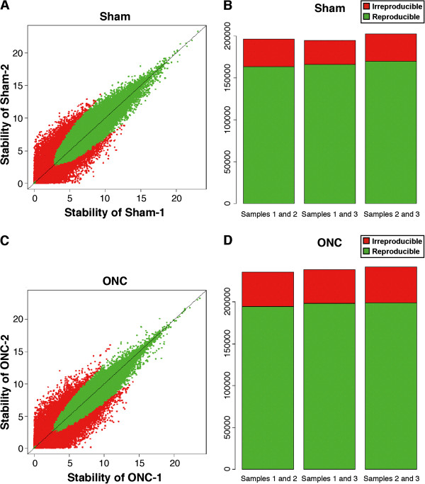
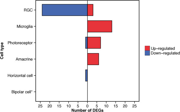
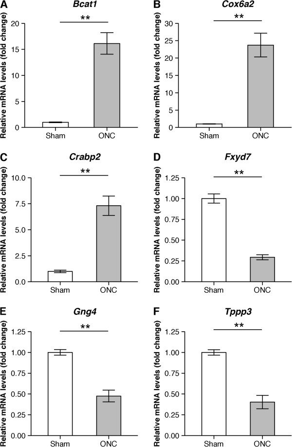
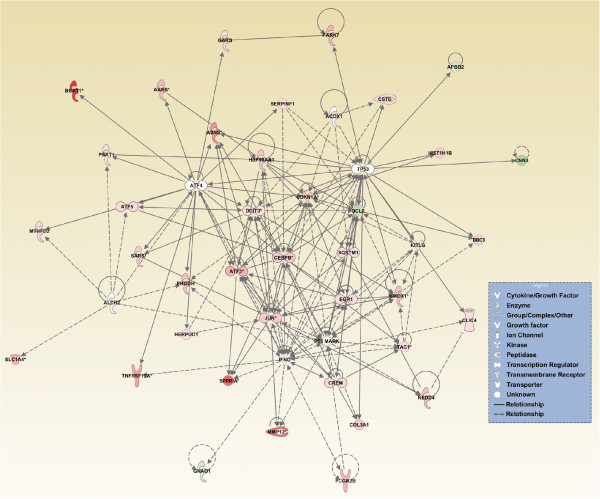
Results: We performed optic nerve crush (ONC) in mice to model axonal injury. Two days after ONC, the retinas were isolated, RNA was extracted, and a CAGE library was constructed and sequenced. CAGE data for ONC eyes and sham-treated eyes was compared, revealing 180 differentially expressed genes. Among them, the Bcat1 gene, involved in the catabolism of branched-chain amino acid transaminase, showed the largest change in expression (log2 fold-change=6.70). In some differentially expressed genes, alternative transcription start sites were observed in the ONC eyes, highlighting the dynamism of transcription initiation in a state of disease. In silico pathway analysis predicted that ATF4 was the most significant upstream regulator orchestrating pathological processes after ONC. Its downstream candidate targets included Ddit3, which is known to induce cell death under endoplasmic reticulum stress. In addition, a regulatory network comprising IFNG, P38 MAPK, and TP53 was predicted to be involved in the induction of cell death.

Conclusion: Through CAGE, we have identified differentially expressed genes that may account for the link between axonal injury and RGC death. Furthermore, an in silico pathway analysis provided a global view of alterations in the networks of key regulators of biological pathways that presumably take place in ONC. We thus believe that our study serves as a valuable resource to understand the molecular processes that define axonal injury-driven RGC death.


Figures

Figure 1
Figure 1
Stability and reproducibility of the biological replicates used for CAGE. The scatter plots show the correlation between expression values in replicates 1 and 2 in both the sham (A) and ONC (C) groups. Stability is a score used for clustering reproducible TSSs in multiple replicates. Green dots indicate high reproducibility and red dots indicate low reproducibility. The bar graphs represent a reproducibility evaluation for each pair of replicates in the sham (B) and ONC (D) groups. Green indicates TSS clusters with high reproducibility between the replicates. Red indicates TSS clusters with low reproducibility between the replicates.
Figure 2
Figure 2
Overall reduction in the RGC-specific transcriptome. The horizontal histogram shows the number of DEGs in each retinal cell type [59]. Red: up-regulated, Blue: down-regulated. *No DEGs specific to bipolar cells were detected in this study.
Figure 3
Figure 3
Validation of selected DEGs with qRT-PCR. Transcriptional changes in six selected DEGs were validated with qRT-PCR (A-F). The graphs show the level of mRNA expression in the ONC group relative to the sham group. The average expression for sham group was normalized as a 1.0-fold change. Values are mean ± SD (n = 5 in each group, **P < 0.01).
Figure 4
Figure 4
Predicted protein interaction networks involved in axonal injury. Prediction of upstream regulators was performed using IPA. The predicted upstream regulators (white nodes) were ATF4, IFNG, TP53, P38MAPK, ALDH2, and ACOX1. The targets of these upstream regulators are also displayed to illustrate the alteration in the interaction networks after ONC. Solid lines represent a direct interaction between two genes, whereas dotted lines represent an indirect relationship. The length of a line reflects the strength of reported evidence supporting the node-to-node relationship. The shapes of the nodes represent the different known biological roles of each of these molecules, as shown in the lower right inset. Red indicates up-regulated genes, green indicates down-regulated genes, and white indicates genes that were not annotated in these CAGE results, but that formed part of the network. *:targets that were duplicated in the dataset.

References

    1. Quigley HA, Broman AT. The number of people with glaucoma worldwide in 2010 and 2020. Br J Ophthalmol. 2006;90:262–267. doi: 10.1136/bjo.2005.081224. - DOI - PMC - PubMed
    1. Pederson JE, Anderson DR. The mode of progressive disc cupping in ocular hypertension and glaucoma. Arch Ophthalmol. 1980;98:490–495. doi: 10.1001/archopht.1980.01020030486010. - DOI - PubMed
    1. Heijl A, Leske MC, Bengtsson B, Hyman L, Bengtsson B, Hussein M. Reduction of intraocular pressure and glaucoma progression: results from the Early Manifest Glaucoma Trial. Arch Ophthalmol. 2002;120:1268–1279. doi: 10.1001/archopht.120.10.1268. - DOI - PubMed
    1. Collaborative Normal-Tension Glaucoma Study Group The effectiveness of intraocular pressure reduction in the treatment of normal-tension glaucoma. Am J Ophthalmol. 1998;126:498–505. doi: 10.1016/S0002-9394(98)00272-4. - DOI - PubMed
    1. Hollands H, Johnson D, Hollands S, Simel DL, Jinapriya D, Sharma S. Do findings on routine examination identify patients at risk for primary open-angle glaucoma? The rational clinical examination systematic review. JAMA. 2013;309:2035–2042. doi: 10.1001/jama.2013.5099. - DOI - PubMed

Publication types

MeSH terms

Substances