. 2014 Nov 18:14:446.
doi: 10.1186/1472-6882-14-446.

Antiproliferative effect of the jararhagin toxin on B16F10 murine melanoma

Affiliations

Antiproliferative effect of the jararhagin toxin on B16F10 murine melanoma

Durvanei Augusto Maria et al. BMC Complement Altern Med. .

Abstract

Background: Malignant melanoma is a less common but highly dangerous form of skin cancer; it starts in the melanocytes cells found in the outer layer of the skin. Jararhagin toxin, a metalloproteinase isolated from Bothrops jararaca snake venom acts upon several biological processes, as inflammation, pain, platelet aggregation, proliferation and apoptosis, though not yet approved for use, may one day be employed to treat tumors.

Methods: B16F10 murine melanoma cells were treated with jararhagin (jara), a disintegrin-like metalloproteinase isolated from Bothrops jararaca snake venom, and jari (catalytic domain inactivated with 1,10-phenanthroline). Viability and adhesion cells were evaluated by MTT assay. The expression of caspase-3 active, phases of the cell cycle and apoptosis were assessed by flow cytometry. We analyze in vivo the effects of jararhagin on melanoma growth, apoptosis and metastasis.

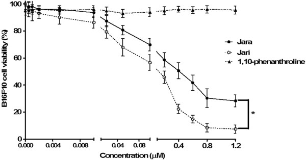
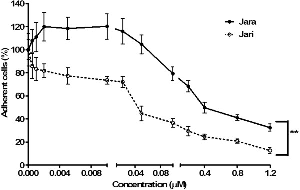
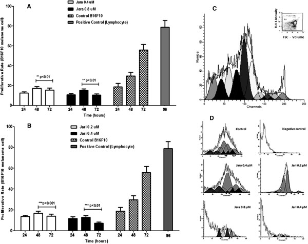
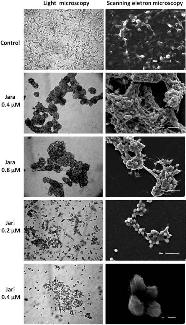
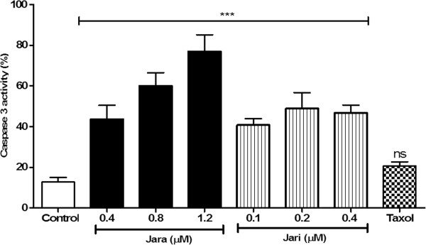
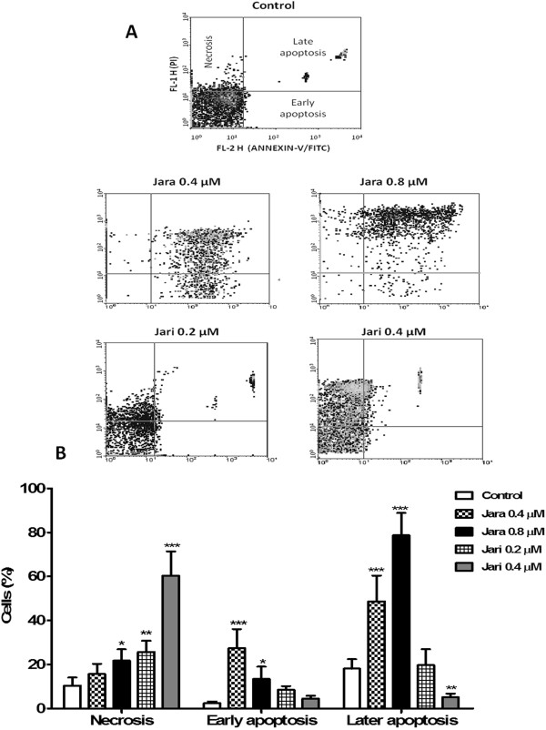
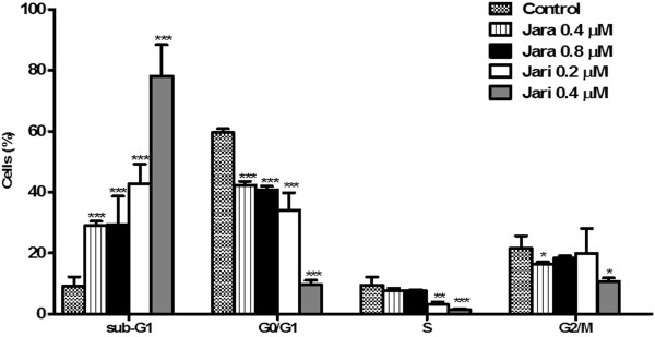
Results: The tumor cells acquired round shapes, lost cytoplasmic expansions, formed clusters in suspension and decreased viability. Jari was almost 20 times more potent toxin than jara based on IC50 values and on morphological changes of the cells, also observed by scanning electron microscopy. Flow cytometry analysis showed 48.3% decrease in the proliferation rate of cells and 47.2% increase in apoptosis (jara) and necrosis (jari), following 1.2 μM jara and 0.1 μM jari treatments. Caspase-3 activity was increased whereas G0/G1 cell cycle phase was on the decline. Proliferative rate was assessed by staining with 5,6-carboxyfluoresceindiacetate succinimidyl ester, showing a significant decrease in proliferation at all concentrations of both toxins.

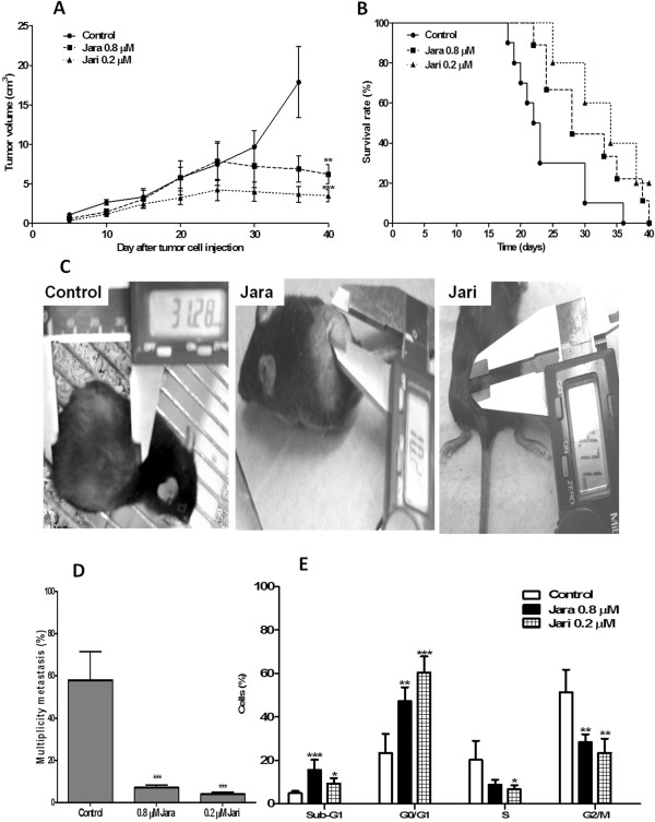
Conclusions: In vivo treatment of the toxins was observed reduction in the incidence of nodules, and metastasis and antiproliferative inhibition capacity. This data strengthens the potential use jararhagin as an anti-neoplastic drug.


Figures

Figure 1
Figure 1
Analysis of cell viability after treatment with toxins in B16F10 melanoma cell. The viability of B16F10 melanoma cells was determined by the MTT assay after 24 h treatment using varying concentrations of jara, jari, and the chelating agent 1,10-phenanthroline. The viability curves regarding jara and jari treatments showed significant differences according to the Student unpaired t test (p < 0.05).
Figure 2
Figure 2
Cell adhesion assay 24 hours after treatment with toxins in B16F10 melanoma cell. The percentage of adherent cells after treatments was significantly different for jara and jari (p < 0.01) using by Student unpaired t test. The results are representative of three independent experiments.
Figure 3
Figure 3
Proliferative rate of B16F10 melanoma cells. Toxins inhibit the proliferation and induce cell cycle arrest in B16F10 cells. Proliferation rates in B16F10 melanoma cells treated with jara and jari toxins for 24, 48 and 72 h determined using the ModFitLT 2.0 software. (A) The means proliferative rate using CFSE-DA assay of jara group; (B) CFSE-DA proliferation jari group; (C) CFSE-DA proliferation lymphocytes positive control (inset – dot plot representative); (D) Histograms represent the flow cytometric of CFSE-DA proliferative assay analyzes the proliferation of B16F10 melanoma cells after exposed to jara and jari toxins. All the experiments were repeated three times. Data are expressed as means ± SD. The statistical difference was obtained between control group non treated and toxins groups.
Figure 4
Figure 4
Morphological features of B16F10 cells exposed to toxins. The morphology of tumor cells was dramatically altered by toxin treatments, as observed by light microscopy. Scanning electron microscopy (SEM) showed the melanoma cells grown as a large, spreading monolayer, and the organization of the extracellular matrix with many protrusions. The cells showed retraction, and aggregates with 0.4 μM and 0.8 μM jara. Cells treated with jari showed detachment of the plate surface and formation of smaller aggregates (0.2 μM jari), and apoptotic bodies and necrosis debris (0.4 μM jari). Magnification 400× (microscopy), scale bar 30 μm and 3 μm (SEM).
Figure 5
Figure 5
Effects of toxins on caspase 3 active in B16F10 cells as evaluated by flow cytometry. Treatments induced significant increase of caspase 3 active as compared to untreated cells or Taxol chemotherapeutic agent. Data were analyzed by the ANOVA one way variance test. The results are representative mean ± SD of three independent experiments, *p < 0.05.
Figure 6
Figure 6
Analysis of apoptosis by flow cytometry. Induction of apoptosis in B16F10 cells by jara (0.4-0.8 μM) and jari (0.2-0.4 μM) for 24 h, as shown through Annexin-V/PI double staining by flow cytometry. The representative dotplot adquisitions showed apoptotic cells and necrosis areas (A). The apoptosis rate was estatistically significant for the percentage of early/late apoptotic cells after jara treatment, while jari showed significant increase in the percentage of necrotic cells (B). Data represent mean ± SD of three independent experiments. *p < 0.05 and ***p < 0.001.
Figure 7
Figure 7
Cell cycle analysis of B16F10 cells treated with jara and jari. Cells were stained with iodide propidium for DNA content analysis by flow cytometry. The bars represent the proportions of G2/M proliferative cells; in phase S synthesis; G0/G1 quiescent cell, and debris in sub-G1. The figure shows a significant decrease in the percentage of cells in G0/G1 phase and a subsequent increase in sub-G1 phase with increased concentration of jara and jari. Data represents mean ± SD from three independent experiments. *Significantly different from control *p < 0.05; **p < 0.01 and ***p < 0.001.
Figure 8
Figure 8
Inhibition of tumor growth and metastasis dorsal treated with jara toxins and jari. C57/Bl6J mice were administered via subcutaneous with B16F10 melanoma cells pretreated with 0.8 μM jara, 0.2 μM jari, and with untreated cells (control group). The growth curve (A) and macroscopic aspect of the melanoma dorsal tumor of the control and treated groups (B) are shown, as well as the Kaplan-Meier survival curve (C), metastasis multiplicity (D) and cell cycle phase of lung metastasis (E). Pre-treatment of tumor cells in vitro with jara or jari reduced the dorsal tumor volume of B16F10 melanoma cells, with statistically significant increase in survival rates with p < 0.05 (Log rank p < 0.018). The number of nodules in lung parenchyma was reduced on jara and jari groups. The distribution of cell cycle phase showed increase of tumor cells in sub-G1, fragmented DNA and decreased number of cells in proliferative response arrest in G2/M, caused by jara and jari on metastasis lung.

References

    1. Beviglia L, Stewart GJ, Niewiarowski S. Effect of four disintegrins on the adhesive and metastatic properties of B16F10 melanoma cells in a murine model. Oncogene Res. 1995;7:7–20. - PubMed
    1. Morris VL, Schmidt EE, Koop S, MacDonald IC, Grattan M, Khokha R, McLane MA, Niewiarowski S, Chambers AF, Groom AC. Effects of the disintegrin eristostatin on individual steps of hematogenous metastasis. Exp Cell Res. 1995;219:571–578. doi: 10.1006/excr.1995.1266. - DOI - PubMed
    1. Danen EHJ, Marcinkiewicz C, Cornelissen IMHA, Van Kraats AA, Pachter JA, Ruiter DJ, Niewiarowski S, Van Muijen GNP. The disintegrin eristostatin interferes with integrin α4β1 function and with experimental metastasis of human melanoma cells. Exp Cell Res. 1998;238:188–196. doi: 10.1006/excr.1997.3821. - DOI - PubMed
    1. Staiano N, Garbi C, Squillacioti C, Espósito S, Di Martino E, Belisario MA, Nitsch L, Di Natale P. Echistatin induces decrease of pp125FAK phosphorilation, disassembly of actin cytoskeleton and focal adhesions, and detachment of fibronectin-adherent melanoma cells. Eur J Cell Biol. 1997;73:298–305. - PubMed
    1. Della Morte R, Squillacioti C, Garbi C, Derkinderen P, Maria A, Belisario MA, Girault JA, Natale PD, Nitsch L, Staiano N. Echistatin inhibits pp125FAK autophosphorylation, paxillin phosphorylation and pp125FAK±paxillin interaction in fibronectin-adherent melanoma cells. Eur J Biochem. 2000;267:5047–5054. doi: 10.1046/j.1432-1327.2000.01561.x. - DOI - PubMed
Pre-publication history
    1. The pre-publication history for this paper can be accessed here: http://www.biomedcentral.com/1472-6882/14/446/prepub

Publication types

MeSH terms