Review
. 2014 Nov 18:9:51.
doi: 10.1186/1750-1326-9-51.

Models of β-amyloid induced Tau-pathology: the long and "folded" road to understand the mechanism

Affiliations
Review

Models of β-amyloid induced Tau-pathology: the long and "folded" road to understand the mechanism

Ilie-Cosmin Stancu et al. Mol Neurodegener. .

Abstract

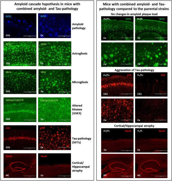
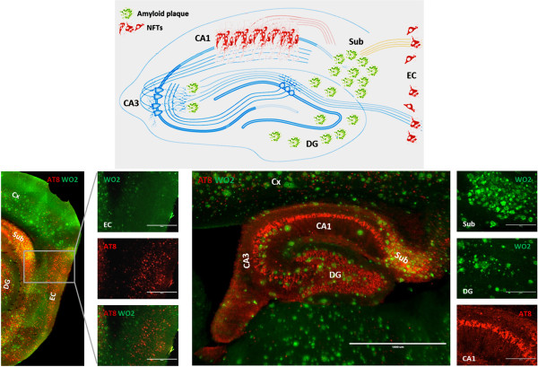
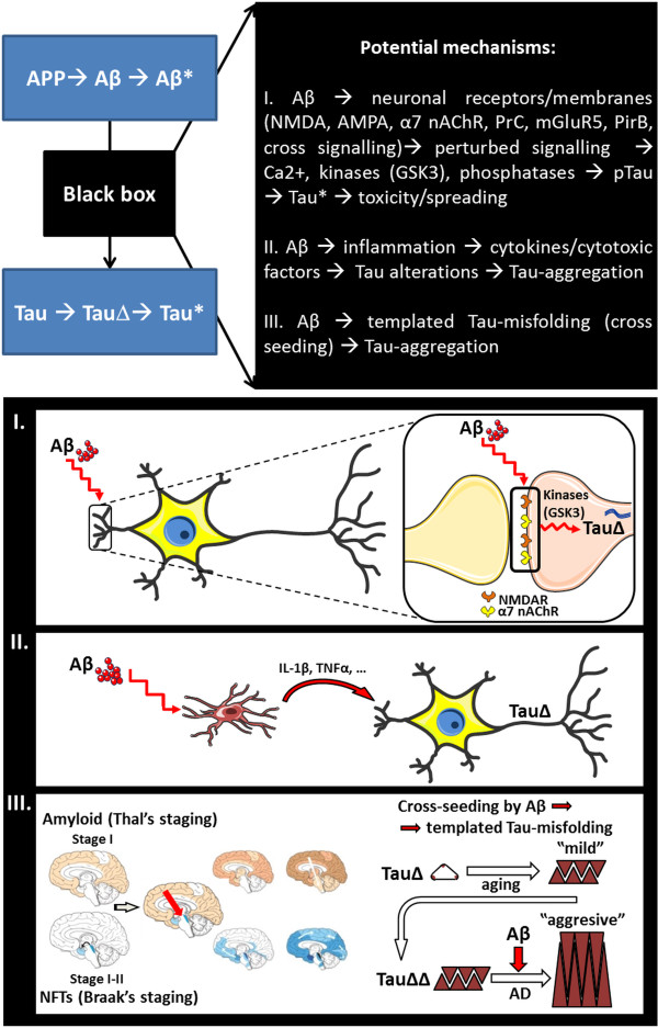
The amyloid cascade hypothesis has been the prevailing hypothesis in Alzheimer's Disease research, although the final and most wanted proof i.e. fully successful anti-amyloid clinical trials in patients, is still lacking. This may require a better in depth understanding of the cascade. Particularly, the exact toxic forms of Aβ and Tau, the molecular link between them and their respective contributions to the disease process need to be identified in detail. Although the lack of final proof has raised substantial criticism on the hypothesis per se, accumulating experimental evidence in in vitro models, in vivo models and from biomarkers analysis in patients supports the amyloid cascade and particularly Aβ-induced Tau-pathology, which is the focus of this review. We here discuss available models that recapitulate Aβ-induced Tau-pathology and review some potential underlying mechanisms. The availability and diversity of these models that mimic the amyloid cascade partially or more complete, provide tools to study remaining questions, which are crucial for development of therapeutic strategies for Alzheimer's Disease.


Figures

Figure 1
Figure 1
Amyloid cascade hypothesis in transgenic mice with combined amyloid and Tau pathology (left panel). Hippocampal (HC; CA1) and cortical (Cx) immunohistochemical staining for amyloid plaques (anti-Aβ, WO2), astrocytes (GFAP), microglia (Iba1), P-GSK3 (GSK3pT216/279), P-Tau (anti-pTau, AT-8) and NeuN for cortical/hippocampal atrophy (all images are 20×, except NeuN at 4×) is presented in transgenic mice expressing mutant APP/PS1 and mutant Tau [55]. These mice were generated by crossing mutant APP/PS1 and mutant Tau mice, respectively denoted as F+/T- (5xFAD; [59]) and F-/T+(TauP301S; [60]). Of note, AT-8 staining was optimized to match Gallyas silver staining patterns, hence representing NFTs (data not shown). Main pathological features found in F+/T+ mice (right panel). Immunolabeling with anti-Aβ (WO2), showing no changes in amyloid plaque load in the cortex of F+/T + and F+/T- mice (upper panel, 4× and 20×); Aggravation of Tau-pathology, anti-pTau (AT-8) staining (NFT), in hippocampal CA1 region and cortex of F+/T+ compared to F-/T+ parental strain transgenic mice (middle panel, 20×); anti-neuronal nuclear staining (NeuN) showing decreased cortical and hippocampal area in F+/T+ compared to F-/T- mice (lower panel, 4×).
Figure 2
Figure 2
Graphical representation of the regional distribution of amyloid plaques and NFTs in hippocampus and entorhinal cortex (upper panel, hippocampal-entorhinal connectivity map modified after Deng et al. [61]). Analysis of the relation between amyloid plaques (anti-Aβ, WO2, green) and Tau-pathology (anti-pTau, AT-8, red) in entorhinal cortex (lower left panel) and hippocampus (lower right panel) of F+/T+ transgenic mice, with details of the regional distribution of amyloid- and Tau-pathology for each of these regions.
Figure 3
Figure 3
Schematic presentation of proposed mechanisms involved in Aβ-induced Tau-aggregation as discussed in this review (elements in panel I. and II. modified from Servier Medical Art; elements in panel III. modified from Jucker and Walker [76] , Thal et al. [75] , Braak and Braak [73] ).

References

    1. Hardy J, Selkoe DJ. The amyloid hypothesis of Alzheimer’s disease: progress and problems on the road to therapeutics. Science. 2002;297(5580):353–356. doi: 10.1126/science.1072994. - DOI - PubMed
    1. Spires-Jones TL, Stoothoff WH, de CA, Jones PB, Hyman BT. Tau pathophysiology in neurodegeneration: a tangled issue. Trends Neurosci. 2009;32(3):150–159. doi: 10.1016/j.tins.2008.11.007. - DOI - PubMed
    1. Perrin RJ, Fagan AM, Holtzman DM. Multimodal techniques for diagnosis and prognosis of Alzheimer’s disease. Nature. 2009;461(7266):916–922. doi: 10.1038/nature08538. - DOI - PMC - PubMed
    1. Selkoe DJ. Alzheimer’s disease. Cold Spring Harb Perspect Biol. 2011;3(7):a004457. doi: 10.1101/cshperspect.a004457. - DOI - PMC - PubMed
    1. Serrano-Pozo A, Frosch MP, Masliah E, Hyman BT. Neuropathological alterations in Alzheimer disease. Cold Spring Harb Perspect Med. 2011;1(1):a006189. doi: 10.1101/cshperspect.a006189. - DOI - PMC - PubMed

Publication types