

The proteome of human retina
- PMID: 25407473
- PMCID: PMC4329094
- DOI: 10.1002/pmic.201400397
The proteome of human retina
Abstract
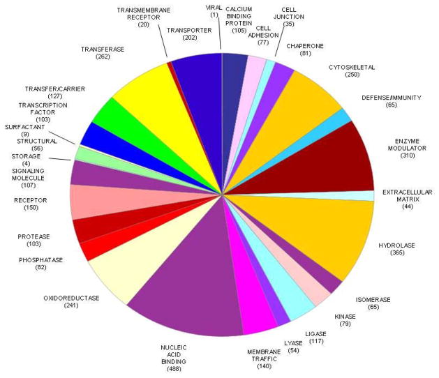
The retina is a delicate tissue that detects light, converts photochemical energy into neural signals, and transmits the signals to the visual cortex of the brain. A detailed protein inventory of the proteome of the normal human eye may provide a foundation for new investigations into both the physiology of the retina and the pathophysiology of retinal diseases. To provide an inventory, proteins were extracted from five retinas of normal eyes and fractionated using SDS-PAGE. After in-gel digestion, peptides were analyzed in duplicate using LC-MS/MS on an Orbitrap Elite mass spectrometer. A total of 3436 nonredundant proteins were identified in the human retina, including 20 unambiguous protein isoforms, of which eight have not previously been demonstrated to exist at the protein level. The proteins identified in the retina included most of the enzymes involved in the visual cycle and retinoid metabolism. One hundred and fifty-eight proteins that have been associated with age-related macular degeneration were identified in the retina. The MS proteome database of the human retina may serve as a valuable resource for future investigations of retinal biology and disease. All MS data have been deposited in the ProteomeXchange with identifier PXD001242 (http://proteomecentral.proteomexchange.org/dataset/PXD001242).
Keywords: Age-related macular degeneration; Biomedicine; Eye; Human; Phototransduction; Retina.
© 2014 WILEY-VCH Verlag GmbH & Co. KGaA, Weinheim.
Conflict of interest statement
Craig Dufresne is an employee of Thermo Fisher Scientific. The remaining authors have declared no conflict of interest.
Figures
Comment in
-
The proteomes of the human eye, a highly compartmentalized organ.Proteomics. 2017 Jan;17(1-2):10.1002/pmic.201600340. doi: 10.1002/pmic.201600340. Epub 2016 Dec 21. Proteomics. 2017. PMID: 27860232 Free PMC article.
References
-
- Jager RD, Meiler WF, Miller JW. Age-related macular degeneration. N Engl J Med. 2008;358:2606–2617. - PubMed
-
- Antonetti DA, Klein R, Gardner TW. Diabetic retinopathy. N Engl J Med. 2012;366:1227–1239. - PubMed
-
- Kumagai K, Ogino N, Hangai M, Larson E. Percentage of fellow eyes that develop full-thickness macular hole in patients with unilateral macular hole. Arch Ophthalmol. 2012;130:393–394. - PubMed
Publication types
MeSH terms
Substances
Grants and funding
LinkOut - more resources
Full Text Sources
Other Literature Sources
Molecular Biology Databases
