. 2014 Nov 17;15(11):21179-201.
doi: 10.3390/ijms151121179.

The stability of G6PD is affected by mutations with different clinical phenotypes

Affiliations

The stability of G6PD is affected by mutations with different clinical phenotypes

Saúl Gómez-Manzo et al. Int J Mol Sci. .

Abstract

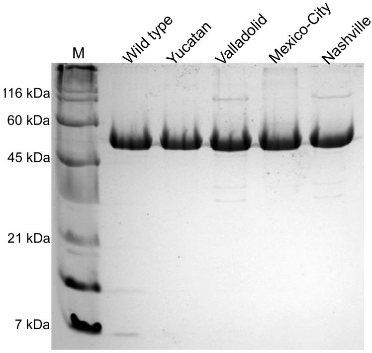
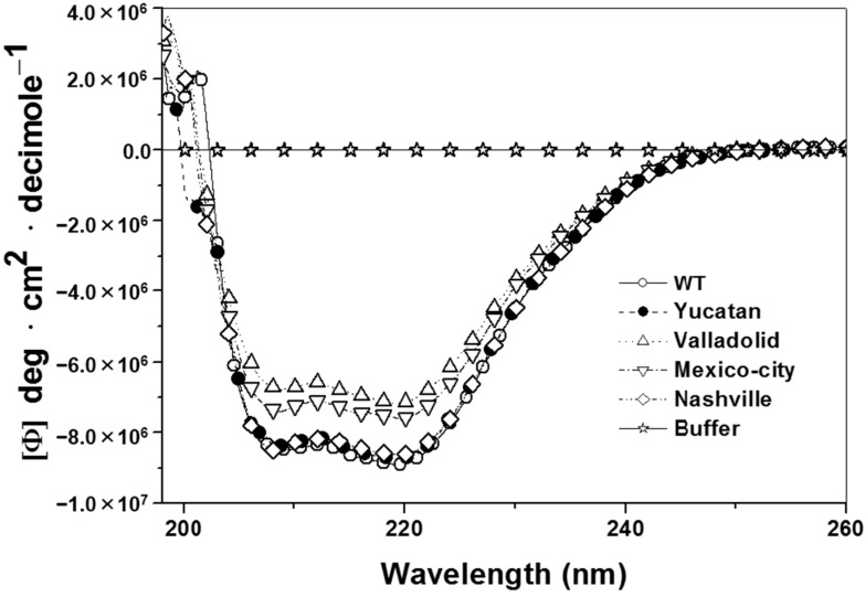
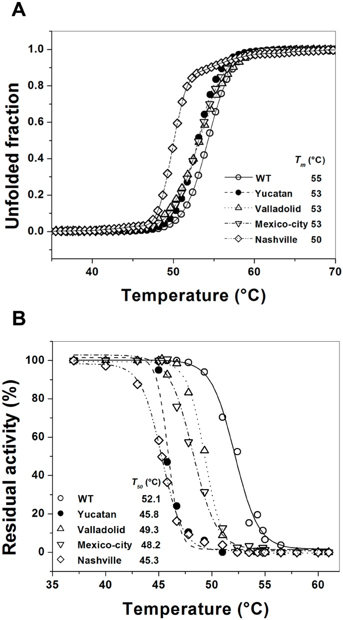
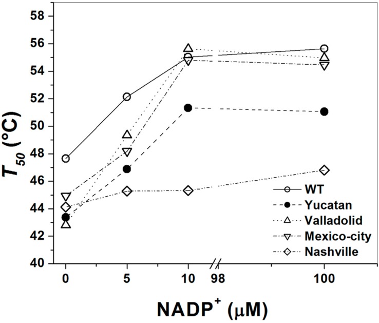
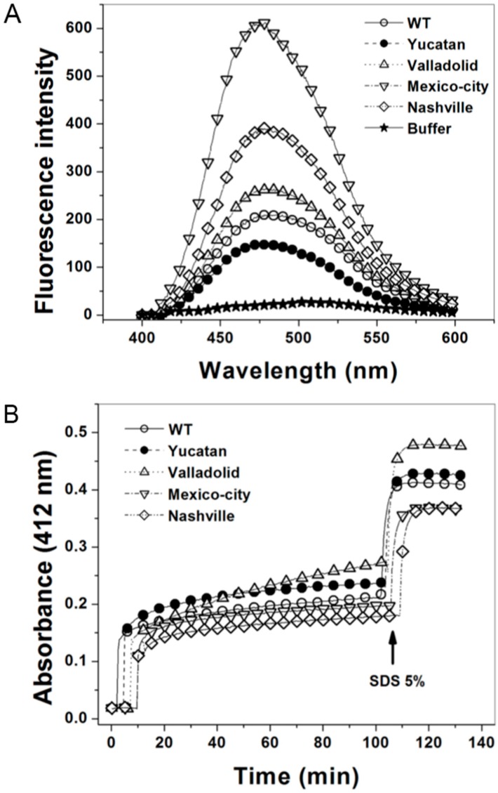
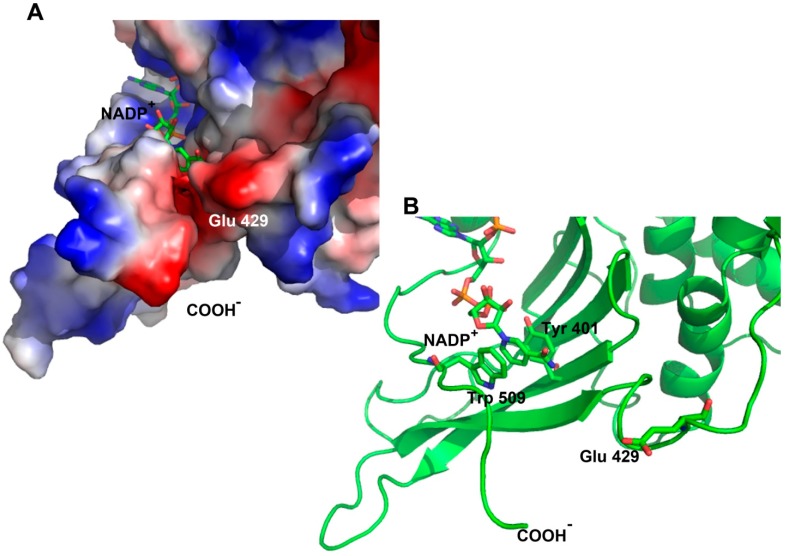
Glucose-6-phosphate dehydrogenase (G6PD) deficiency is the most common enzyme deficiency worldwide, causing a wide spectrum of conditions with severity classified from the mildest (Class IV) to the most severe (Class I). To correlate mutation sites in the G6PD with the resulting phenotypes, we studied four naturally occurring G6PD variants: Yucatan, Nashville, Valladolid and Mexico City. For this purpose, we developed a successful over-expression method that constitutes an easier and more precise method for obtaining and characterizing these enzymes. The k(cat) (catalytic constant) of all the studied variants was lower than in the wild-type. The structural rigidity might be the cause and the most evident consequence of the mutations is their impact on protein stability and folding, as can be observed from the protein yield, the T50 (temperature where 50% of its original activity is retained) values, and differences on hydrophobic regions. The mutations corresponding to more severe phenotypes are related to the structural NADP+ region. This was clearly observed for the Classes III and II variants, which became more thermostable with increasing NADP+, whereas the Class I variants remained thermolabile. The mutations produce repulsive electric charges that, in the case of the Yucatan variant, promote increased disorder of the C-terminus and consequently affect the binding of NADP+, leading to enzyme instability.


Figures

Figure 1
Figure 1
Construction of E. coli BL21(DE3)Δzwf::kanr mutant. (A) Representation of gene arrangement in E. coli BL21(DE3)Δzwf::kanr generated by homologous recombination. The primers used for mutant analysis and the PCR probe amplified for each construct are shown. The specific primers were located upstream and downstream from the zwf gene (−100 bp zwf and +100 bp zwf), and the primers for kanamycin resistance gene amplification (K1 and K2) were internal; (B) Generation of E. coli BL21(DE3)Δzwf::kanr by knockout of the zwf gene was confirmed by PCR. M: Molecular weight marker; Lane 1 shows the PCR product using gDNA from E. coli BL21(DE3) as a template and the primers −100 bp zwf Forward (Fw) and +100 bp zwf Reverse (Rv); Lane 2 shows the PCR product with gDNA from E. coli BL21(DE3)Δzwf::kanr and the primers −100 bp zwf Fw and +100 bp zwf Rv; Lane 3 corresponds to the PCR product using gDNA from E. coli BL21(DE3)Δzwf::kanr and the specific primers +100 bp zwf Rv and K1; Lane 4 corresponds to the PCR product using gDNA from E. coli BL21(DE3)Δzwf::kanr and the primers K2 and +100 bp zwf Rv. In all cases, 100 ng of gDNA was used as template; and (C) Specific activity of glucose-6-phosphate dehydrogenase (G6PD) measured in crude extract of both E. coli BL21(DE3) and E. coli BL21(DE3)Δzwf::kanr cells. The reaction was started with 10 µL of crude extract in the mixture reaction containing 1 mM G6P and 2 mM NADP+ in 50 mM Tris-HCl buffer, pH 8.0.
Figure 1
Figure 1
Construction of E. coli BL21(DE3)Δzwf::kanr mutant. (A) Representation of gene arrangement in E. coli BL21(DE3)Δzwf::kanr generated by homologous recombination. The primers used for mutant analysis and the PCR probe amplified for each construct are shown. The specific primers were located upstream and downstream from the zwf gene (−100 bp zwf and +100 bp zwf), and the primers for kanamycin resistance gene amplification (K1 and K2) were internal; (B) Generation of E. coli BL21(DE3)Δzwf::kanr by knockout of the zwf gene was confirmed by PCR. M: Molecular weight marker; Lane 1 shows the PCR product using gDNA from E. coli BL21(DE3) as a template and the primers −100 bp zwf Forward (Fw) and +100 bp zwf Reverse (Rv); Lane 2 shows the PCR product with gDNA from E. coli BL21(DE3)Δzwf::kanr and the primers −100 bp zwf Fw and +100 bp zwf Rv; Lane 3 corresponds to the PCR product using gDNA from E. coli BL21(DE3)Δzwf::kanr and the specific primers +100 bp zwf Rv and K1; Lane 4 corresponds to the PCR product using gDNA from E. coli BL21(DE3)Δzwf::kanr and the primers K2 and +100 bp zwf Rv. In all cases, 100 ng of gDNA was used as template; and (C) Specific activity of glucose-6-phosphate dehydrogenase (G6PD) measured in crude extract of both E. coli BL21(DE3) and E. coli BL21(DE3)Δzwf::kanr cells. The reaction was started with 10 µL of crude extract in the mixture reaction containing 1 mM G6P and 2 mM NADP+ in 50 mM Tris-HCl buffer, pH 8.0.
Figure 2
Figure 2
Purification of human recombinant glucose-6-phosphate dehydrogenase (G6PD) enzymes. SDS-PAGE of purified G6PD enzymes. M: molecular weight marker; Lanes 25: WT G6PD and G6PD variants. Each lane was loaded with 10 µg of protein and visualized using coomassie brilliant blue.
Figure 3
Figure 3
Structural characterization of human recombinant wild-type (WT) G6PD and mutants. Far-UV circular dichroism (CD) spectra of WT G6PD and mutants are shown. The protein concentration was 0.8 mg/mL in all cases. The experiments were performed in duplicate; standard errors were less than 5%.
Figure 4
Figure 4
Thermal stability of human G6PD enzymes. (A) Thermal unfolding of WT G6PD and the mutants (0.8 mg/mL) in 25 mM NaPO4 pH 7.4 was monitored by recording the change in CD signal at 222 nm at different temperatures ranging from 20 to 90 °C. The unfolded fraction of protein and the Tm (melting temperature midpoint of the transition values) (inset) were calculated as previously reported [22]; and (B) Thermal inactivation assays of WT G6PD and the mutants after incubation for 20 min at the indicated temperature. The T50 (temperature where 50% of its original activity is retained) after incubation at different temperatures for 20 min is shown. In all cases, 200 ng of total protein was used. The assays were performed in duplicate; standard errors were lower than 5%.
Figure 5
Figure 5
Effect of NADP+ on the thermoinactivation of recombinant G6PD enzymes. T50 is plotted against the NADP+ concentration. The WT and mutant enzymes were incubated at different NADP+ concentrations. In all cases, 200 ng of total protein was used.
Figure 6
Figure 6
8-Anilinonaphthalene-1-sulphonate (ANS) and 5,5'-dithiobis-2-nitrobenzoic acid (DTNB) accessibility assays of WT G6PD and the four clinical variants. (A) ANS fluorescence spectra of WT G6PD and the four clinical mutants were obtained using an excitation wavelength of 395 nm and recording emission spectra from 400 to 600 nm; the final concentrations of ANS and enzyme were 165 μM and 400 μg/mL, respectively. Values obtained from buffer containing ANS without protein (open stars) were subtracted from the recordings with protein; and (B) The derivatization of Cys residues in G6PD (200 µg/mL) in 1 mM DTNB was followed spectrophotometrically at 412 nm. The arrow indicates the time of addition of 5% SDS, after which the number of Cys per monomer was calculated for the enzymes. Each trace is the average of two independent experiments.
Figure 7
Figure 7
Schematic representation of human G6PD enzyme (PDB code 2BH9). (A) Surface electrostatic potential of the crystallographic structure of the G6PD; and (B) G6PD in ribbon format, shown are π–π interactions between the amino acid residues Try 509 and Tyr 401 with the pyramidal ring of NADP+. Modeled with PyMOL [30].

References

    1. Luzzatto L., Mehta A., Vulliamy T.J. Glucose-6-phosphate dehydrogenase deficiency. In: Scriver C.R., Beaudet A.L., et al., editors. Metabolic and Molecular Bases of Inherited Disease. 8th ed. Volume 179. McGraw-Hill; New York, NY, USA: 2001. pp. 4517–4553.
    1. Gaetani G.F., Galiano S., Canepa L., Ferraris A.M., Kirkman H.N. Catalase and glutathione peroxidase are equally active in detoxification of hydrogen peroxide in human erythrocytes. Blood. 1989;73:334–339. - PubMed
    1. Vulliamy T., Luzzatto L., Hirono A., Beutler E. Hematologically important mutations: Glucose-6-phosphate dehydrogenase. Blood Cells Mol. Dis. 1997;23:302–313. doi: 10.1006/bcmd.1997.0147. - DOI - PubMed
    1. Mason P.J., Bautista J.M., Gilsanz F. G6PD deficiency: The genotype-phenotype association. Blood Rev. 2007;5:267–283. doi: 10.1016/j.blre.2007.05.002. - DOI - PubMed
    1. Minucci B., Giardina C., Zuppi E. Glucose-6-phosphate dehydrogenase laboratory assay: How, when, and why? IUBMB Life. 2009;61:27–34. doi: 10.1002/iub.137. - DOI - PubMed

Publication types

MeSH terms

Substances

LinkOut - more resources