. 2014 Nov 19:11:102.
doi: 10.1186/s12977-014-0102-z.

Human semen contains exosomes with potent anti-HIV-1 activity

Affiliations

Human semen contains exosomes with potent anti-HIV-1 activity

Marisa N Madison et al. Retrovirology. .

Abstract

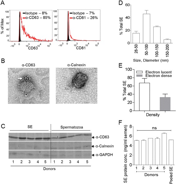
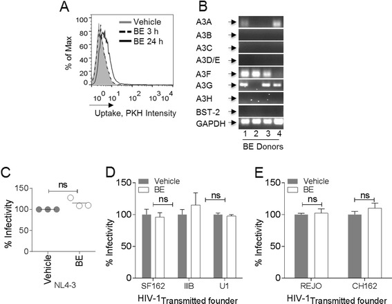
Background: Exosomes are membranous nanovesicles secreted into the extracellular milieu by diverse cell types. Exosomes facilitate intercellular communication, modulate cellular pheno/genotype, and regulate microbial pathogenesis. Although human semen contains exosomes, their role in regulating infection with viruses that are sexually transmitted remains unknown. In this study, we used semen exosomes purified from healthy human donors to evaluate the role of exosomes on the infectivity of different strains of HIV-1 in a variety of cell lines.

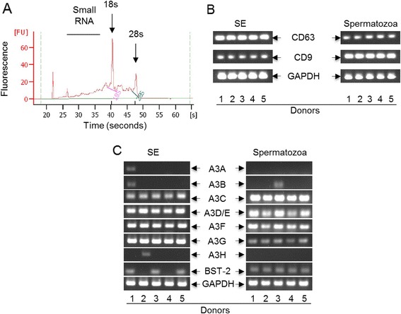
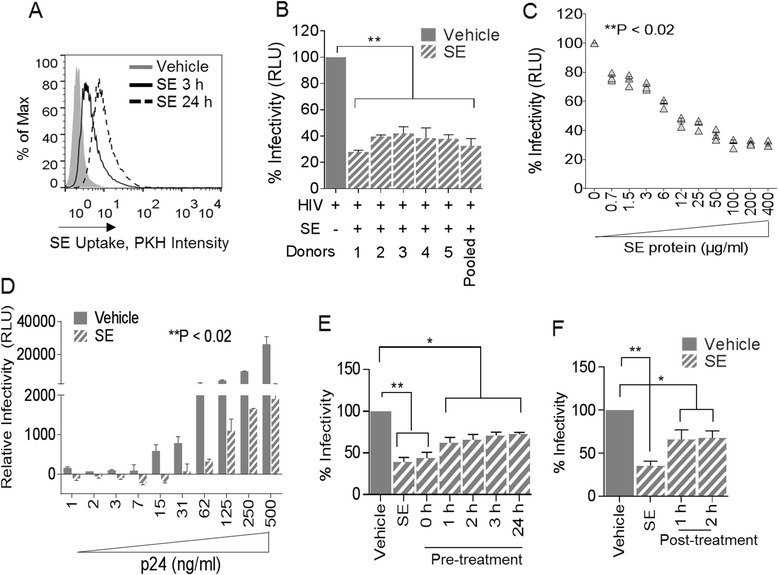
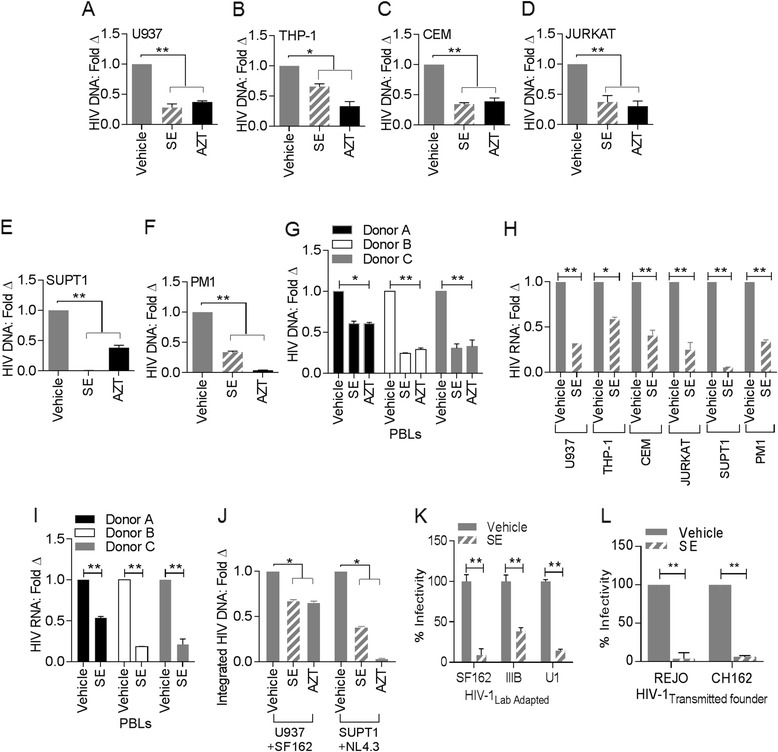
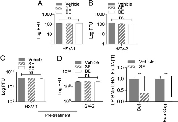
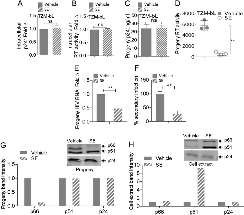
Results: We show that human semen contains a heterologous population of exosomes, enriched in mRNA encoding tetraspanin exosomal markers and various antiviral factors. Semen exosomes are internalized by recipient cells and upon internalization, inhibit replication of a broad array of HIV-1 strains. Remarkably, the anti-HIV-1 activity of semen exosomes is specific to retroviruses because semen exosomes blocked replication of the murine AIDS (mAIDS) virus complex (LP-BM5). However, exosomes from blood had no effect on HIV-1 or LP-BM5 replication. Additionally, semen and blood exosomes had no effect on replication of herpes simplex virus; types 1 and 2 (HSV1 and HSV2). Mechanistic studies indicate that semen exosomes exert a post-entry block on HIV-1 replication by orchestrating deleterious effects on particle-associated reverse transcriptase activity and infectivity.

Conclusions: These illuminating findings i) improve our knowledge of the cargo of semen exosomes, ii) reveal that semen exosomes possess anti-retroviral activity, and iii) suggest that semen exosome-mediated inhibition of HIV-1 replication may provide novel opportunities for the development of new therapeutics for HIV-1.


Figures

Figure 1
Figure 1
Characteristics of human semen exosomes. (A) Fluorescence-activated cell sorting (FACS) analysis of pooled (n =20) SE incubated with α-MHC-II antibody coated latex beads (4 μm) and stained for CD63 (Left, red histogram) and CD81 (Right, red histogram). Isotype control antibody is represented by black histograms. (B) Left, transmission electron microscopy (TEM) of pooled (n =20) semen exosome (SE) stained for CD63 (α-CD63) and detected by 6 nm gold-labeled secondary antibody (arrow). Right, TEM of pooled (n =20) SE stained for Calnexin (α- Calnexin) and counterstained with 10 nm gold-labeled secondary antibody. Calnexin was not detected. (C) Western blot of SE (n =5) and paired spermatozoa protein extracts, stained for CD63, Calnexin, and GAPDH. Pooled SE population (n =89) were categorized based on (D) size and (E) electron density. (F) SE protein concentrations from different donors range between 5.3 – 6.8 mg/ml of semen.
Figure 2
Figure 2
Human semen contains exosomes loaded with coding mRNA. (A) RNA analysis with the Bioanalyzer revealed that SE contains different RNA species. Arrows show peaks for 18 s, 28 s, and the horizontal line represents small RNA. (B and C) PCR analysis of tetraspanin (CD9 and CD63) mRNA and host restriction factors (HRFs) mRNA present in paired SE and spermatozoa samples. Numbers 1 to 5 represent different donors. GAPDH is present in SE and spermatozoa and was used as loading control.
Figure 3
Figure 3
Semen exosomes inhibit HIV-1 replication. (A) TZM-bl cells were evaluated for cellular uptake of green fluorochrome-labelled SE by FACS. PKH67Green-labelled PBS (gray histogram) was used as control. (B) TZM-bl indicator cells challenged with HIV-1NL4.3 pre-incubated with PBS Vehicle or with 100 μg/ml individual donor SE, or pooled (n =20) SE. Infectivity was determined by luminescence output with control set at 100% infection. (C) TZM-bl indicator cells challenged with HIV-1NL4.3 pre-incubated with increasing concentrations (0–400 μg/ml) of SE and analyzed for infectivity by luminescence output. (D) TZM-bl cells were challenged with 100 μg/ml of SE pre-incubated with increasing concentrations (1 – 500 ng/ml p24) of HIV-1NL4.3. Infectivity was determined by luminescence output. (E) HIV-1NL4.3 was pre-incubated for 1 h with vehicle (gray bar, vehicle) or with SE (SE), or added simultaneously with SE (0 h), or added prior (1 h, 2 h, 3 h, and 24 h) to HIV-1 challenge. Infectivity on TZM-bl cells was measured by luminescence emission with vehicle set at 100% infection. (F) HIV-1NL4.3 was pre-incubated for 1 h with vehicle (gray bar, vehicle) or with SE (SE), or added directly to TZM-bl indicator cells at 1 h or 2 h post-infection. Infectivity was measured by luminescence emission with vehicle set at 100%. Student’s t test was used for all samples, significance was taken at *P <0.05, **P <0.02. Experiments were repeated at least 3 times with similar results.
Figure 4
Figure 4
Cell type- and virus strain- independent restriction of HIV-1 replication by semen exosomes. (A and B) Monocytic cell lines (A) U937 and (B) THP1; (C to F) T lymphocyte cell lines; (C) Rev-CEM, (D) JURKAT, (E) SupT1, (F) PM1, and (G) PBL (from 3 different healthy donors) pretreated ± AZT for 2 h were exposed to HIV-1SF-162 or HIV-1NL4-3 pre-incubated with PBS or SE for 1 h. Total DNA isolated from cells was examined for levels of HIV-1 Gag DNA 24 h later by qPCR. (H) U937, THP1, Rev-CEM, JURKAT, SupT1, and PM1 were exposed to HIV-1SF-162 or HIV-1NL4-3 pre-incubated with PBS or SE for 1 h. Total RNA was examined for levels of HIV-1 Gag RNA 24 h later by RT-qPCR. (I) PBL from 3 healthy donors were pretreated ± AZT for 2 h and exposed to HIV-1NL4-3 pre-incubated with PBS or SE. Infectivity was assessed by qPCR and RT-qPCR for detection of HIV-1 Gag DNA or RNA respectively. (J) Total DNA was examined for integrated HIV-1 DNA in U937 cells exposed to HIV-1SF-162 or SupT1 cells exposed to HIV-1NL4-3 in the presence or absence of SE or AZT by Alu-PCR. (K) TZM-bl cells exposed to R5-monotropic clade B (SF162) or X4-monotropic clade B (IIIB and U1) pre-incubated with PBS or SE. HIV-1 pre-incubated with PBS was set at 1. (L) TZM-bl cells exposed to transmitted/founder (T/F) molecular infectious clones (REJO and CH162) of HIV-1 pre-incubated with PBS or SE were examined for infectivity by luminescence output. HIV-1 pre-incubated with PBS was set at 1. **P < 0.02 and Student's t test was used for all samples. Data are expressed as mean ± SD and presented as fold change of vehicle. Experiments were repeated at least three times with similar results.
Figure 5
Figure 5
HIV-1 is not sensitive to blood exosomes. (A) TZM-bl cells were evaluated for cellular uptake of green fluorochrome-labelled blood serum exosome (BE) by FACS. PKH67Green-labelled PBS (gray histogram) was used as control. (B) PCR analysis of host restriction factor (HRF) mRNA present in BE donor samples. Numbers 1 to 4 represent different donors. GAPDH is present in BE and was used as loading control. (C) TZM-bl indicator cells challenged with HIV-1NL4-3 pre-incubated with PBS or with 100 μg/ml donor (n =3) BE. Infectivity was determined by luminescence output with control set at 100% infection. (D) TZM-bl cells exposed to R5- and X4-tropic lab-adapted HIV-1 isolates, including R5-monotropic clade B (SF162), X4-monotropic clade B (IIIB and U1) pre-incubated with PBS or BE. Infectivity was assessed by luminescence expression. Vehicle infectivity was set at 100%. (E) TZM-bl cells exposed to transmitted founder (T/F) molecular infectious clones (REJO and CH162) of HIV-1 pre-incubated with PBS or BE were examined for infectivity by luminescence output. Differences between treatments were not significant (ns). Experiments were repeated at least three times with similar results.
Figure 6
Figure 6
HSV −1 and HSV −2 are refractory to the antiviral activity of semen exosomes. (A and B) Infectivity of HSV-1 or HSV-2 on HEp-2 cells was determined by counting plaque forming units (PFU) two days post infection with 2 X 10⁵ PFU of HSV-1 or HSV-2 pre incubated for 1 h at 37°C with PBS vehicle (gray bar), 100 μg/ml semen exosomes or 100 μg/ml blood exosomes (white bar). Virus titre is shown as particle forming unit (PFU) per ml. (C and D) HEp-2 cells were preincubated at 37°C for 1 h with PBS vehicle (gray bar), 100 μg/ml SE or 100 μg/ml BE (white bar) followed by challenge with HSV-1 or HSV-2 (MOI =5) at 37°C for 1 h. Viral replication was allowed to ensue for 18 h. Progeny virus was collected and titered on Vero cells for determination of virus titre by plaque assay. Data are mean ± SD and are reported as plaque forming units (PFU). Student’s t test was used for all samples; significance was taken at P ≤0.05. ns = not significant. Experiments were repeated at least 3 times with similar results. (E) Murine splenocytes are susceptible to SE-mediated inhibition of LP-BM5 replication ex vivo: Naïve splenocytes were infected with LP-BM5 pre-incubated with vehicle or SE. 96 h post challenge, total DNA was isolated and used to evaluate viral load by quantitative PCR detection of proviral DNA. Results were normalized for GAPDH. Data are mean ± SD expressed as fold change of vehicle set at 1. *P <0.02, Student’s t test was used for all samples. Experiments were repeated at least three times with similar results.
Figure 7
Figure 7
Post-entry effect of semen exosomes on HIV infectivity. Intracellular HIV-1NL4.3 (A) p24 ELISA and (B) RT activity in TZM-bl cells 3 h post-challenge. Data ± SD are plotted as fold change of vehicle set at 1. (C) Cell free supernatant progeny of HIV-1NL4-3 produced from TZM-bl 24 h post-challenge depicting levels of p24 Gag as assessed by p24 ELISA. (D) Intravirion RT activity of HIV-1 progeny produced from TZM-bl 24 h post-infection. Data ± SD are expressed as fold change of vehicle set at 1 for all graphs. (E) Intravirion Gag RNA expression in cell-free supernatant progeny of HIV-1NL4-3 produced from TZM-bl reporter cells 24 h post-challenge with HIV-1 pre-incubated with vehicle or SE. Total progeny viral RNA was isolated from cell-free supernatant, converted to cDNA and examined for HIV-1 Gag expression by RT-qPCR. Results were normalized for GAPDH. Data ± SD are expressed as fold change of vehicle set at 1. (F) Secondary infectivity of HIV-1 viral progeny isolated from cell free supernatant of TZM-bl reporter cells 24 h post-challenge with HIV-1 pre incubated with vehicle or SE. Input was normalized for p24, added to fresh TZM-bl reporter cells and infectivity was evaluated by luminescence output 24 h post-challenge. Data ± SD are shown as % secondary infectivity with progeny generated in the presence of vehicle set at 100%. For panels A-F, ns = not significant, **P < 0.02, Student's t test was used for all samples. (G and H) Immunoblot using 30 μg of viral protein (progeny) or cellular protein from TZM-bl cells challenged for 24 h with HIV-1NL4-3 ±SE and probed with antibody against HIV-1 RT or p24 Gag to detect (G) intravirion or (H) intracellular HIV-1 RT and p24 Gag proteins.

References

    1. Simpson RJ, Jensen SS, Lim JW. Proteomic profiling of exosomes: current perspectives. Proteomics. 2008;8(19):4083–4099. doi: 10.1002/pmic.200800109. - DOI - PubMed
    1. Schorey JS, Bhatnagar S. Exosome function: from tumor immunology to pathogen biology. Traffic. 2008;9(6):871–881. doi: 10.1111/j.1600-0854.2008.00734.x. - DOI - PMC - PubMed
    1. Alvarez-Erviti L, Seow Y, Yin H, Betts C, Lakhal S, Wood MJ. Delivery of siRNA to the mouse brain by systemic injection of targeted exosomes. Nat Biotechnol. 2011;29(4):341–345. doi: 10.1038/nbt.1807. - DOI - PubMed
    1. Caby MP, Lankar D, Vincendeau-Scherrer C, Raposo G, Bonnerot C. Exosomal-like vesicles are present in human blood plasma. Int Immunol. 2005;17(7):879–887. doi: 10.1093/intimm/dxh267. - DOI - PubMed
    1. Pisitkun T, Shen RF, Knepper MA. Identification and proteomic profiling of exosomes in human urine. Proc Natl Acad Sci U S A. 2004;101(36):13368–13373. doi: 10.1073/pnas.0403453101. - DOI - PMC - PubMed

Publication types

Substances