. 2015 Jul;75(7):778-90.
doi: 10.1002/dneu.22247. Epub 2014 Nov 26.

Differential FoxP2 and FoxP1 expression in a vocal learning nucleus of the developing budgerigar

Affiliations

Differential FoxP2 and FoxP1 expression in a vocal learning nucleus of the developing budgerigar

Osceola Whitney et al. Dev Neurobiol. 2015 Jul.

Abstract

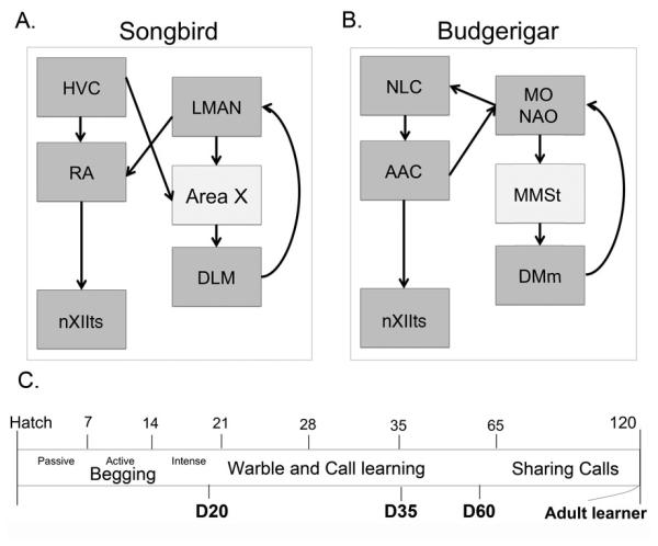
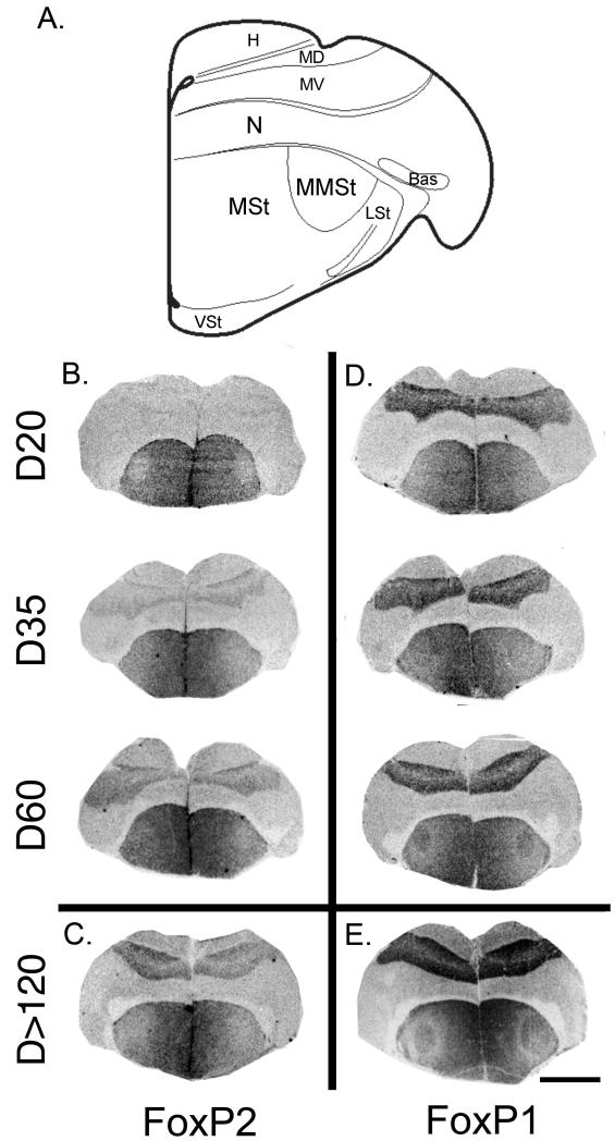
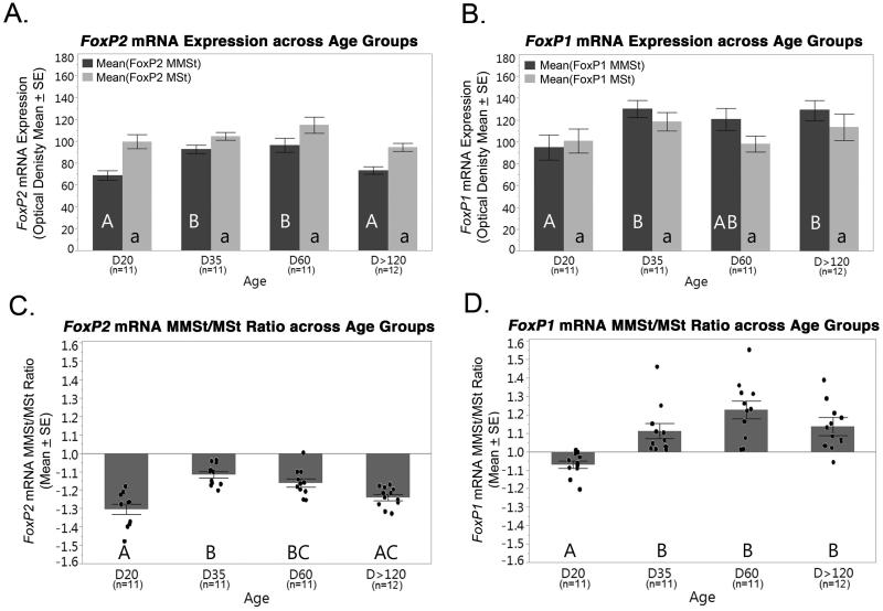
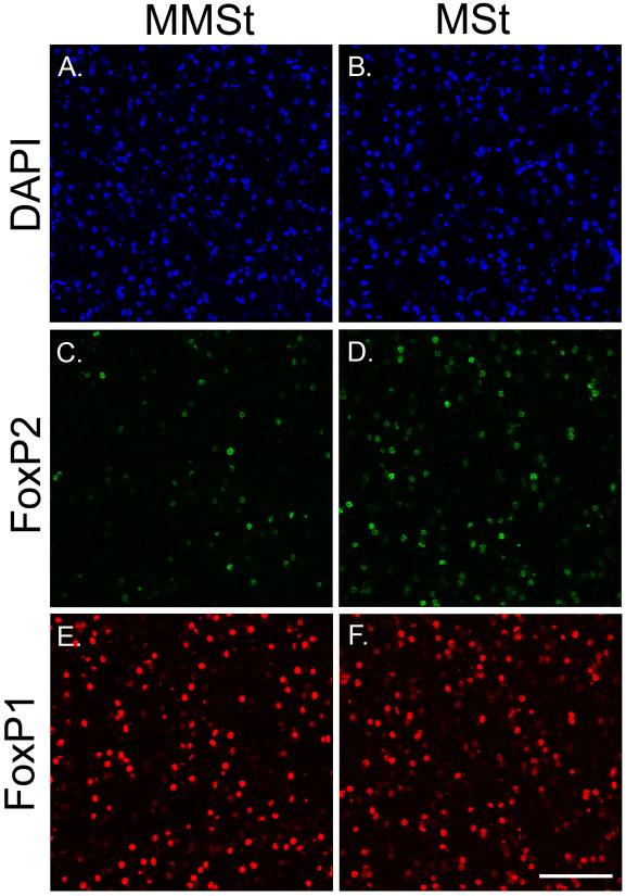
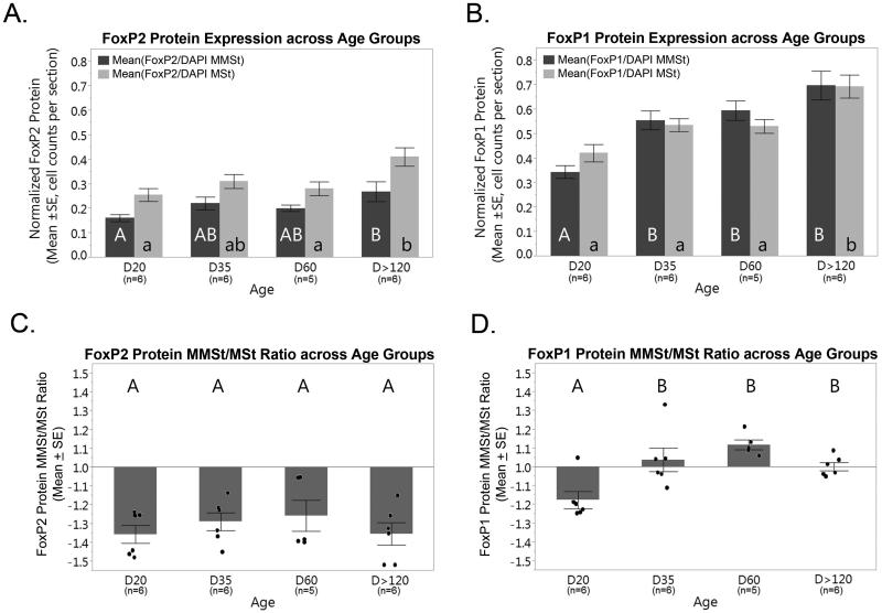
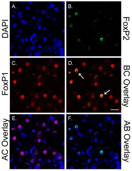
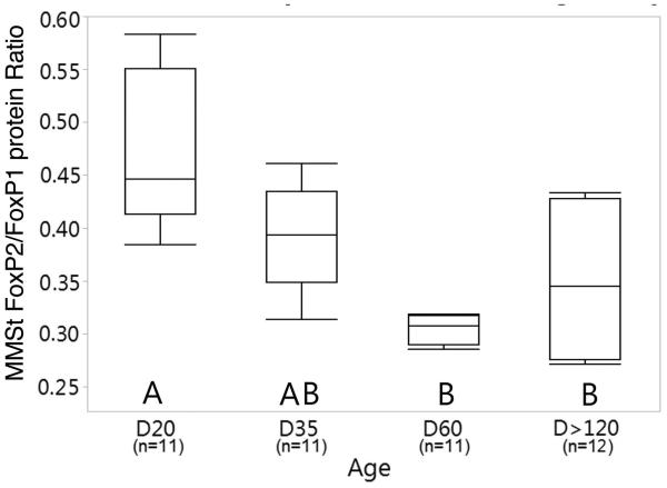
The forkhead domain FOXP2 and FOXP1 transcription factors are implicated in several cognitive disorders with language deficits, notably autism, and thus play a central role in learned vocal motor behavior in humans. Although a similar role for FoxP2 and FoxP1 is proposed for other vertebrate species, including songbirds, the neurodevelopmental expression of these genes are unknown in a species with lifelong vocal learning abilities. Like humans, budgerigars (Melopsittacus undulatus) learn new vocalizations throughout their entire lifetime. Like songbirds, budgerigars have distinct brain nuclei for vocal learning, which include the magnocellular nucleus of the medial striatum (MMSt), a basal ganglia region that is considered developmentally and functionally analogous to Area X in songbirds. Here, we used in situ hybridization and immunohistochemistry to investigate FoxP2 and FoxP1 expression in the MMSt of juvenile and adult budgerigars. We found FoxP2 mRNA and protein expression levels in the MMSt that were lower than the surrounding striatum throughout development and adulthood. In contrast, FoxP1 mRNA and protein had an elevated MMSt/striatum expression ratio as birds matured, regardless of their sex. These results show that life-long vocal plasticity in budgerigars is associated with persistent low-level FoxP2 expression in the budgerigar MMSt, and suggests the possibility that FoxP1 plays an organizational role in the neurodevelopment of vocal motor circuitry. Thus, developmental regulation of the FoxP2 and FoxP1 genes in the basal ganglia appears essential for vocal mimicry in a range of species that possess this relatively rare trait.

Keywords: FoxP1; FoxP2; basal ganglia; budgerigar; gene expression; vocal learning.


Figures

Figure 1
Figure 1
Shown here is a general schematic of interconnected vocal control nuclei in (A) the songbird brain and (B) the budgerigar brain (Nottebohm et al., 1976; Striedter, 1994). Area X and MMSt in the basal ganglia are part of a cortico-basal ganglia-thalamo-cortical loop important for learning acoustic gestures (Petkov and Jarvis, 2012). The songbird and budgerigar CNS via nXIIts projects to the syringeal muscles that produce sound. (C). Vocal development of budgerigars begins after hatch with food begging calls followed by a transitional period around D20 when socially-learned vocalizations first appear. Adult like vocalizations begin to emerge around D35, and at around D60 birds begin to join social groups and imitate conspecifics (Brittan-Powell et al., 1997; Hall et al., 1999). Vocal learning in new social groups occurs frequently in adults. The present study used birds that were isolated and recorded at the ages shown in bold, D20, D35, D60 and adults D>120. Abbreviations: Songbird: Area X and HVC are used as proper names: DLM, medial portion of the dorsolateral thalamic nucleus; LMAN, lateral magnocellular nucleus of the anterior nidopallium; RA, robust nucleus of the archipallium. Budgerigar: AAc, central nucleus of the anterior arcopallium; MO, oval nucleus of the mesopallium; MMSt, magnocellular nucleus of the medial striatum; NAO, oval nucleus of the anterior nidopallium; DMm magnocellular nucleus of the dorsomedial thalamus; nXIIts, tracheosyringeal motor nucleus, a portion of the 12th (hypoglossal) nucleus.
Figure 2
Figure 2
A. Location of the MMSt and adjoining striatum (MSt) in a schematic section from the budgerigar brain atlas at http://www.brauthlab.umd.edu/atlas.htm (Brauth SE; Jarvis et al., 2013). (B) In situ hybridized FoxP2 mRNA in which the MMSt can be found at (B) D20, D35, D60, and (C) in adults, D>120 (all birds are male). Sections of similar male brains show in situ hybridized FoxP1 mRNA in (D) and (E). Scale bar in (E) = 4 mm. Abbreviations: H, Hyperpallium; MD, dorsal Mesopallium; MV, ventral Mesopallium; N, Nidopallium; Bas, Basorostral pallial nucleus; MMSt, Magnocellular nucleus of the medial striatum; MSt, Medial striatum; LSt, Lateral striatum; VSt, Ventral striatum.
Figure 3
Figure 3
(A) FoxP2 mRNA expression in the MMSt (dark grey) and MSt (light grey) across age groups. No significant differences were found between groups in the MSt (p<0.05). (B) FoxP1 mRNA expression in the MMSt and MSt across age groups. No significant differences were found between age groups for both the MSt. FoxP1 mRNA expression in the MMSt was significantly lower at D20 compared to D35 and D>120. (C, D) FoxP2 and FoxP1 mRNA expression ratios (MMSt/MSt) show significant differences between age groups (p<0.05). Points in (C) and (D) represent individual birds. For all graphs, significant mRNA expression differences in the MMSt, MSt, (A, B, respectively) or a ratio thereof (C, D), between the four age groups is denoted with different letters. Bars with different letters are significantly different (p<05). The letter case is used to denote significant differences between age groups separately for the MMSt (uppercase) and MSt (lowercase). Error bars = SE.
Figure 4
Figure 4
Confocal images taken with a 40X objective to detect FoxP2 and FoxP1 protein within the MSt and MMSt. Example images are shown from a female animal at D35. From top to bottom: (A, B) DAPI stained cells in 405 nm within the MMSt and MSt; (C, D) FoxP2 protein expressing neurons in 488 nm within the MMSt and MSt; (E, F) FoxP1 expressing neurons in 594 nm within the MMSt and MSt; Scale bar in H = 50 μM.
Figure 5
Figure 5
(A) DAPI normalized FoxP2 protein expression across age groups in the MMSt (dark grey) and MSt (light grey). Significant differences between age groups in the MMSt and MSt. are shown with bars not connected by the same letter (p<0.05). (B) DAPI normalized FoxP1 protein expression across age groups in the MMSt (dark grey) and MSt (light grey) show significant differences in D20 and adult birds using bars with different letters (p<0.05). (C) FoxP2 and (D) FoxP1 MMSt/MSt protein expression ratios across age groups. No significant differences in FoxP2 expression ratios were found between groups. Ratios were <1 for all age groups. (D) MMSt/MSt FoxP1 protein expression ratios were significantly lower at D20 (p<0.05). Points in (C) and (D) represent individual birds. Error bars = SE.
Figure 6
Figure 6
Representative confocal images taken with a 63X objective show overlapping FoxP2 and FoxP1 protein expression within the MMSt of a female animal at D20. A, B and C show DAPI, FoxP2 and FoxP1 labeled cells. D, E, and F show different combinations of overlapping expression between A, B, and C. In (D) arrows indicate example cells co-expressing FoxP2 and FoxP1; scale bar = 20 μM.
Figure 7
Figure 7
Box plots show a ratio of FoxP2/FoxP1 protein expression in the MMSt. FoxP expression is DAPI normalized. Top and bottom whisker lines, and box line represent maximum, minimum and median values, respectively.

References

    1. Bacon C, Rappold GA. The distinct and overlapping phenotypic spectra of FOXP1 and FOXP2 in cognitive disorders. Hum Genet. 2012;131:1687–1698. - PMC - PubMed
    1. Belton E, Salmond CH, Watkins KE, Vargha-Khadem F, Gadian DG. Bilateral brain abnormalities associated with dominantly inherited verbal and orofacial dyspraxia. Hum Brain Mapp. 2003;18:194–200. - PMC - PubMed
    1. Bolhuis JJ, Okanoya K, Scharff C. Twitter evolution: converging mechanisms in birdsong and human speech. Nat Rev Neurosci. 2010;11:747–759. - PubMed
    1. Bowers JM, Perez-Pouchoulen M, Edwards NS, McCarthy MM. Foxp2 mediates sex differences in ultrasonic vocalization by rat pups and directs order of maternal retrieval. J Neurosci. 2013;33:3276–3283. - PMC - PubMed
    1. Brauth SEHJ, Roberts TF, Liang W. Budgerigar Brain Atlas. http://www.brauthlab.umd.edu/atlas.htm. accessed Nov 2014.

Publication types