. 2014 Sep;8(9-10):E715-20.
doi: 10.5489/cuaj.1978.

Circulating tumour cells in patients with urothelial tumours: Enrichment and in vitro culture

Affiliations

Circulating tumour cells in patients with urothelial tumours: Enrichment and in vitro culture

Katarina Kolostova et al. Can Urol Assoc J. 2014 Sep.

Abstract

Introduction: Results of clinical trials have demonstrated that circulating tumour cells (CTCs) are frequently detected in patients with urothelial tumours. The monitoring of CTCs has the potential to improve therapeutic management at an early stage and also to identify patients with increased risk of tumour progression or recurrence before the onset of clinically detected metastasis. In this study, we report a new effectively simplified methodology for a separation and in vitro culturing of viable CTCs from peripheral blood.

Method: We include patients diagnosed with 3 types of urothelial tumours (prostate cancer, urinary bladder cancer, and kidney cancer). A size-based separation method for viable CTC - enrichment from unclothed peripheral blood has been introduced (MetaCell, Ostrava, Czech Republic). The enriched CTCs fraction was cultured directly on the separation membrane, or transferred from the membrane and cultured on any plastic surface or a microscopic slide.

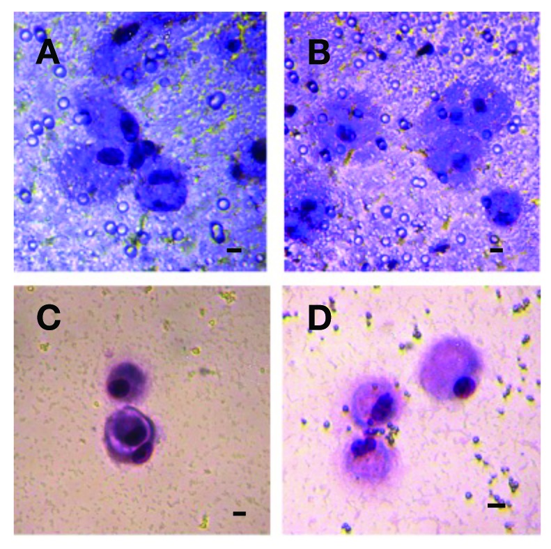
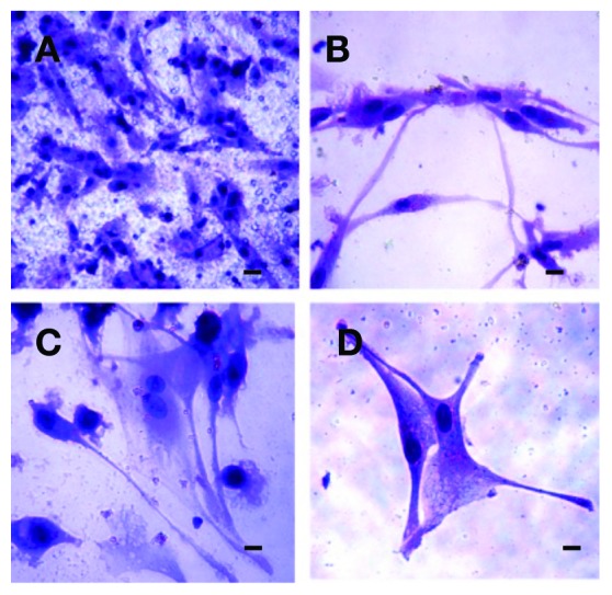
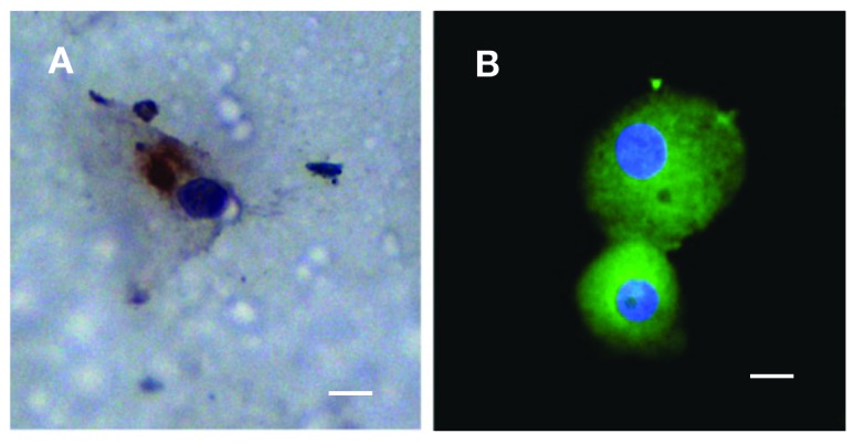
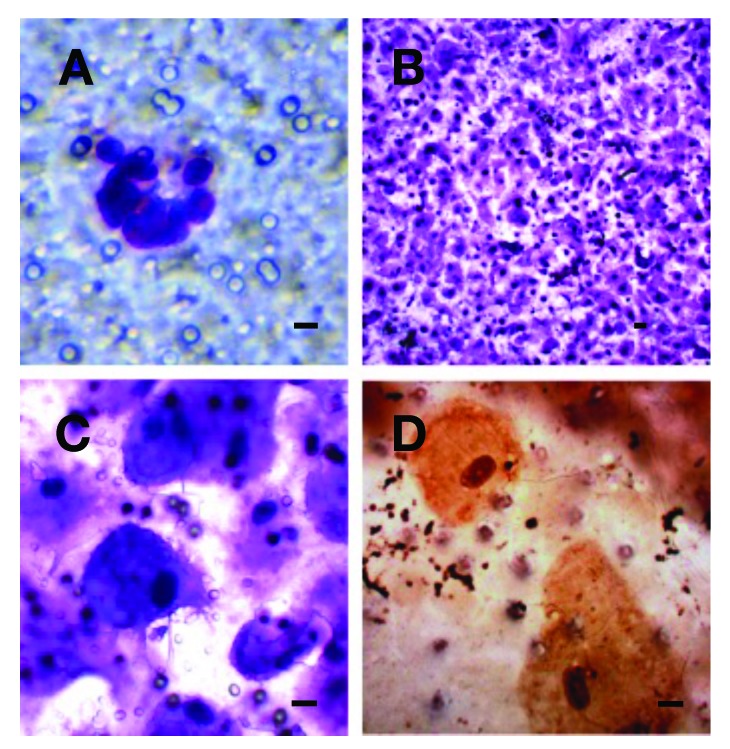
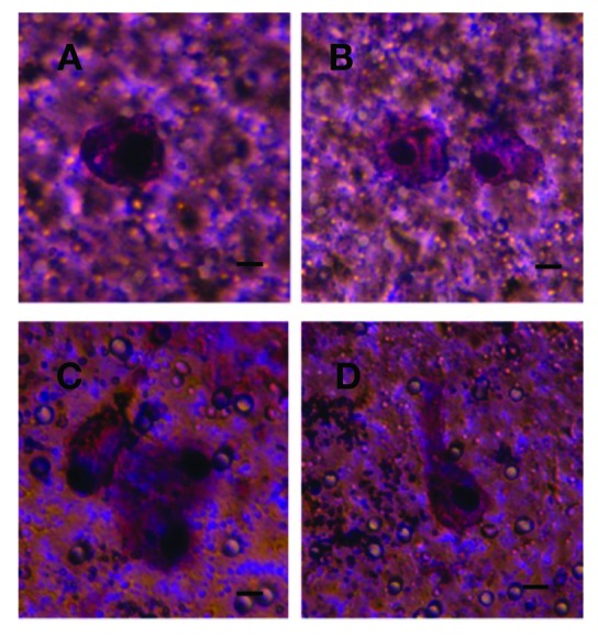
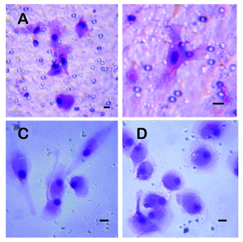
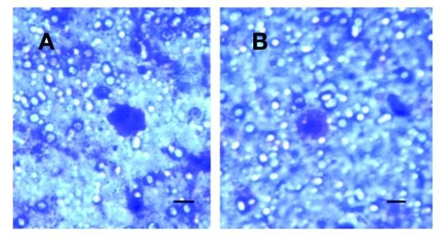
Results: We report a successful application of a CTCs isolation procedure in patients with urothelial cancers. The CTCs captured on the membrane are enriched with a remarkable proliferation potential. This has enabled us to set up in vitro cell cultures from the viable CTCs unaffected by any fixation buffers, antibodies or lysing solutions. Next, the CTCs were cultured in vitro for a minimum of 10 to 14 days to enable further downstream analysis (e.g., immunohistochemistry).

Conclusion: We demonstrated an efficient CTCs capture platform, based on a cell size separation principle. Furthermore, we report an ability to culture the enriched cells - a critical requirement for post-isolation cellular analysis.


Figures

Fig. 1.
Fig. 1.
Circulating tumour cells (CTCs) isloated from a patient (P1) with prostate cancer grown in vitro 10 days. A, B: The CTCs detected on the membrane. C, D: The CTCs growing on the bottom of the culture plate after their escape from the separating membrane through the pores/counterstained by MGG-stain. A bar represents 10 μm.
Fig. 2.
Fig. 2.
Circulating tumour cells (CTCs) isloated from a patient (P2) with prostate cancer grown in vitro 14 days. A: The CTCs growing on the membrane. B, C, D: The cells growing through the membrane to the bottom of the culture plate showing mesenchymal features/counterstained by MGG-stain. A bar represents 10 μm.
Fig. 3.
Fig. 3.
Circulating tumour cells (CTCs) captured from a patient (P1) with prostate cancer grown in vitro on the membrane. A: The CTCs counterstained by Pan-cytokeratin antibody (DAB-visualized). B: The CTCs captured through enrichment and subsequent cytospin procedure, counterstained by FITCPancytokeratin and DAP1. A bar represents 10 μm.
Fig. 4.
Fig. 4.
Circulating tumour cells (CTCs) from a patient (B1) with bladder cancer. A: The CTCs immediately after the separations. B: The CTCs on the separation membrane grown in vitro for 14 days creating a cell layer. C: The CTCs growing on the membrane. D: The CTCs growing on the membrane counterstained by CK-7 antibody (DAB-visualized). A bar represents 10 μm.
Fig. 5.
Fig. 5.
Circulating tumour cells captured from a patient (B2) with bladder cancer on the separation membrane grown in vitro for 14 days. A bar represents 10 μm.
Fig. 6.
Fig. 6.
Circulating tumour cells (CTCs) captured from a patient (B3) with bladder cancer grown in vitro for 14 days. A, B: The CTCs growing on the separation membrane. C, D: The CTCs growing on the culture flask bottom after their escape from the membrane through the pores. A bar represents 10 μm.
Fig. 7.
Fig. 7.
Circulating tumour cells captured from patients with kidney cancer (A: patient K1. B: patient K2) on the separating membrane, grown in vitro for 7 days. A bar represents 10 μm.

References

    1. Msaouel P, Koutsilieris M. Diagnostic value of circulating tumor cell detection in bladder and urothelial cancer: Systematic review and meta-analysis. BMC Cancer. 2011;4:336. doi: 10.1186/1471-2407-11-336. - DOI - PMC - PubMed
    1. de Bono JS, Scher HI, Montgomery RB, et al. Circulating tumor cells predict survival benefit from treatment in metastatic castration-resistant prostate cancer. Clin Cancer Res. 2008;14:6302–9. doi: 10.1158/1078-0432.CCR-08-0872. - DOI - PubMed
    1. Rink M, Chun FK, Minner S, et al. Detection of circulating tumour cells in peripheral blood of patients with advanced non-metastatic bladder cancer. BJU Int. 2011;107:1668–75. doi: 10.1111/j.1464-410X.2010.09562.x. - DOI - PubMed
    1. Fidler IJ, Kripke ML. Metastasis results from preexisting variant cells within a malignant tumor. Science. 1977;197:893–5. - PubMed
    1. Fidler IJ. Metastasis: Guantitative analysis of distribution and fate of tumor emboli labeled with 125 I-5-iodo-2’-deoxyuridine. J. Natl. Cancer Inst. 1970;45:773–82. - PubMed