. 2014 Dec 9;53(48):7680-92.
doi: 10.1021/bi501011m. Epub 2014 Nov 26.

Electrostatic properties of complexes along a DNA glycosylase damage search pathway

Affiliations

Electrostatic properties of complexes along a DNA glycosylase damage search pathway

Shannen L Cravens et al. Biochemistry. .

Abstract

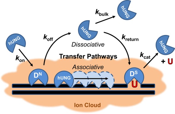
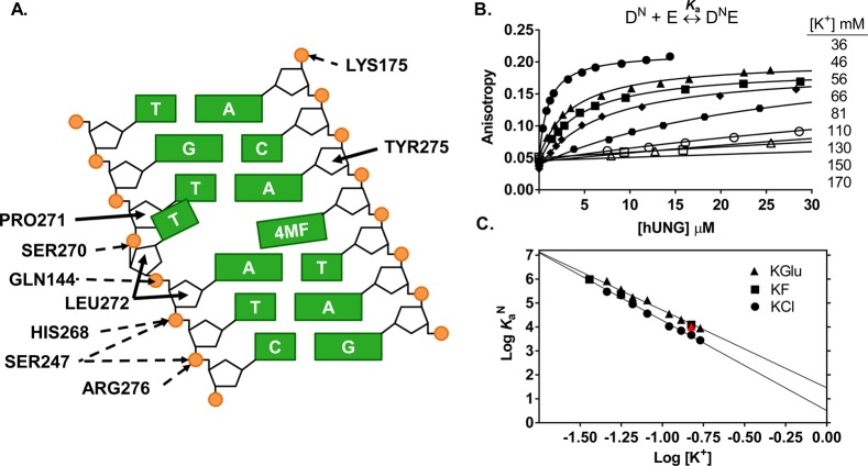
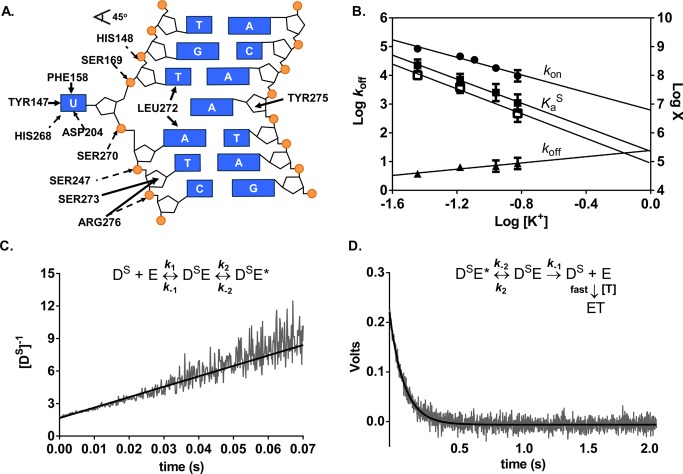
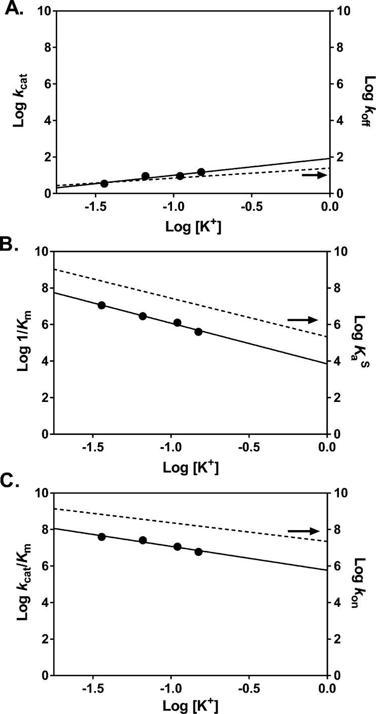
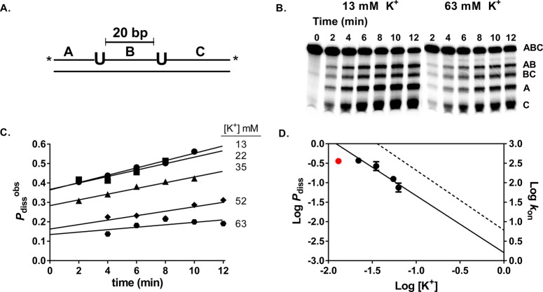
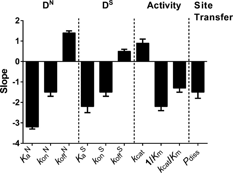
Human uracil DNA glycosylase (hUNG) follows an extended reaction coordinate for locating rare uracil bases in genomic DNA. This process begins with diffusion-controlled engagement of undamaged DNA, followed by a damage search step in which the enzyme remains loosely associated with the DNA chain (translocation), and finally, a recognition step that allows the enzyme to efficiently bind and excise uracil when it is encountered. At each step along this coordinate, the enzyme must form DNA interactions that are highly specialized for either rapid damage searching or catalysis. Here we make extensive measurements of hUNG activity as a function of salt concentration to dissect the thermodynamic, kinetic, and electrostatic properties of key enzyme states along this reaction coordinate. We find that the interaction of hUNG with undamaged DNA is electrostatically driven at a physiological concentration of potassium ions (ΔGelect = -3.5 ± 0.5 kcal mol(-1)), with only a small nonelectrostatic contribution (ΔGnon = -2.0 ± 0.2 kcal mol(-1)). In contrast, the interaction with damaged DNA is dominated by the nonelectrostatic free energy term (ΔGnon = -7.2 ± 0.1 kcal mol(-1)), yet retains the nonspecific electrostatic contribution (ΔGelect = -2.3 ± 0.2 kcal mol(-1)). Stopped-flow kinetic experiments established that the salt sensitivity of damaged DNA binding originates from a reduction of kon, while koff is weakly dependent on salt. Similar findings were obtained from the salt dependences of the steady-state kinetic parameters, where the diffusion-controlled kcat/Km showed a salt dependence similar to kon, while kcat (limited by product release) was weakly dependent on salt. Finally, the salt dependence of translocation between two uracil sites separated by 20 bp in the same DNA chain was indistinguishable from that of kon. This result suggests that the transition-state for translocation over this spacing resembles that for DNA association from bulk solution and that hUNG escapes the DNA ion cloud during translocation. These findings provide key insights into how the ionic environment in cells influences the DNA damage search pathway.


Figures

Figure 1
Figure 1
Human UNG (hUNG) DNA search and repair pathway is composed of four transient states: stationary states where the enzyme is engaged with nonspecific (DN) or specific uracilated sequences (DS), and two mobile states where the enzyme can translocate along DNA via associative or dissociative pathways of facilitated diffusion. Nonspecific and specific complexes must have distinct interactions that facilitate efficient recognition and repair (see text). The DN complex is characterized primarily by contacts with the phosphate backbone, while the DS complex involves additional nonpolar and hydrogen bonding interactions with the uracil base (see Figures 2A and 3A). The overall transfer probability between two uracil lesions is defined as the sum of two pathways: Ptrans = Passoc + Pdiss, where Passoc and Pdiss are the probabilities of transfer via the associative and dissociative pathways, respectively. When uracils are spaced far enough apart such that all successful transfers occur via at least one dissociation event, this equation reduces to Ptrans = Pdiss. Kinetically, Pdiss is defined as the product of two probabilities: Pdiss = [koff/(kassoc + koff)][kreturn/(kbulk + kreturn)]. The first term describes the probability that hUNG will dissociate from a nonspecific DNA site as opposed to making an associative step along the DNA, and the second term gives the likelihood that the enzyme, once dissociated, escapes to the bulk solvent (kbulk) rather than reassociating with the DNA chain (kreturn) to complete transfer by the dissociative pathway.
Figure 2
Figure 2
Salt dependence of the nonspecific DNA (DN) equilibrium binding affinity. (A) Schematic of electrostatic interactions implicated in ion release (dashed arrows) and the nonelectrostatic (solid arrows) interactions between hUNG and nonspecific DNA (Protein Data Bank entry 2OXM, 4MF = 4-methylindole). Using mutagenesis and NMR imino exchange methods, the partially extruded thymine residue and its interactions with the enzyme have been substantiated in solution using an hUNG-nonspecific DNA complex with central T/A base pair., These studies showed that normal T/A base pairs (not G/C) undergo enhanced imino proton exchange when bound to hUNG and involve the residues depicted in the graphic. Electrostatic interactions are defined by nitrogen and oxygen atoms <3.3 Å apart from DNA phosphate oxygens, while nonelectrostatic interactions are all carbon–carbon pairs <3.9 Å apart. An additional hydrogen bond between hUNG and the O2 of the partially extruded thymine across from 4MF was omitted in the diagram for clarity. (B) Changes in fluorescence anisotropy of DN (100 nM) as a function of hUNG concentration at varying potassium ion concentrations (36–170 mM). Full binding curves for 81–170 mM K+ are provided in Supplementary Figure S1, Supporting Information. (C) Dependence of Ka on the concentrations of KGlu (triangles), KCl (circles), KF (squares). Inclusion of 500 μM MgCl2 at physiological K+ (150 mM) had a negligible effect on the observed Ka (red triangle, see text).
Figure 3
Figure 3
Salt dependences of the association and dissociation constants and equilibrium binding affinity for specific DNA (DS) determined by stopped-flow fluorescence measurements at 20 °C. (A) Schematic of electrostatic interactions implicated in ion release (dashed arrows) and the nonelectrostatic (solid arrows) interactions between hUNG and specific DNA (Protein Data Bank entry 1EMH(45)). Insertion of the side chain of Leu272 into the DNA duplex and movement of uracil into the hUNG active site results in numerous nonelectrostatic contacts not present in the nonspecific complex. (B) Dependence on KGlu concentration of konS (circles), koffS (triangles), the calculated KaS obtained from the ratio koffS/konS (solid squares), and the measured KaS from equilibrium fluorescence titrations using KF (open squares). Log koffS is plotted on the left y-axis and the remaining parameters are plotted on the right y-axis [X = KaS (M–1) and kon (M–1 s–1)]. (C) Linearized kinetic trace of the second-order association of DS (600 nM) with hUNG (600 nM) at 150 mM K+. Equal volume solutions of DS and hUNG of equal concentration (400–600 nM) were mixed and the time dependent increase in 2-AP fluorescence was followed (λex= 310 nm). The line is the best-fit to a second-order rate equation. (D) Kinetic trace of the dissociation of hUNG from DS at 150 mM K+. Abasic site-containing DNA (aDNA, 5 μM) was mixed with an equal volume solution containing 0.8 μM hUNG and 0.2 μM DS and the time dependent decrease in 2-AP fluorescence was followed (λex = 310 nm). The line is the best-fit to a single exponential decay. Controls established that the observed rate was zero-order with respect to DNA trap.
Figure 4
Figure 4
Salt dependences of kcat, Km, and kcat/Km. (A) The value for kcat (circles) is limited by product release and is minimally dependent on KGlu concentration. This is similar to the behavior observed for koff (dashed line). (B) The dependence of 1/Km (circles) on KGlu concentration is very similar to that observed for specific DNA binding KaS (dashed line). (C) The dependence of kcat/Km (circles) on KGlu concentration is identical to the dependence observed for kon (dashed line).
Figure 5
Figure 5
Salt dependence of the intramolecular dissociative transfer probability of hUNG between two uracil sites spaced 20 bp apart (Pdiss). (A) Schematic of the substrate (S20) used. The asterisk denotes the location of the 32P end labels. (B) Phosphorimages of the gel-resolved site transfer products derived from S20 in the presence of 13 mM and 63 mM K+. (C) Determination of Pdiss at varying K+ levels in the range 13–63 mM. The observed site transfer probability (Pdissobs, eq 3) is calculated at each time point and then linearly extrapolated to time zero to determine the true value (Pdiss). (D) Comparison of the dependences of Pdiss (circles) and kon (dashed line) on K+ concentration. The Pdiss value at 13 mM (red circle) deviated negatively from the linear correlation and was omitted from the linear regression analysis.
Figure 6
Figure 6
Summary of the salt dependences of each measured thermodynamic and kinetic parameter (X) (see Discussion). The dependences are represented as the slopes of the respective log [salt] vs log X plots. Positive slopes indicate dissociation processes resulting in ion condensation, which are facilitated by high ionic strength (kcat, koff). Negative slopes result from processes that involve ion displacement and are hindered by high salt concentrations.

References

    1. Schonhoft J. D.; Kosowicz J. G.; Stivers J. T. (2013) DNA Translocation by Human Uracil DNA Glycosylase: Role of DNA Phosphate Charge. Biochemistry 52, 2526–2535. - PMC - PubMed
    1. Rowland M. M.; Schonhoft J. D.; McKibbin P. L.; David S. S.; Stivers J. T. (2014) Microscopic mechanism of DNA damage searching by hOGG1. Nucleic Acids Res. 42, 9295–9303. - PMC - PubMed
    1. Schonhoft J. D.; Stivers J. T. (2013) DNA translocation by human uracil DNA glycosylase: the case of single-stranded DNA and clustered uracils. Biochemistry 52, 2536–2544. - PMC - PubMed
    1. Schonhoft J. D.; Stivers J. T. (2012) Timing facilitated site transfer of an enzyme on DNA. Nat. Chem. Biol. 8, 205–210. - PMC - PubMed
    1. Stivers J. T.; Jiang Y. L. (2003) A Mechanistic Perspective on the Chemistry of DNA Repair Glycosylases. Chem. Rev. 103, 2729–2760. - PubMed

Publication types

LinkOut - more resources