. 2015 Feb 1;38(2):241-50.
doi: 10.5665/sleep.4406.

Cerebrovascular reactivity in young subjects with sleep apnea

Affiliations

Cerebrovascular reactivity in young subjects with sleep apnea

John Buterbaugh et al. Sleep. .

Abstract

Study objectives: Regional brain alterations may be involved in the pathogenesis and adverse consequences of obstructive sleep apnea (OSA). The objectives for the current study were to (1) determine cerebrovascular reactivity in the motor areas that control upper airway musculature in patients with OSA, and (2) determine whether young patients with OSA have decreased cerebrovascular reactivity in response to breath holding.

Design: Case-control study.

Setting: Academic center.

Participants: Twelve subjects with OSA (age 24-42 y; apnea-hypopnea index 17; interquartile range [IQR] 9, 69 per hour) and control subjects (n = 10; age 29-44 y; AHI 2; IQR 1, 3 per hour).

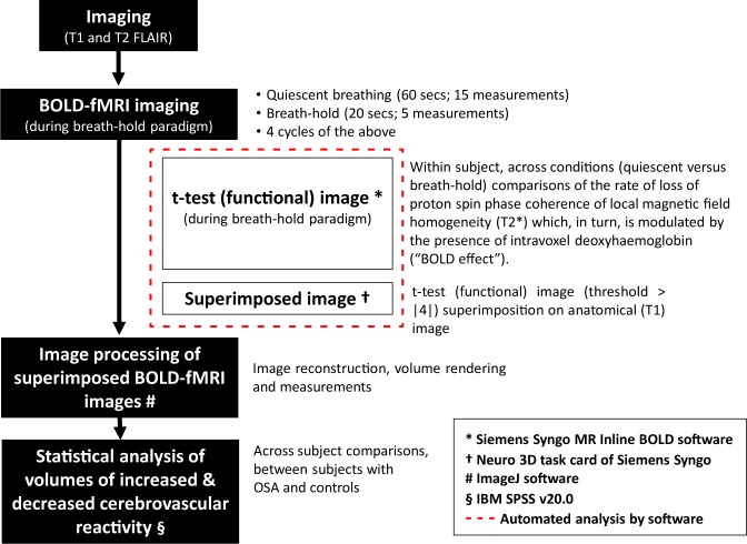
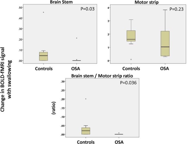
Measurements and results: Subjects underwent blood oxygen level-dependent functional magnetic resonance imaging (BOLD-fMRI) while awake, swallowing, and breath holding. In subjects with OSA, during swallowing, there was less activity in the brainstem than in controls (P = 0.03) that remained reduced after adjusting for cortical motor strip activity (P = 0.036). In OSA subjects, brain regions of increased cerebrovascular reactivity (38; IQR 17, 96 cm(3)) was smaller than that in controls (199; IQR 5, 423 cm(3); P = 0.01). In OSA subjects, brain regions of decreased cerebrovascular reactivity during breath hold was greater (P = 0.01), and the ratio of increased-to-decreased brain regions was lower than that of controls (P = 0.006). Adjustment for cerebral volumes, body mass index, and white matter lesions did not change these results substantively.

Conclusions: In patients with obstructive sleep apnea (OSA), diminished change in brainstem activity during swallowing and reduced cerebrovascular reactivity may contribute to the etiopathogenesis and adverse cerebrovascular consequences, respectively. We speculate that decreased cerebral auto-regulation may be causative of gray matter loss in OSA.

Keywords: cerebral autoregulation; functional magnetic resonance imaging; obstructive sleep apnea; sleep apnea; stroke.


Figures

Figure 1
Figure 1
Blood oxygen level-dependent functional magnetic resonance imaging (BOLD-fMRI) was performed during breath-holding maneuvers each lasting for 20 sec that were aimed at assessing cerebrovascular reactivity in response to a uniform stimulation: hypercapnia induced by breath holding (top panel). BOLD-fMRI imaging was performed using task paradigms for swallowing (bottom panel) that were designed to preferentially activate upper airway motoneurons involved in controlling voluntary upper airway muscle contractions involved with swallowing. During such task paradigms including the time periods of quiescence or calm breathing, the BOLD-fMRI scans were performed every 4 sec.
Figure 2
Figure 2
Schematic diagram of image acquisition and processing. The software used for such analysis is provided in the legend. The automated analyses are shown within the red box. BOLD-fMRI, blood oxygen level-dependent functional magnetic resonance imaging.
Figure 3
Figure 3
During swallowing, regions of increased brain stem activity in patients with obstructive sleep apnea (OSA) was smaller than that in controls (top panel, left). However, the regions of increased cortical motor strip activity in patients with OSA were not different than those in controls (top panel, right). The ratio of volume of increased activity in brain stem to motor strip was lower in OSA patients than controls (bottom panel).
Figure 4
Figure 4
Blood oxygen level-dependent functional magnetic resonance imaging (BOLD-fMRI) images of the upper medulla oblongata obtained during swallowing paradigm in a control subject (left, upper panel). Increased BOLD-fMRI activity was present bilaterally in the region of the nucleus ambiguus of the control subject (blue arrows in inset). In contrast, notice that there was no activity in the same region of the subject with obstructive sleep apnea (OSA) (top panel, right and inset). BOLD-fMRI images of the hemispheres during swallowing paradigm in a control subject (bottom panel, left) and control subject (blue arrows in inset). Note that the motor strip activity was similar in the patient with OSA and the control subject during the swallowing maneuver.
Figure 5
Figure 5
During breath hold, regions of increased cerebrovascular reactivity were smaller in subjects with OSA than controls (top panel, left). Conversely, in subjects with OSA, regions of decreased cerebrovascular reactivity was greater than that in controls (top panel, right), and the ratio of increased-to-decreased cerebrovascular reactivity in brain regions was lower in patients with OSA than controls (bottom panel).
Figure 6
Figure 6
Blood oxygen level-dependent functional magnetic resonance imaging (BOLD-fMRI) of the cerebral hemispheres in a control subject (top panel) and a subject with OSA (bottom panel). Regions of increased cerebrovascular reactivity are shown in red and decreased cerebrovascular reactivity are shown in green. Note the increased cerebrovascular reactivity in the control subject and the scant increase in cerebrovascular reactivity in the patient with OSA. Also, note the increase in cerebrovascular reactivity in the control subject is more marked in the gray matter when compared to that in the white matter.
Figure 7
Figure 7
Bland-Altman plot of reproducibility measurements of blood oxygen level-dependent functional magnetic resonance imaging (BOLD-fMRI) changes in response to breath holding. The difference in the ratio of volume of increased-to-decreased brain regions between the two measurements performed at different points in time is plotted against the mean values of the two measurements of volume of increased-to-decreased brain regions (Bland-Altman plot). The bias (dashed line) and precision error (dotted lines) are shown. Please note that twice the magnitude of the precision error (12) is still less than the magnitude of the observed mean difference between patients with OSA and controls (40.5). See Figure 5.
Figure 8
Figure 8
Least misclassification plots of change in blood oxygen level-dependent functional magnetic resonance imaging (BOLD-fMRI) signal with breath holding versus apnea-hypopnea index (AHI) with vertical threshold lines at AHI of 5 per hour and horizontal threshold lines placed to yield least data points in the indeterminate (left lower and right upper) quadrants of the scatterplot. Only two (8%) or four participants' values (8% and 16%, respectively) fell in the indeterminate areas (top right and bottom left quadrants) that was not explained by the hypothesis that in subjects with obstructive sleep apnea (OSA) performing breath hold, cerebrovascular reactivity measured by BOLD-fMRI would be diminished in comparison to that in controls.

Similar articles

Cited by

References

    1. Young T, Peppard PE, Gottlieb DJ. Epidemiology of obstructive sleep apnea: a population health perspective. Am J Respir Crit Care Med. 2002;165:1217–39. - PubMed
    1. Punjabi NM. The epidemiology of adult obstructive sleep apnea. Proc Am Thorac Soc. 2008;5:136–43. - PMC - PubMed
    1. Younes M, Ostrowski M, Thompson W, Leslie C, Shewchuk W. Chemical control stability in patients with obstructive sleep apnea. Am J Respir Crit Care Med. 2001;163:1181–90. - PubMed
    1. Schwartz AR, Schneider H, Smith PL, McGinley BM, Patil SP, Kirkness JP. Physiologic phenotypes of sleep apnea pathogenesis. Am J Respir Crit Care Med. 2011;184:1105–6. - PMC - PubMed
    1. Dempsey JA, Veasey SC, Morgan BJ, O'Donnell CP. Pathophysiology of sleep apnea. Physiol Rev. 2010;90:47–112. - PMC - PubMed

Publication types

LinkOut - more resources