. 2014 Nov 19;9(11):e113499.
doi: 10.1371/journal.pone.0113499. eCollection 2014.

Hepatitis C virus nonstructural protein 5A inhibits thapsigargin-induced apoptosis

Affiliations

Hepatitis C virus nonstructural protein 5A inhibits thapsigargin-induced apoptosis

Xia Jiang et al. PLoS One. .

Abstract

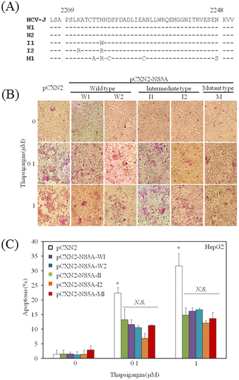
Background: We previously reported that the hepatitis C virus (HCV) nonstructural protein 5A (NS5A) down-regulates TLR4 signaling and lipopolysaccharide-induced apoptosis of hepatocytes. There have been several reports regarding the association between HCV infection and endoplasmic reticulum (ER) stress. Here, we examined the regulation of HCV NS5A on the apoptosis of hepatocytes induced by thapsigargin, an inducer of ER stress.

Methods: The apoptotic response to thapsigargin and the expression of molecules involved in human hepatocyte apoptotic pathways were examined in the presence or absence of HCV NS5A expression.

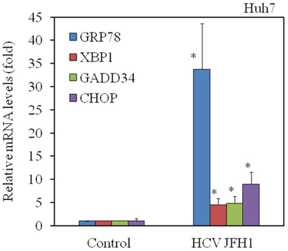
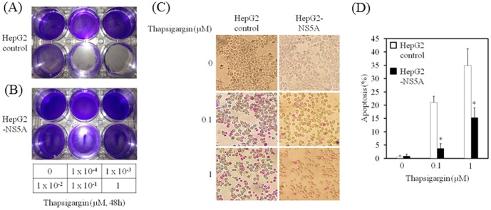
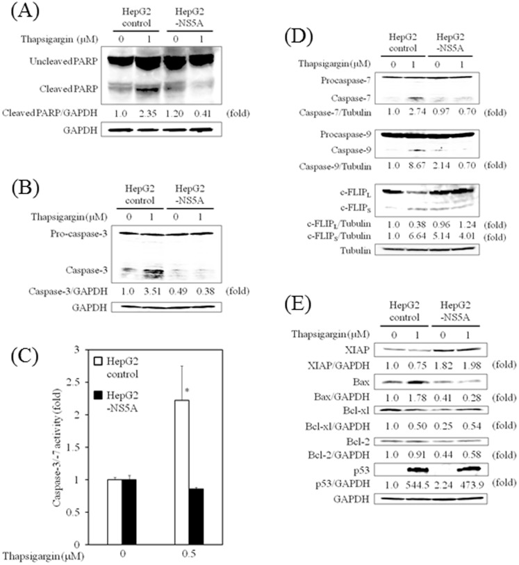
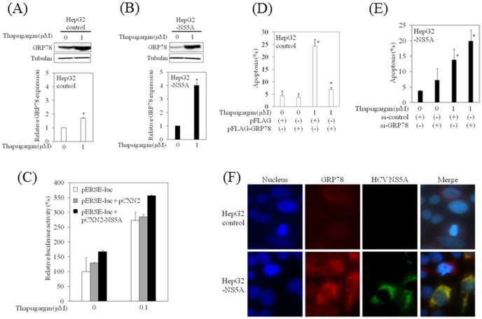
Results: HCV JFH1 infection induced ER stress in the Huh7 cell line. HCV NS5A protected HepG2 cells against thapsigargin-induced apoptosis, the effect of which was linked to the enhanced expression of the 78-kDa glucose-regulated protein/immunoglobulin heavy-chain binding protein (GRP78). Consistent with a conferred pro-survival advantage, HCV NS5A reduced poly(adenosine diphosphate-ribose) polymerase cleavage and activation of caspases-3, -7 and -9, and Bax expression, while increasing the expressions of the anti-apoptotic molecules XIAP and c-FLIP. HCV NS5A weakly interacts with GRP78 and enhances GRP78 expression in hepatocytes.

Conclusion: HCV NS5A enhances GRP78 expression, resulting in the inhibition of apoptotic properties, and inhibits thapsigargin-induced apoptotic pathways in human hepatocytes, suggesting that disruption of ER stress-mediated apoptosis may have a role in the pathogenesis of HCV infection. Thus, HCV NS5A might engender the survival of HCV-infected hepatocytes contributing to the establishment of persistent infection.


Conflict of interest statement

Competing Interests: Drs. XJ, SW, SN, TW and HS have no conflict of interest to declare. Dr. TK reports receiving lecture fees from Chugai Pharmaceutical, MSD, Tanabe-Mitsubishi, Daiichi-Sankyo, and Bristol-Myers Squibb, and Prof. OY reports receiving grant support from Chugai Pharmaceutical, Bayer, MSD, Daiichi-Sankyo, Tanabe-Mitsubishi, and Bristol-Myers Squibb. These relationships do not alter the authors' adherence to all the PLOS ONE policies on sharing data and materials. Dr. TK, a PLOS ONE Editorial Board member, is listed as a co-author of the manuscript. This does not alter the authors' adherence to PLOS ONE policies on sharing data and materials.

Figures

Figure 1
Figure 1. Hepatitis C virus (HCV) infection induces ER stress in hepatocytes.
HCV JFH1 infection of Huh7 cells up-regulates mRNA expression of GRP78, XBP1, GADD34 and CHOP. Total cellular RNA was isolated from cells 72 h after infection with HCV. Intracellular gene expression levels of GRP78, XBP1, GADD34, CHOP and GAPDH were measured by real-time RT-PCR. The ratios of GRP78/GAPDH, XBP1/GAPDH, GADD34/GAPDH and CHOP/GAPDH are presented as induction (n-fold) relative to the levels observed in mock-infected control. Data are expressed as mean ± standard deviation. *P<0.05.
Figure 2
Figure 2. Hepatitis C virus (HCV) nonstructural protein 5A (NS5A) protects hepatocytes from thapsigargin-induced cell death.
HepG2 control (A) and HepG2-NS5A (B) cell lines were cultured for 48 h with thapsigargin at 0, 1×10⁻⁴, 1×10⁻³, 1×10⁻², 1×10⁻¹, and 1 µM. Cells were washed and stained with crystal violet. (C), (D) HCV NS5A protects hepatocytes from thapsigargin-induced apoptosis. HepG2 control and HepG2-NS5A cells were cultured for 24 h with thapsigargin at 0, 1×10⁻¹, and 1 µM. Apoptosis was evaluated using the APOPercentage Apoptosis Assay. Purple-red stained cells were identified as apoptotic cells by light microscopy. The number of purple-red cells/300 cells was counted. Data are expressed as mean ± standard deviation. *P<0.05.
Figure 3
Figure 3. Hepatitis C virus (HCV) nonstructural protein 5A (NS5A) protects hepatocytes from thapsigargin-induced apoptosis.
(A) HCV NS5A inhibits PARP cleavage in HepG2 cells. Western blot analysis shows the expression of PARP and cleaved PARP in HepG2 control and HepG2-NS5A cells treated for 24 h with or without thapsigargin (1 µM). Blots were reprobed with GAPDH-specific antibodies to assess equivalent protein loading. (B) HCV NS5A inhibits caspase-3 expression in HepG2 cells. Western blot analysis shows the expression of procaspase-3 and caspase-3 in HepG2 control and HepG2-NS5A cells treated for 24 h with or without thapsigargin (1 µM). (C) HCV NS5A inhibits the caspase-3/-7 activity in HepG2 cells. The Caspase-Glo 3/7 assay (Promega, Madison, WI, USA) shows the caspase-3/-7 activity in HepG2 control and HepG2-NS5A cells treated for 6 h with or without thapsigargin (0.5 µM). Data are expressed as mean ± standard deviation. *P<0.05. (D) HCV NS5A inhibits caspase-7/-9 and enhances cellular FADD-like interleukin-1beta-converting enzyme (FLICE)-like inhibitory protein, long form (c-FLIPL) expression in HepG2 cells. Western blot analysis shows the expression of procaspase-7 and caspase-7 (upper panel), procaspase-9 and caspase-9 (middle panel) and c-FLIPL and c-FLIP, short form (c-FLIPS), in HepG2 control and HepG2-NS5A cells treated for 24 h with or without thapsigargin (1 µM). Blots were reprobed with tubulin-specific antibodies to assess equivalent protein loading. (E) HCV NS5A enhances XIAP expression and inhibits Bax expression in HepG2 cells. Western blot analysis shows the expression of XIAP, Bax, Bcl-xl, Bcl-2 and p53 in HepG2 control and HepG2-NS5A cells treated for 24 h with or without thapsigargin (1 µM). Blots were reprobed with GAPDH-specific antibodies to assess equivalent protein loading. Densitometric analyses were performed using ImageJ software.
Figure 4
Figure 4. Hepatitis C virus (HCV) nonstructural protein 5A (NS5A) up-regulates GRP78 expression in hepatocytes.
(A), (B) HCV NS5A enhances GRP78 expression in HepG2 cells. Western blot analysis shows the expression of GRP78 in HepG2 control (A) and HepG2-NS5A cells (B) treated for 24 h with or without thapsigargin (1 µM). Blots were reprobed with tubulin-specific antibodies to assess equivalent protein loading. Densitometric analyses were performed using ImageJ software. Data are expressed as mean ± standard deviation. *P<0.05. (C) HepG2 cells were transiently transfected with pCXN2 or pCXN2-NS5A and an ER stress response element (ERSE)-directed luciferase reporter construct (pERSE-luc). Luciferase assays were performed 48 h after transfection. Data are expressed as mean ± standard deviation of triplicate determinations from 1 experiment representative of 3 independent experiments. (D) The effects of overexpression of GRP78 on thapsigargin-induced apoptosis in HepG2 control cells. Apoptosis was evaluated using the APOPercentage Apoptosis Assay. Purple-red stained cells were identified as apoptotic cells using light microscopy. The number of purple-red cells/300 cells was counted. Data are expressed as mean ± standard deviation. *P<0.05. (E) The effects of GRP78 knockdown on thapsigargin-induced apoptosis in HepG2-NS5A cells. (F) HCV NS5A specifically co-localizes with GRP78. HepG2 cells were transiently co-transfected with 0.1 µg pCXN2 or pCXN2-NS5A. HCV NS5A expression was probed for an anti-HCV NS5A primary antibody and Alexa-Fluor-488 secondary antibody. Endogenous GRP78 was detected by an anti-GRP78 primary antibody and Alexa-Fluor-555 secondary antibody.
Figure 5
Figure 5. Mutations in the hepatitis C virus (HCV) nonstructural protein 5A (NS5A) interferon-sensitivity determining region (ISDR) do not have impact on thapsigargin-induced apoptosis in hepatocytes.
(A) Amino acid sequences of the HCV NS5A ISDRs of pCXN2-NS5A in the present study. W, wild type; I, intermediate type; M, mutant type. The sequence of HCV-J was reported by Kato et al. . (B), (C) No effect of HCV NS5A ISDR sequences on apoptosis was observed by thapsigargin. HepG2 cells were transfected with 0.3 µg of each vector as indicated. 24 h post-transfection cells were treated with thapsigargin at the indicated concentrations. Apoptosis was evaluated at 48 h post-transfection by APOPercentage Apoptosis Assay. Purple-red stained cells were identified as apoptotic cells by light microscopy. The number of purple-red cells/300 cells was counted. Data are expressed as mean ± standard deviation. *P<0.05.

Similar articles

Cited by

References

    1. Di Bisceglie AM (1997) Hepatitis C and hepatocellular carcinoma. Hepatology 26: 34S–38S 10.1002/hep.510260706. PubMed: 9305661 - DOI - PubMed
    1. Saito I, Miyamura T, Ohbayashi A, Harada H, Katayama T, et al. (1990) Hepatitis C virus infection is associated with the development of hepatocellular carcinoma. Proc Natl Acad Sci U S A 87: 6547–6549 PubMed: 2168552 - PMC - PubMed
    1. Banerjee A, Ray RB, Ray R (2010) Oncogenic potential of hepatitis C virus proteins. Viruses 2: 2108–2133 10.3390/v2092108. PubMed: 21994721 - DOI - PMC - PubMed
    1. Simmonds P (2001) The origin and evolution of hepatitis viruses in humans. J Gen Virol 82: 693–712 PubMed: 11257174 - PubMed
    1. Gale M Jr, Blakely CM, Kwieciszewski B, Tan SL, Dossett M, et al. (1998) Control of PKR protein kinase by hepatitis C virus nonstructural 5A protein: molecular mechanisms of kinase regulation. Mol Cell Biol 18: 5208–5218 PubMed: 9710605 - PMC - PubMed

Publication types

MeSH terms