. 2014 Nov 19;9(11):e113416.
doi: 10.1371/journal.pone.0113416. eCollection 2014.

Amyotrophic lateral sclerosis-linked mutant VAPB inclusions do not interfere with protein degradation pathways or intracellular transport in a cultured cell model

Affiliations

Amyotrophic lateral sclerosis-linked mutant VAPB inclusions do not interfere with protein degradation pathways or intracellular transport in a cultured cell model

Paola Genevini et al. PLoS One. .

Abstract

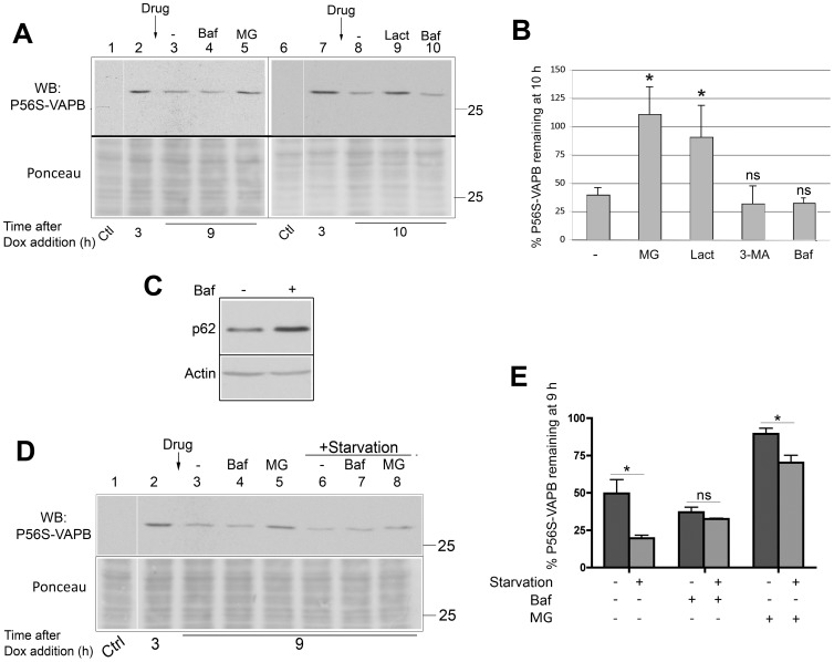
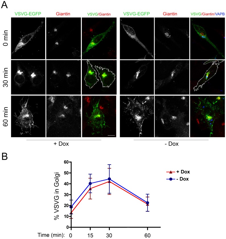
VAPB is a ubiquitously expressed, ER-resident adaptor protein involved in interorganellar lipid exchange, membrane contact site formation, and membrane trafficking. Its mutant form, P56S-VAPB, which has been linked to a dominantly inherited form of Amyotrophic Lateral Sclerosis (ALS8), generates intracellular inclusions consisting in restructured ER domains whose role in ALS pathogenesis has not been elucidated. P56S-VAPB is less stable than the wild-type protein and, at variance with most pathological aggregates, its inclusions are cleared by the proteasome. Based on studies with cultured cells overexpressing the mutant protein, it has been suggested that VAPB inclusions may exert a pathogenic effect either by sequestering the wild-type protein and other interactors (loss-of-function by a dominant negative effect) or by a more general proteotoxic action (gain-of-function). To investigate P56S-VAPB degradation and the effect of the inclusions on proteostasis and on ER-to-plasma membrane protein transport in a more physiological setting, we used stable HeLa and NSC34 Tet-Off cell lines inducibly expressing moderate levels of P56S-VAPB. Under basal conditions, P56S-VAPB degradation was mediated exclusively by the proteasome in both cell lines, however, it could be targeted also by starvation-stimulated autophagy. To assess possible proteasome impairment, the HeLa cell line was transiently transfected with the ERAD (ER Associated Degradation) substrate CD3δ, while autophagic flow was investigated in cells either starved or treated with an autophagy-stimulating drug. Secretory pathway functionality was evaluated by analyzing the transport of transfected Vesicular Stomatitis Virus Glycoprotein (VSVG). P56S-VAPB expression had no effect either on the degradation of CD3δ or on the levels of autophagic markers, or on the rate of transport of VSVG to the cell surface. We conclude that P56S-VAPB inclusions expressed at moderate levels do not interfere with protein degradation pathways or protein transport, suggesting that the dominant inheritance of the mutant gene may be due mainly to haploinsufficiency.


Conflict of interest statement

Competing Interests: The authors have declared that no competing interests exist.

Figures

Figure 1
Figure 1. P56S-VAPB is degraded by the proteasome and by activated, but not basal, autophagy.
A: Immunoblotting analysis of degradation of P56S-VAPB in the presence or absence of proteasome or autophagy inhibitors. 3 h after the inhibition of transcription of the P56S-VAPB transgene by addition of Dox to the media (lanes 2 and 7), cells were either left untreated (lanes 3 and 8), treated with the autophagy inhibitor Bafilomycin (Baf) or with the proteasome inhibitors MG132 (MG) or Lactacystin (Lact) for 6–7 h, as indicated. Control (Ctl) cells were grown in the presence of Dox. Equal aliquots of each sample were loaded (see Methods). The lower panel shows Ponceau staining of the blotted gel region, as loading control. The vertical white line (here and in panel D) juxtaposes lanes deriving from the same blot exposure. The position of the 25 kDa size marker is indicated. B: Quantification (means from 2–5 experiments +SEM) of P56S-VAPB remaining at 10 h after Dox addition in the presence or absence of drugs, as indicated, compared to levels measured at 3 h *: p = 0.013 and 0.025 for MG132 and lactacystin treated samples vs untreated by Student's t test. respectively. The difference between 3-MA or bafilomycin-treated samples and untreated was non-significant (ns). C: Equal amounts of protein of the samples of lanes 3 and 4 of panel A were analyzed for p62 by immunoblotting, to control for inhibition of autophagy by bafilomycin. Actin was probed as loading control. D: Effect of starvation on clearance of P56S-VAPB. 3 h after addition of Dox to the media (lane 2), cells were either left untreated (lane 3), or treated with bafilomycin (Baf) or MG132 (MG), as indicated, for 6 h; the samples of lanes 6–8 were also starved during the incubation with or without the drugs. Control (Ctl) cells were cultured in presence of Dox. Ponceau staining of the blotted region is shown in the lower panel. E: Quantification of three experiments (means +S.E.M.) of P56S-VAPB remaining 9 h after Dox addition under the indicated conditions compared to levels measured before drug treatment and/or starvation at 3 h after Dox addition. *: p = 0.036 by Student's t test; ns, non significant.
Figure 2
Figure 2. Lack of interference of P56S-VAPB inclusions with general proteostasis.
A: Immunoblotting analysis of the degradation of the ERAD substrate CD3δ. Induced or not induced cells, co-transfected with plasmids specifying HA-CD3δ and EGFP, were treated with CHX for 3 h as indicated. Equal amounts of protein (30 µg) were loaded. B: Quantification of three experiments (means+SEM) of CD3δ remaining 3 h after CHX addition compared to untreated samples. Values were normalized to EGFP. By two-way Anova, the presence of Dox had no significant effect on CD3δ, while the effect of CHX was very significant (p = 0.0014). C: Immunofluorescence analysis of induced P56S-VAPB-Tet-Off cells co-transfected with HA-CD3δ and EGFP. The arrows in the merge panel indicate EGFP positive cells containing P56S-VAPB inclusions, revealed with anti-myc antibodies (left panel). Approximately equal proportions of cells with or without detectable inclusions were transfected (see text). The arrowhead indicates a non-transfected cell positive for P56S-VAPB. Asterisks indicate non-transfected cells negative also for VAPB. Nuclei were stained with DAPI (blue). Scale bar, 10 µm. D: Immunoblotting analysis of the effect of P56S-VAPB inclusions on autophagic flux. Cells expressing or not expressing P56S-VAPB where either left untreated or treated for 3 h with Torin1 or starvation medium (EBSS), as indicated. The levels of p62, as percentage of the values in untreated cells are indicated below the lanes. Values were normalized to actin content. E: Quantification of three experiments (means+SEM) of LC3II/LC3I ratio of cells treated either with Torin 1 or with starvation medium, in comparison to untreated cells. Two-way Anova analysis reported that the source of variation between samples was due to autophagocytosis induction (non-treated vs Torin 1: p<0.01 and <0.05 for non-induced and induced cells, respectively) and not to P56S-VAPB expression.
Figure 3
Figure 3. P56S-VAPB inclusions in a model motoneuronal cell line are degraded by the proteasome.
A: Immunofluorescence analysis of NSC34 Tet-Off cells induced to express myc-wt-VAPB (left) or myc-P56S-VAPB (right). The upper panel shows anti-myc immunofluorescence, the lower one the superposition of myc staining with phase contrast. The inset of the upper left panel shows a 2 fold enlargement of the boxed area, and illustrates the web-like distribution of wt VAPB typical of an ER protein. Scale bar: 15 µm. B: Degradation of P56S-VAPB stably expressed in NSC34 cells. Induced cells were supplemented with Dox; 3 h thereafter the cells were either left untreated or treated with MG132 (MG) or Bafilomycin (Baf) for 7 h. Control (Ctl) cells were grown in the presence of Dox. Equal aliquots of each sample were loaded. The lower panel shows Ponceau staining of the blotted gel region; the positions of the 25 and 37 kDa size marker are indicated. The vertical white line indicates removal of irrelevant lanes form the image. The levels of P56S-VAPB, as percentage of values in untreated cells at 3 h after Dox addition, are indicated below the lanes. p62 immunoblotting was performed to check the efficacy of bafilomycin to inhibit autophagy (upper). C: Confocal analysis (single sections are shown) of P56S-VAPB inclusions stained with anti-myc antibody (red) at 3 h after Dox addition (left) and 7 h later in the presence or absence of the indicated drugs. Nuclei were stained with DAPI. The number and size of the inclusions decreased in the absence of drugs or in the presence of Bafilomycin, but remained similar to the 3 h cells when MG132 was present. Scale bar, 10 µm.
Figure 4
Figure 4. Close relationship between P56S-VAPB inclusions and the Golgi Complex.
Induced HeLa Tet-Off cells were doubly immunostained with anti-myc antibodies, to reveal P56S-VAPB, and antibodies against the Golgi proteins GM130 or giantin, as indicated. Nuclei, stained with DAPI, are shown in the merge panel. Shown are maximum intensity projections of z-stacks. Scale bars: upper row, 10 µm; middle and lower row 5 µm.
Figure 5
Figure 5. Transport of VSVG to the Golgi Complex occurs normally in cells expressing P56S-VAPB inclusions.
A: HeLa-TetOff cells, induced (−Dox, right) or not induced (+Dox, left) to express myc-P56S-VAPB, were transfected with VSVG-EGFP at 39.3°C. After 24 h, one coverslip of each sample was fixed (0 min), while the others were shifted to 32°C and fixed after incubation for the indicated times. Cells were stained with anti-Giantin (red) and anti-myc (blue) antibodies. Maximum intensity projections of z-stacks are shown. The cell boundaries at the 30 min time point are indicated by the white line in the merge panel. Acquisition parameters were the same in all images. Scale bar, 10 µm. B: Time course (means ± SD) of VSVG transport through the Golgi. Significant differences between induced or non-induced samples were not detected by Student's t-test.
Figure 6
Figure 6. Transport of VSVG to the cell surface occurs normally in cells expressing P56S-VAPB inclusions.
A: HeLa-TetOff cells, induced (−Dox) or not induced (+Dox) to express myc-P56S-VAPB, were transfected with VSVG-EGFP at 39.3°C. After 24 h, cells were shifted to 32°C. At the indicated times, the cells were chilled and incubated with anti-lumenal domain of VSVG under non-permeabilizing conditions (red). The cells were then permeabilized and stained with anti-VAPB antibodies (blue in merge panel - see Methods). Total VSVG (intracellular+surface) was revealed by GFP fluorescence (green). Maximum intensity projections of z-stacks are shown. The acquisition parameters were the same in all images. Scale bar, 10 µm. B: Time course (means ± SD) of VSVG surface labeling normalized to total EGFP fluorescence. Significant differences between induced or non-induced samples were not detected by Student's t-test.

Similar articles

Cited by

References

    1. Lev S, Ben Halevy D, Peretti D, Dahan N (2008) The VAP protein family: from cellular functions to motor neuron disease. Trends Cell Biol 18: 282–290. - PubMed
    1. Yang Z, Huh SU, Drennan JM, Kathuria H, Martinez JS, et al. (2012) Drosophila Vap-33 is required for axonal localization of Dscam isoforms. J Neurosci 32: 17241–17250. - PMC - PubMed
    1. Kuijpers M, Yu KL, Teuling E, Akhmanova A, Jaarsma D, et al. (2013) The ALS8 protein VAPB interacts with the ER-Golgi recycling protein YIF1A and regulates membrane delivery into dendrites. EMBO J 32: 2056–2072. - PMC - PubMed
    1. Kawano M, Kumagai K, Nishijima M, Hanada K (2006) Efficient trafficking of ceramide from the endoplasmic reticulum to the Golgi apparatus requires a VAMP-associated protein-interacting FFAT motif of CERT. J Biol Chem 281: 30279–30288. - PubMed
    1. Peretti D, Dahan N, Shimoni E, Hirschberg K, Lev S (2008) Coordinated lipid transfer between the endoplasmic reticulum and the Golgi complex requires the VAP proteins and is essential for Golgi-mediated transport. Mol Biol Cell 19: 3871–3884. - PMC - PubMed

Publication types

MeSH terms