. 2014 Nov 20:14:852.
doi: 10.1186/1471-2407-14-852.

Fas Activated Serine-Threonine Kinase Domains 2 (FASTKD2) mediates apoptosis of breast and prostate cancer cells through its novel FAST2 domain

Affiliations

Fas Activated Serine-Threonine Kinase Domains 2 (FASTKD2) mediates apoptosis of breast and prostate cancer cells through its novel FAST2 domain

Sharmistha Das et al. BMC Cancer. .

Abstract

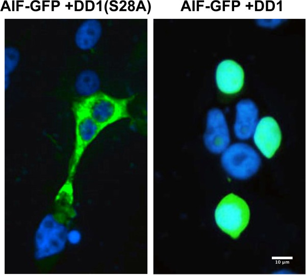
Background: Expression of NRIF3 (Nuclear Receptor Interacting Factor-3) rapidly and selectively leads to apoptosis of breast cancer cells. This occurs through binding of NRIF3 or its 30 amino acid Death Domain-1 (DD1) region to the transcriptional repressor, DIF-1 (DD1 Interacting Factor-1). DIF-1 acts in a wide variety of breast cancer cells but not other cell types to repress the pro-apoptotic gene, FASTKD2. Expression of NRIF3 or DD1 inactivates the DIF-1 repressor leading to rapid derepression of FASTKD2, which initiates apoptosis within 5-8 h of expression. Although FASTKD2 is an inner mitochondrial membrane protein, it does not require mitochondrial localization to initiate apoptosis.

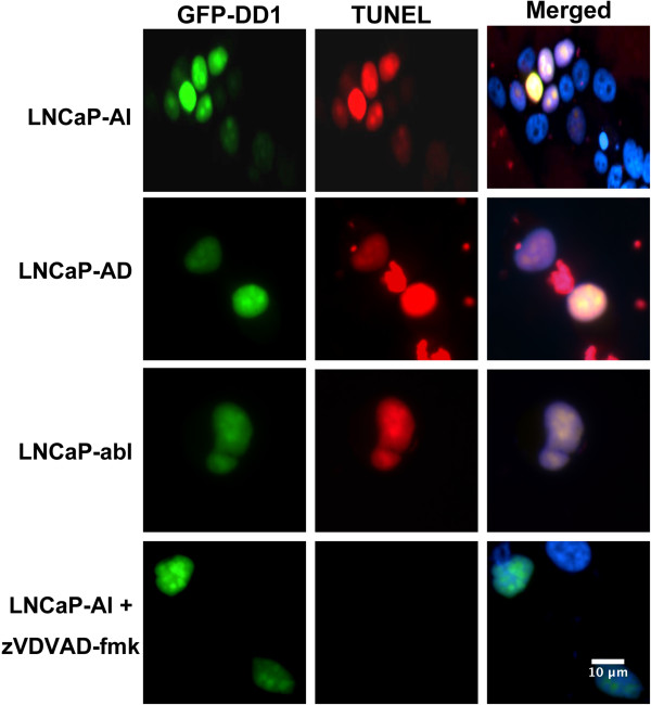
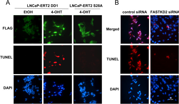
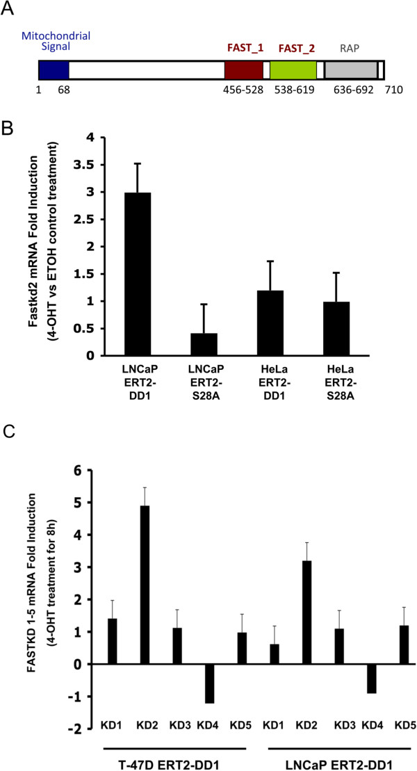
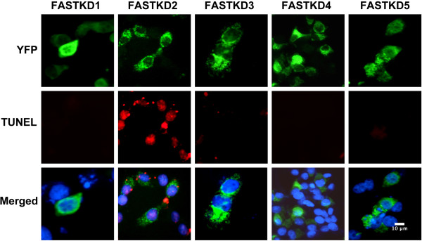
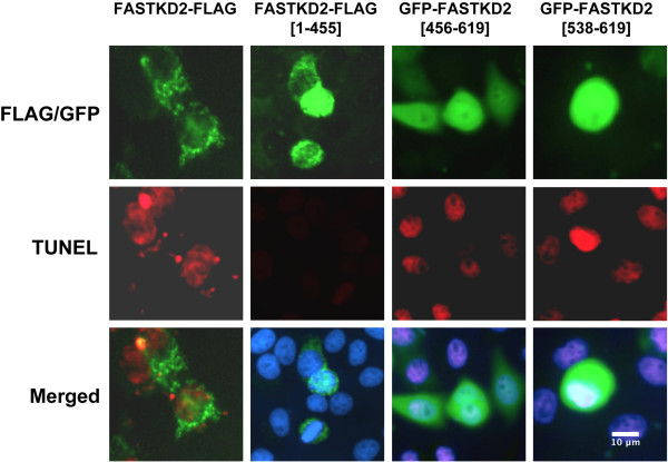
Methods: Androgen dependent LNCaP cells as well as two androgen independent LNCaP cell lines (LNCaP-AI and LNCaP-abl) were studied and LNCaP-AI cells were engineered to conditionally express DD1 or the inactive DD1-S28A with 4-hydroxytamoxifen. Apoptosis was assessed by TUNEL assay. FASTKD2 is related to 4 other proteins encoded in the human genome (FASTKD1, 3, 4, 5). All contain a poorly conserved putative bipartite kinase domain designated as FAST1_FAST2. We examined whether expression of any of the other FASTKD isoforms leads to apoptosis and sought to identify the region of FASTKD2 necessary to initiate the apoptotic pathway.

Results: Of the FASTKD1-5 isoforms only expression of FASTKD2 leads to apoptosis. Although, the NRIF3/DD1/DIF-1 pathway does not mediate apoptosis of a wide variety of non-breast cancer cell lines, because of certain similarities and gene signatures between breast and prostate cancer we explored whether the NRIF3/DD1/DIF-1/FASTKD2 pathway mediates apoptosis of prostate cancer cells. We found that the pathway leads to apoptosis in LNCaP cells, including the two androgen-independent LNCaP cell lines that are generally resistant to apoptosis. Lastly, we identified that FASTKD2-mediated apoptosis is initiated by the 81 amino acid FAST2 region.

Conclusions: The NRIF3/DIF-1/FASTKD2 pathway acts as a "death switch" in breast and prostate cancer cells. Deciphering how this pathway is regulated and how FASTKD2 initiates the apoptotic response will allow for the development of therapeutic agents for the treatment of androgen-independent prostate cancer or Tamoxifen-unresponsive Estrogen Receptor negative tumors as well as metastatic breast or prostate cancer.


Figures

Figure 1
Figure 1
Expression of DD1 leads to apoptosis in androgen dependent and androgen independent LNCaP cell lines. LNCaP-AI, LNCaP-abl and LNCaP-AD cells were transfected with 50 ng of GFP-DD1 using Lipofectamine 2000. Fifteen h later the cells were fixed and permeablized for analysis of GFP fluorescence (green) or TUNEL assay (red). The Merged panel on the right also shows nuclei (blue) stained with DAPI. In the bottom panels LNCaP-AI cells were treated with 20 uM zVDVAD-fmk prior to transfection with of GFP-DD1.
Figure 2
Figure 2
DD1-mediated apoptosis in LNCaP cells leads to changes in mitochondrial membrane permeability and release of AIF which translocates to the cell nucleus. LNCaP-AI cells were first transfected with a plasmid to express AIF-GFP. Twenty-four h later cells were transfected to express DD1 which leads to apoptosis, or DD1(S28A) which is inactive [DD1 and DD1(S28A) were expressed as GAL4 fusion proteins to ensure nuclear localization]. Fifteen h after the second transfection cells were fixed and permeablized for AIF-GFP fluorescence (green). Nuclei were stained with DAPI (blue). The Figure shows an extra-nuclear mitochondrial distribution of AIF-GFP in cells expressing DD1(S28A) while in cells expressing DD1, AIF-GFP is localized to the nucleus.
Figure 3
Figure 3
Activation of DD1 leads to expression of FASTKD2 which mediates the apoptotic response. (A) LNCaP-AI cells stably expressing DD1-ERT2 or DD1(S28A)-ERT2 were incubated with 1 uM 4-OHT for 8 h and then analyzed for DD1 and DD1(S28A) expression by immunofluorescense using FLAG-M2 antibody (green) and apoptosis by TUNEL assay (red). Nuclei were stained with DAPI (blue). (B) LNCaP-AI cells stably expressing DD1-ERT2 were transfected with a Control siRNA or an siRNA known to knock down expression of FASTKD2 [5]. Twenty-four h later the cells were transfected again with the siRNAs as described in the Methods section. Twenty h after the second siRNA transfection cells were incubated with 4-OHT for 15 h. Cells were then fixed and permeablized for TUNEL assay (red). Nuclei are stained with DAPI (blue). Cells treated with the FASTKD2 siRNA exhibited essentially no apoptosis after 4-OHT incubation while cells treated with the Control siRNA showed extensive apoptosis.
Figure 4
Figure 4
FASTKD2 expression is enhanced by DD1 activation in LNCaP cells and is the only member of the FASTKD gene family that is enhanced by DD1. (A) Domain structure of FASTKD2. (B) LNCaP-AI cells and HeLa cells stably expressing DD1-ERT2 or DD1(S28A)-ERT2 were first incubated with 20 uM zVDVAD-fmk to block apoptosis and then treated with 4-OHT (1 uM) or EtOH vehicle for 8 h. FASTKD2 mRNA expression was examined by qRT-PCR. Fold induction represents the FASTKD2 expression values of 4-OHT treated cells relative to those of EtOH treated cells. Data represents the mean +/- SEM from three representative experiments and the p values are given in the text. (C) LNCaP-AI and T-47D breast cancer cells stably expressing DD1-ERT2 were incubated with 4-OHT (1 uM) or EtOH vehicle for 8 h (cells were pretreated with 20 uM zVDVAD-fmk to block apoptosis). The expression of the 5 FASTKD mRNAs (indicated as KD1, KD2, KD3, KD4, and KD5) was examined by qRT-PCR using the primers indicated in the Methods section and in [12]. Fold induction represents the FASTKD expression values of 4-OHT treated cells relative to those of EtOH treated cells. Data represents the mean +/- SEM from three representative experiments and the p values are given in the text.
Figure 5
Figure 5
Of the FASTKD isoforms only FASTKD2 mediates apoptosis. Hela cells were transfected to express the 5 FASTKD isoforms as YFP chimeras with YFP at the C-terminus. Fifteen h later cells were fixed and permeablized for analysis of YFP fluorescence (green) and apoptosis by TUNEL assay (red). Nuclei were stained with DAPI (blue). The FASTKD-YFP chimeras localize in a peri-nuclear fashion which was previously documented to reflect mitochondrial localization [12]. Only expression of FASTKD2 leads to apoptosis.
Figure 6
Figure 6
The FAST2 domain mediates the apoptotic effect of FASTKD2. HeLa cells were transfected with 50 ng of vectors that express either FASTKD2, FASTKD2(1–455) lacking the FASTKD2 FAST1_FAST2 and RAP domains, FASTKD2(456–619) which contains the FAST1_FAST2 domain or FASTKD2(538–619) which contains just the FAST2 domain. Fifteen h later cells were fixed and permeablized for TUNEL assay (red) and fluorescence studies (green). Nuclei were stained with DAPI (blue). FASTKD2 and FASTKD2(1–455) were detected by immunofluorescence using FLAG-M2 antibody while expression of the other FASTKD2 domains were detected as GFP chimeras.

References

    1. Tan TT, White E. Therapeutic targeting of death pathways in cancer: mechanisms for activating cell death in cancer cells. Adv Exp Med Biol. 2008;615:81–104. doi: 10.1007/978-1-4020-6554-5_5. - DOI - PubMed
    1. Li D, Das S, Yamada T, Samuels HH. The NRIF3 family of transcriptional coregulators induces rapid and profound apoptosis in breast cancer cells. Mol Cell Biol. 2004;24:3838–3848. doi: 10.1128/MCB.24.9.3838-3848.2004. - DOI - PMC - PubMed
    1. Das S, Nwachukwu JC, Li D, Vulin AI, Martinez-Caballero S, Kinnally KW, Samuels HH. The nuclear receptor interacting factor-3 transcriptional coregulator mediates rapid apoptosis in breast cancer cells through direct and bystander-mediated events. Cancer Res. 2007;67:1775–1782. doi: 10.1158/0008-5472.CAN-06-4034. - DOI - PubMed
    1. Tinnikov AA, Yeung KT, Das S, Samuels HH. Identification of a novel pathway that selectively modulates apoptosis of breast cancer cells. Cancer Res. 2009;69:1375–1382. doi: 10.1158/0008-5472.CAN-08-2896. - DOI - PMC - PubMed
    1. Yeung KT, Das S, Zhang J, Lomniczi A, Ojeda SR, Xu CF, Neubert TA, Samuels HH. A Novel Transcription Complex That Selectively Modulates Apoptosis of Breast Cancer Cells through Regulation of FASTKD2. Mol Cell Biol. 2011;31:2287–2298. doi: 10.1128/MCB.01381-10. - DOI - PMC - PubMed
Pre-publication history
    1. The pre-publication history for this paper can be accessed here:http://www.biomedcentral.com/1471-2407/14/852/prepub

Publication types

MeSH terms

LinkOut - more resources