Review
. 2014 Dec;8(4):432-44.
doi: 10.1007/s12105-014-0590-0. Epub 2014 Nov 20.

Fibro-osseous lesions of the craniofacial skeleton: an update

Affiliations
Review

Fibro-osseous lesions of the craniofacial skeleton: an update

Samir K El-Mofty. Head Neck Pathol. 2014 Dec.

Abstract

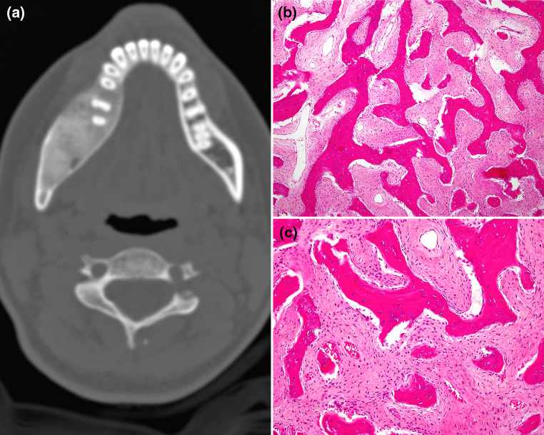
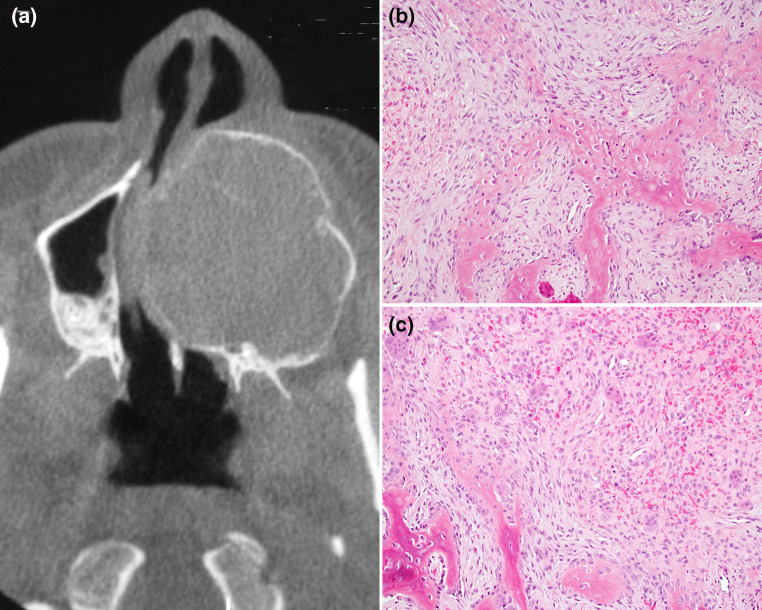
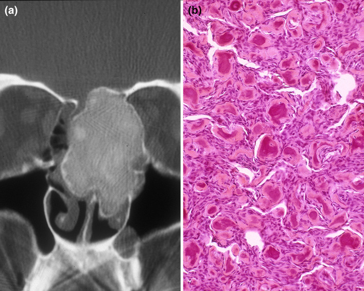
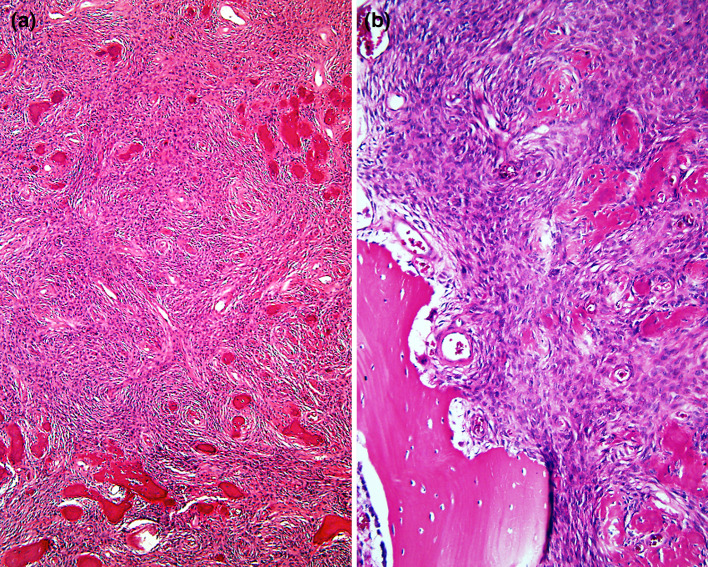
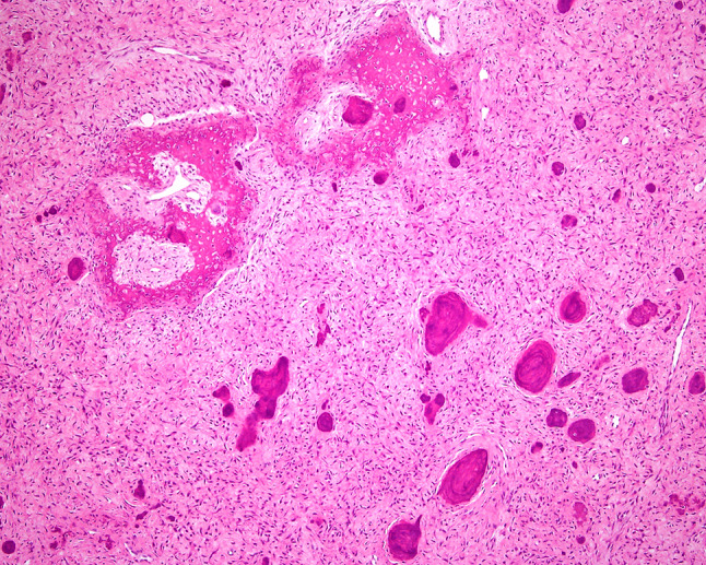
Benign fibro-osseous lesions of the craniofacial skeleton (BFOL) are a variant group of intraosseous disease processes that share similar microscopic features characterized by hypercellular fibroblastic stroma containing various combinations of bone or cementum-like tissue and other calcified structures [1-6]. Whereas some are diagnosable histologically, most require a combined assessment of clinical, microscopic and radiologic features. Some BFOL of the craniofacial complex are unique to that location whereas others are encountered in bones from other regions. Reactive, neoplastic, developmental and dysplastic pathologic processes are included under the rubric of BFOL and treatment varies from disease to disease. This review will discuss the clinical, microscopic and radiologic aspects of the more important types of BFOL of the craniofacial complex with updated information on underlying genetic and molecular pathogenic mechanisms of disease. Four main groups of BFOLs will be addressed.


Figures

Fig. 1
Fig. 1
a CT scan of fibrous dysplasia showing expansion of the mandible by a uniformly sclerotic lesion that blends with the surrounding bone; b immature woven bone trabeculae with no osteoblastic rimming, forming irregular structures resembling Chinese letters, in a fibrous stroma (×200); c higher magnification (×400)
Fig. 2
Fig. 2
a Panoramic radiograph of ossifying fibroma of the mandible. The tumor is expansive with well defined, corticated borders; b bone trabeculae and cementum-like structures dispersed in hypercellular fibrous stroma (×400); c Cementum-like calcified tissue and a well defined border with subjacent cortical bone (×200)
Fig. 3
Fig. 3
a CT scan of trabecular juvenile ossifying fibroma of the maxilla. The lesion is expansive with ground glass appearance and corticated border; b trabecular juvenile ossifying fibroma showing cellular osteoid trabeculae in spindle cell rich stroma (×400); c aggregates of osteoclast-like giant cells (×400)
Fig. 4
Fig. 4
a Periorbital psammomatoid juvenile ossifying fibroma. CT scan shows expansive, well defined but incompletely corticated sclerotic lesion; b psommomatoid juvenile ossifying fibroma composed of uniform, small round ossicles (psammomatoid bodies) in a cellular stroma (×400)
Fig. 5
Fig. 5
a Dental radiographs showing progression of calcification in periapical cemento-osseous dysplasia over a period of several years (left to right); b florid cemento-osseous dysplasia. Panoramic radiograph showing bilateral involvement of mandible and maxilla
Fig. 6
Fig. 6
a Cemento-osseous dysplasia, hypocellular bone trabeculae and cementum-like structures (×200); b with polarized light microscopy the lesion appears well demarcated from the mature lamellar bone of the Jaw (upper left corner). Dense sclerotic masses form towards the center of the lesion
Fig. 7
Fig. 7
a Hyperparathyroidism-Jaw tumor, microscopically identical to cemento-ossyfynig fibroma; b higher magnification showing well defined but unencapsulated tumor abutting cortical bone of the mandible
Fig. 8
Fig. 8
a CT scan of the skull of a patient with familial gigantiform cementoma. Massive expansive masses of the maxilla and mandible with well circumscribed borders presenting as radiolucent areas containing radiopaque calcifications; b microscopic features of FGC are analogous to those of cemento-ossifying fibroma. Cementum-like tissue formed of hypocellular basophilic and curvilinear structures and immature bone trabeculae; c are dispersed in hypercellular fibroblastic stroma composed of monomorphic appearing spindle shaped fibroblasts and collagen fibers
Fig. 9
Fig. 9
Jaw lesions in gnathodiaphysial dysplasia are analogous to those described in FGC (see Fig. 8)

References

    1. Waldron CA, Giansanti JS. Benign fibro-osseous lesions of the jaws: a clinical radiologic-histologic review of sixty-five cases I. Oral Surg Oral Med Oral Pathol. 1973;35:190–201. doi: 10.1016/0030-4220(73)90285-5. - DOI - PubMed
    1. Waldron CA, Giansanti JS. Benign fibro-osseous lesions of the jaws: a clinical radiologic-histologic review of sixty-five cases II. Oral Surg Oral Med Oral Pathol. 1973;35:340–350. doi: 10.1016/0030-4220(73)90072-8. - DOI - PubMed
    1. Hamner JE III, Scofield HH, Cornyn J (1968) Benign fibro-osseous lesions of periodontal membrane origin. An analysis of 249 cases. Cancer. 1968;22:861–78. - PubMed
    1. Waldron CA. Fibro-osseous lesions of the jaws. J Oral Maxillofac Surg. 1993;51(8):828–835. doi: 10.1016/S0278-2391(10)80097-7. - DOI - PubMed
    1. El-Mofty S. Bone lesions. In: Gnepp D, editor. Diagnostic surgical pathology of the head and neck, 2nd ed. Philadelphia: Saunders, Elsevier; 2009. p. 729–84.

MeSH terms

LinkOut - more resources