Review
. 2014 Dec;8(4):463-74.
doi: 10.1007/s12105-014-0591-z. Epub 2014 Nov 20.

Metastatic tumors to the jaws and mouth

Affiliations
Review

Metastatic tumors to the jaws and mouth

Abraham Hirshberg et al. Head Neck Pathol. 2014 Dec.

Abstract

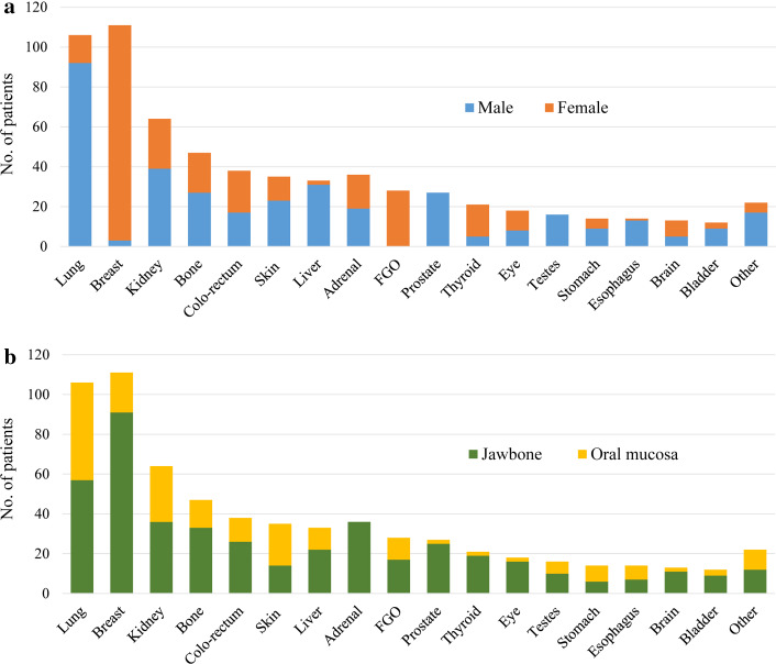
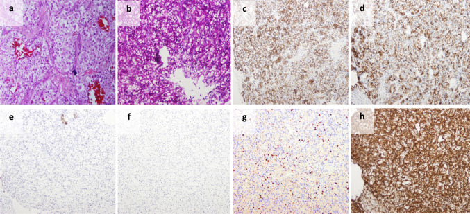
Metastatic dissemination to the oral cavity is rare and is usually the evidence of a wide spread disease with an average survival rate of 7 months. In almost a quarter of the cases, oral metastasis was found to be the first indication of an occult malignancy at a distant site. Metastatic lesions can be found anywhere in the oral cavity, however, the jaw bones with the molar area is the most frequently involved site. In the oral soft tissues, the gingiva is the most common site, suggesting the possible role of inflammation in the attraction of metastatic deposits. The most common primary malignancies presenting oral metastases were the lung, kidney, liver, and prostate for men, and breast, female genital organs, kidney, and colo-rectum for women. Most patients with jawbone metastasis complain of swelling, pain, and paresthesia. An exophytic lesion is the most common clinical presentation of metastatic lesions in the oral soft tissues. Early lesions, mainly those located in the gingiva, may resemble a hyperplastic or reactive lesion. Once a lesion is recognized as metastasis, the primary tumor site should be identified following clinical, radiological and histopathological investigations. If standardized diagnostic workup fails to detect the site of origin, then the term carcinoma of unknown primary is applied. Personalized medicine tools such as tissue-of-origin assays should be applied, either by immunohistochemical testing or by molecular-profiling methods as these may lead to a more favorable outcome.


Figures

Fig. 1
Fig. 1
The metastatic cascade. A successful metastatic colonization represents the end products of a complex sequential events including a invasion through the extracellular matrix (ECM); b proliferation and angiogenesis; c intravasation into the blood vessels; d survival in the circulation. e Circulating cancer cells that survive preferentially adhere to the endothelium in the microvasculature of the target organ and f extravasate through the vessel wall; g tumor cells settle at the target organ, which consists of a permissive microenvironment
Fig. 2
Fig. 2
a The microenvironment present in the chronically inflamed gingiva may provide a favorable niche for metastatic cells to colonize and proliferate (circle). Soluble cytokines such as IL-1 and TNF-α are known to facilitate metastatic progression by stimulating angiogenesis and accelerating the generation of extracellular matrix necessary for tumor stroma and also may attract or induce the tumor associated macrophages. b Tumor cells recruit resident cells in the bone marrow mainly osteoclast and osteoblast to promote the ‘vicious cycle’ of bone metastases. (1) Tumor cells stimulate bone resorption which result in release of growth factors [transforming growth factor-β (TGFβ), insulin-like growth factor I (IGF-I)] and ionized calcium from the mineralized bone matrix. In addition, osteoclasts directly produce many protumorigenic factors, including growth factors (such as platelet-derived growth factor, IL-1, TNF), angiogenic factors [vascular endothelial growth factor (VEGF), basic fibroblast growth factor (bFGF)], and matrix metalloproteinases. These factors increase tumor cell proliferation and induce further production of osteolytic and osteoblastic factors creating a positive feedback loop. (2) Metastatic tumor cells express many factors such as transforming growth factor-β (TGFβ), bone morphogenetic proteins (BMPs) and WNT proteins promoting osteoblastic activity. (3) RANKL (receptor activator of NF-κB ligand) functions via its receptor RANK, and is inhibited by the soluble osteoprotegerin (OPG). (4) The ratio of RANKL to OPG is upregulated leading to increased osteoclast-mediated bone destruction
Fig. 3
Fig. 3
Distribution of the common primary tumors metastasizing the oral cavity correlated with gender (a) and oral site (b). Based on Hirshberg et al. [8] total 655 cases after omitting the cases of unknown gender. Skin, including malignant melanoma; FGO, female genital organs, including uterus, ovaries, cervix, fallopian tubes; Adrenal, cases of pheochromocytoma and neuroblastoma including those from the retroperitoneum and mediastinum
Fig. 4
Fig. 4
a A metastasis to the mouth exhibited clear cells with moderate atypia, arranged in closely packed groups surrounded by a vascular matrix; b PAS stained the clear cytoplasm. The tumor cells were positive for EMA (c), CD10 (d) and PAX8 (not shown), negative for cytokeratin 7 (e) and cytokeratin 20 (f); g Ki67 is positive in less than 10 % of cells. This pattern of immunohistochemistry is typical for renal cell carcinoma (RCC). h Vimentin was also positive. RCC is one of few carcinomas that stain positive for this general mesenchyme marker. S-100, GFAP and SMA were negative, which ruled-out salivary gland origin. Synaptophysin, calcitonin and chromogranin were negative as well, eliminating the possibility of endocrine carcinoma origin

References

    1. Valastyan S, Weinberg RA. Tumor metastasis: molecular insights and evolving paradigms. Cell. 2011;147(2):275–292. doi: 10.1016/j.cell.2011.09.024. - DOI - PMC - PubMed
    1. Nguyen DX, Bos PD, Massague J. Metastasis: from dissemination to organ-specific colonization. Nat Rev Cancer. 2009;9(4):274–284. doi: 10.1038/nrc2622. - DOI - PubMed
    1. Hanahan D, Weinberg RA. Hallmarks of cancer: the next generation. Cell. 2011;144(5):646–674. doi: 10.1016/j.cell.2011.02.013. - DOI - PubMed
    1. Servato JP, de Paulo LF, de Faria PR, Cardoso SV, Loyola AM. Metastatic tumours to the head and neck: retrospective analysis from a Brazilian tertiary referral centre. Int J Oral Maxillofac Surg. 2013;42(11):1391–1396. doi: 10.1016/j.ijom.2013.05.020. - DOI - PubMed
    1. Hashimoto N, Kurihara K, Yamasaki H, Ohba S, Sakai H, Yoshida S. Pathological characteristics of metastatic carcinoma in the human mandible. J Oral Pathol. 1987;16(7):362–367. doi: 10.1111/j.1600-0714.1987.tb00709.x. - DOI - PubMed

LinkOut - more resources