

Homing and restorative effects of bone marrow-derived mesenchymal stem cells on cisplatin injured ovaries in rats
- PMID: 25410907
- PMCID: PMC4275703
- DOI: 10.14348/molcells.2014.0145
Homing and restorative effects of bone marrow-derived mesenchymal stem cells on cisplatin injured ovaries in rats
Abstract
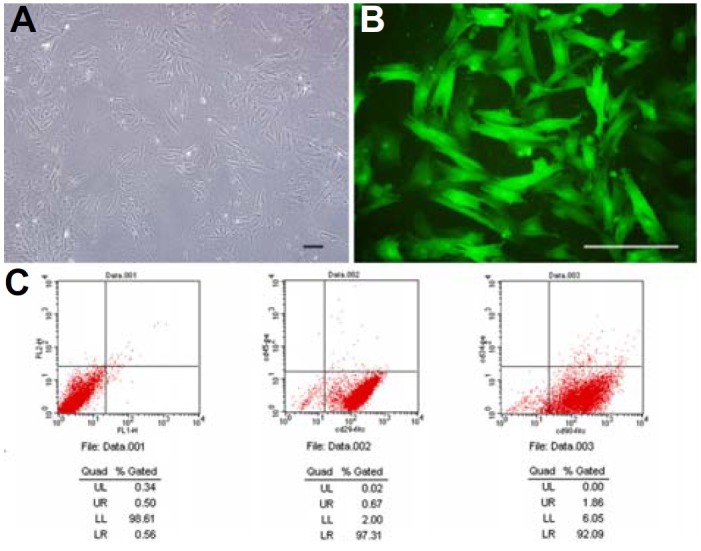
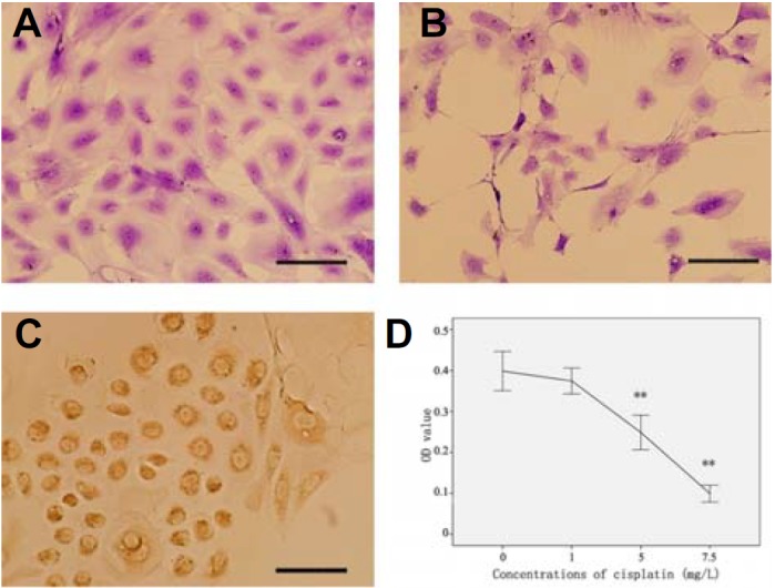
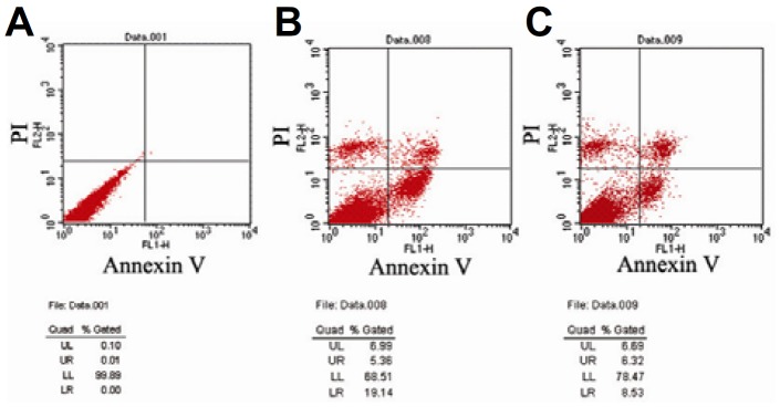
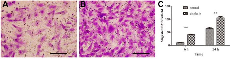
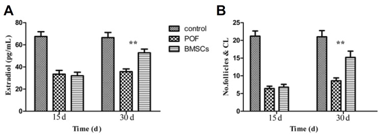
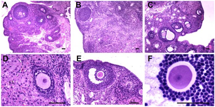
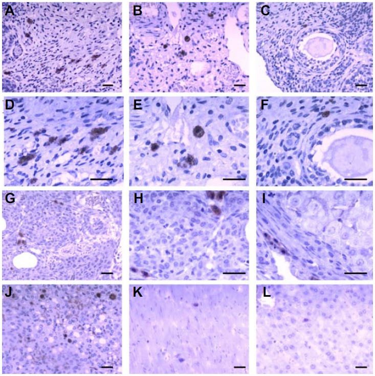
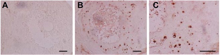
Premature ovarian failure (POF) is a long-term adverse effect of chemotherapy treatment. However, current available treatment regimens are not optimal. Emerging evidence suggests that bone marrow-derived mesenchymal stem cells (BMSCs) could restore the structure and function of injured tissues, but the homing and restorative effects of BMSCs on chemotherapy injured ovaries are still not clear. In this study, we found that granulosa cell (GC) apoptosis induced by cisplatin was reduced when BMSCs were migrated to granulosa cells (GCs) in vitro. Chemotherapy-induced POF was induced by intraperitoneal injection of cisplatin in rats. BMSCs labeled with enhanced green fluorescent protein (EGFP) were injected into the rats via the tail vein to investigate the homing and distribution of BMSCs in vivo. The number of BMSCs in the ovarian hilum and medulla was greater than in the cortex, but no BMSCs were found in the follicles and corpus lutea. In addition, the BMSCs treatment group's antral follicle count and estradiol levels increased after 30 days, compared with the POF group. Hence, our study demonstrates that intravenously delivered BMSCs can home to the ovaries, and restore its structure and function in POF model rats.
Keywords: bone marrow-derived mesenchymal stem cells; cisplatin; homing; intravenous injection; premature ovarian failure.
Figures
Similar articles
-
Heat shock pretreatment of mesenchymal stem cells for inhibiting the apoptosis of ovarian granulosa cells enhanced the repair effect on chemotherapy-induced premature ovarian failure.Stem Cell Res Ther. 2018 Sep 26;9(1):240. doi: 10.1186/s13287-018-0964-4. Stem Cell Res Ther. 2018. PMID: 30257708 Free PMC article.
-
Clusterin-carrying extracellular vesicles derived from human umbilical cord mesenchymal stem cells restore the ovarian function of premature ovarian failure mice through activating the PI3K/AKT pathway.Stem Cell Res Ther. 2024 Sep 13;15(1):300. doi: 10.1186/s13287-024-03926-7. Stem Cell Res Ther. 2024. PMID: 39272156 Free PMC article.
-
The effects of human menstrual blood stem cells-derived granulosa cells on ovarian follicle formation in a rat model of premature ovarian failure.Microsc Res Tech. 2019 Jun;82(6):635-642. doi: 10.1002/jemt.23120. Epub 2018 Dec 23. Microsc Res Tech. 2019. PMID: 30582244
-
Intraovarian injection of autologous human mesenchymal stem cells increases estrogen production and reduces menopausal symptoms in women with premature ovarian failure: two case reports and a review of the literature.J Med Case Rep. 2020 Jul 18;14(1):108. doi: 10.1186/s13256-020-02426-5. J Med Case Rep. 2020. PMID: 32680541 Free PMC article. Review.
-
The therapeutic potential of bone marrow mesenchymal stem cells in premature ovarian failure.Stem Cell Res Ther. 2018 Oct 4;9(1):263. doi: 10.1186/s13287-018-1008-9. Stem Cell Res Ther. 2018. PMID: 30286808 Free PMC article. Review.
Cited by
-
Stem cell-based therapeutic potential in female ovarian aging and infertility.J Ovarian Res. 2024 Aug 24;17(1):171. doi: 10.1186/s13048-024-01492-3. J Ovarian Res. 2024. PMID: 39182123 Free PMC article. Review.
-
miR-644-5p carried by bone mesenchymal stem cell-derived exosomes targets regulation of p53 to inhibit ovarian granulosa cell apoptosis.Stem Cell Res Ther. 2019 Nov 29;10(1):360. doi: 10.1186/s13287-019-1442-3. Stem Cell Res Ther. 2019. PMID: 31783913 Free PMC article.
-
Important role of the SDF-1/CXCR4 axis in the homing of systemically transplanted human amnion-derived mesenchymal stem cells (hAD-MSCs) to ovaries in rats with chemotherapy-induced premature ovarian insufficiency (POI).Stem Cell Res Ther. 2022 Feb 23;13(1):79. doi: 10.1186/s13287-022-02759-6. Stem Cell Res Ther. 2022. PMID: 35197118 Free PMC article.
-
New Solutions for Old Problems: How Reproductive Tissue Engineering Has Been Revolutionizing Reproductive Medicine.Ann Biomed Eng. 2023 Oct;51(10):2143-2171. doi: 10.1007/s10439-023-03321-y. Epub 2023 Jul 19. Ann Biomed Eng. 2023. PMID: 37468688 Review.
-
Human mesenchymal stem cell treatment of premature ovarian failure: new challenges and opportunities.Stem Cell Res Ther. 2021 Mar 3;12(1):161. doi: 10.1186/s13287-021-02212-0. Stem Cell Res Ther. 2021. PMID: 33658073 Free PMC article. Review.
References
-
- Abd-Allah S.H., Shalaby S.M., Pasha H.F., El-Shal A.S., Raafat N., Shabrawy S.M., Awad H.A., Amer M.G., Gharib M.A., El Gendy E.A., et al. Mechanistic action of mesenchymal stem cell injection in the treatment of chemically induced ovarian failure in rabbits. Cytotherapy. 2013;15:64–75. - PubMed
-
- Agung M., Ochi M., Yanada S., Adachi N., Izuta Y., Yamasaki T., Toda K. Mobilization of bone marrow-derived mesenchymal stem cells into the injured tissues after intraarticular injection and their contribution to tissue regeneration. Knee Surg. Sports Traumatol. Arthrosc. 2006;14:1307–1314. - PubMed
-
- Ai H., Zhang H., Zhang Y., Liu Y. Mouse ovarian-related gene expression profiles change with intraperitoneal injection of cisplatin. African J. Pharm. Pharm. 2012;6:2119–2122.
-
- Antoine C., Ameye L., Paesmans M., Rozenberg S. Systematic review about breast cancer incidence in relation to hormone replacement therapy use. Climacteric. 2013;17:116–135. - PubMed
-
- Bi B., Schmitt R., Israilova M., Nishio H., Cantley L.G. Stromal cells protect against acute tubular injury via an endocrine effect. J. Am. Soc. Nephrol. 2007;18:2486–2496. - PubMed
Publication types
MeSH terms
Substances
LinkOut - more resources
Full Text Sources
Other Literature Sources
Medical
Miscellaneous
