Review
. 2015 Dec;227(6):746-56.
doi: 10.1111/joa.12257. Epub 2014 Nov 20.

Advances in 3D cell culture technologies enabling tissue-like structures to be created in vitro

Affiliations
Review

Advances in 3D cell culture technologies enabling tissue-like structures to be created in vitro

Eleanor Knight et al. J Anat. 2015 Dec.

Abstract

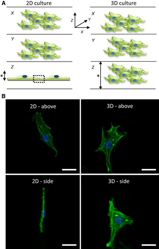
Research in mammalian cell biology often relies on developing in vitro models to enable the growth of cells in the laboratory to investigate a specific biological mechanism or process under different test conditions. The quality of such models and how they represent the behavior of cells in real tissues plays a critical role in the value of the data produced and how it is used. It is particularly important to recognize how the structure of a cell influences its function and how co-culture models can be used to more closely represent the structure of real tissue. In recent years, technologies have been developed to enhance the way in which researchers can grow cells and more readily create tissue-like structures. Here we identify the limitations of culturing mammalian cells by conventional methods on two-dimensional (2D) substrates and review the popular approaches currently available that enable the development of three-dimensional (3D) tissue models in vitro. There are now many ways in which the growth environment for cultured cells can be altered to encourage 3D cell growth. Approaches to 3D culture can be broadly categorized into scaffold-free or scaffold-based culture systems, with scaffolds made from either natural or synthetic materials. There is no one particular solution that currently satisfies all requirements and researchers must select the appropriate method in line with their needs. Using such technology in conjunction with other modern resources in cell biology (e.g. human stem cells) will provide new opportunities to create robust human tissue mimetics for use in basic research and drug discovery. Application of such models will contribute to advancing basic research, increasing the predictive accuracy of compounds, and reducing animal usage in biomedical science.

Keywords: 3D cell culture; in vitro; technology; tissue structure.


Figures

Figure 1
Figure 1
Impact of the physical environment on cell structure. (A) Visualisation of cells for each of the three dimensions (X,Y,Z). In simple terms, X and Y symbolize the length and width of a cell, and Z describes the height. In conventional 2D culture, cells grow as monolayers on a solid substrate; they flatten and possess a low vertical height (left). In contrast, cells cultured in a 3D model maintain a more natural 3D structure and possess more normal dimensions all round (right). Furthermore, the overall height (*) of a conventional 2D monolayer culture is relatively fixed, whereas that of a 3D culture is more versatile, depending on the 3D cell technology used, and can be built up to form multi‐layered tissue‐like structures. Interactions between adjacent cells cultured in 2D are restricted to the periphery of the cells within a single plane (left, dotted box), whereas in 3D models the scope of intercellular contact is all around. (B) Confocal images of a single fibroblast grown in 2D or 3D culture. The cell has been stained with phalloidin to visualize the primary structural elements of the F‐actin cytoskeleton and 4',6‐diamidino‐2‐phenylindole (DAPI) for the nucleus. The images show the shape of a typical cell when visualized from above (top panels) or from the side (bottom panels). Note how thin a cell can become when cultured on a flat substrate as in conventional 2D culture (left) compared with the more normal structure of a cell in a 3D culture model (right). Scale bars: 10 μm. (Images courtesy of Dr. F. Tholozan, Durham University).
Figure 2
Figure 2
Primary technologies used to culture cells in 3D. (A) Formation of 3D micro‐tissues using hanging drop approach. Droplets of cell suspension are placed on the lid of a Petri dish, which is gently inverted and placed on top of the dish containing media to maintain a humid atmosphere. Suspended cells come together in the apex of the droplet, forming a compact 3D aggregate. (B) 3D culture using hydrogel technology. The cartoon shows cells within a matrix of protein molecules that create a nano‐scale micro‐environment mimicking the structure of the extracellular matrix. Cells are embedded within the proteinous 3D framework within an aqueous‐based gel. (C) Porous solid scaffold supporting 3D cultured cells. Cells enter the porous framework of the solid scaffold where they do not flatten, they maintain their 3D structure, and they bind to one another forming 3D tissue‐like masses.
Figure 3
Figure 3
Co‐culture offers an exciting opportunity to create novel 3D tissue‐like models in vitro. Each of the three primary technologies developed for 3D culture can be customized for alternative applications containing different cell types. The cartoons show cells of different colors representing alternative cell types within each technology. (A) Cells of different types can be co‐seeded in suspended hanging drops to create micro‐tissue aggregates composed of more than one cell type. The addition of an alternative cell type to a pre‐formed cell aggregate can produce a concentric layered structure. (B) Mixing cells of different types is relatively straightforward using hydrogel technology. Cells can self‐organize and form tissue‐like structures as they migrate within the gel. (C) Similarly, a mixture of different cells can be added directly to the porous scaffold where they self‐arrange within the matrix.
Figure 4
Figure 4
Construction of tissue‐like structures composed of layers of alternative cell types. The presentation of a solid porous scaffold as a membrane provides a suitable platform to construct layers of alternative cell types. Many types of tissue are composed of alternate layers of cells and such tissue architecture can be replicated by seeding cells sequentially onto supporting porous scaffolds. (A) An example of two cell types: those in blue were seeded first and allowed to grow and establish a 3D culture inside the scaffold; those in green were seeded second onto the surface of the scaffold to establish a 3D layer on top. (B) Example of a human skin construct produced by first seeding human primary dermal fibroblasts into a 200‐μm‐thick porous polystyrene scaffold for 7 days, followed by seeding human primary keratinocytes onto the surface and maintaining the culture for a further 21 days at the air–liquid interface. The skin construct matures over time in culture consisting of differentiated epidermal (epi) and supporting dermal (der) components. Scale bar: 50 μm. (Image courtesy of N. Robinson, Durham University).

References

    1. Abe K, Niwa H, Iwase K, et al. (1996) Endoderm‐specific gene expression in embryonic stem cells differentiated to embryoid bodies. Exp Cell Res 229, 27–34. - PubMed
    1. Alayoubi A, Alqahtani S, Kaddoumi A, et al. (2013) Effect of PEG surface conformation on anticancer activity and blood circulation of nanoemulsions loaded with tocotrienol‐rich fraction of palm oil. AAPS J 15, 1168–1179. - PMC - PubMed
    1. Amatangelo MD, Garipov A, Li H, et al. (2013) Three‐dimensional culture sensitizes epithelial ovarian cancer cells to EZH2 methyltransferase inhibition. Cell Cycle 12, 2113–2119. - PMC - PubMed
    1. Antonchuk J (2013) Formation of embryoid bodies from human pluripotent stem cells using AggreWell plates. Methods Mol Biol, 946, 523–533. - PubMed
    1. Awad HA, Wickham MQ, Leddy HA, et al. (2004) Chondrogenic differentiation of adipose‐derived adult stem cells in agarose, alginate, and gelatin scaffolds. Biomaterials 25, 3211–3222. - PubMed

Publication types