Review
. 2015 Jan 15;308(2):R79-95.
doi: 10.1152/ajpregu.00351.2014. Epub 2014 Nov 19.

Role of renal sensory nerves in physiological and pathophysiological conditions

Affiliations
Review

Role of renal sensory nerves in physiological and pathophysiological conditions

Ulla C Kopp. Am J Physiol Regul Integr Comp Physiol. .

Abstract

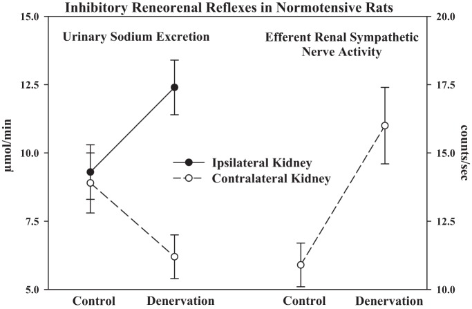
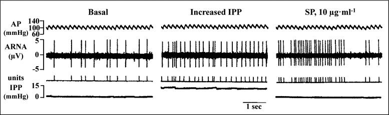
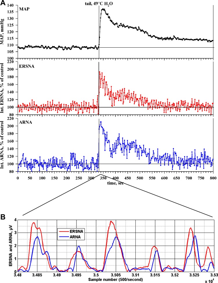
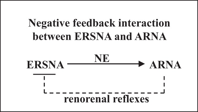
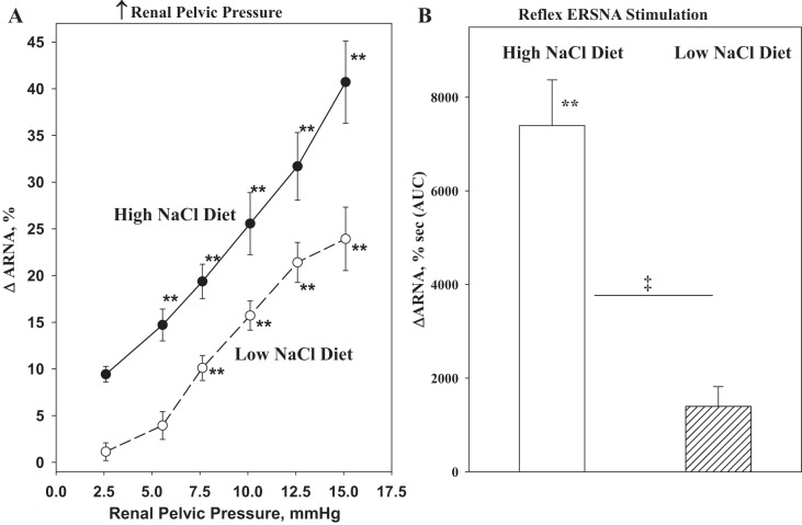
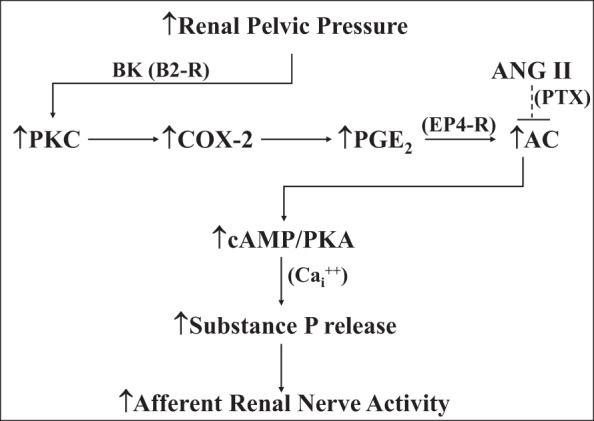
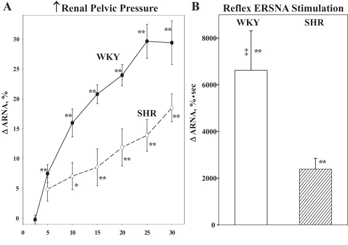
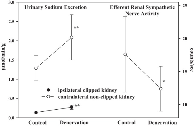
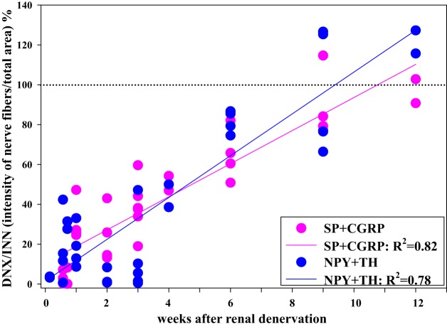
Whether activation of afferent renal nerves contributes to the regulation of arterial pressure and sodium balance has been long overlooked. In normotensive rats, activating renal mechanosensory nerves decrease efferent renal sympathetic nerve activity (ERSNA) and increase urinary sodium excretion, an inhibitory renorenal reflex. There is an interaction between efferent and afferent renal nerves, whereby increases in ERSNA increase afferent renal nerve activity (ARNA), leading to decreases in ERSNA by activation of the renorenal reflexes to maintain low ERSNA to minimize sodium retention. High-sodium diet enhances the responsiveness of the renal sensory nerves, while low dietary sodium reduces the responsiveness of the renal sensory nerves, thus producing physiologically appropriate responses to maintain sodium balance. Increased renal ANG II reduces the responsiveness of the renal sensory nerves in physiological and pathophysiological conditions, including hypertension, congestive heart failure, and ischemia-induced acute renal failure. Impairment of inhibitory renorenal reflexes in these pathological states would contribute to the hypertension and sodium retention. When the inhibitory renorenal reflexes are suppressed, excitatory reflexes may prevail. Renal denervation reduces arterial pressure in experimental hypertension and in treatment-resistant hypertensive patients. The fall in arterial pressure is associated with a fall in muscle sympathetic nerve activity, suggesting that increased ARNA contributes to increased arterial pressure in these patients. Although removal of both renal sympathetic and afferent renal sensory nerves most likely contributes to the arterial pressure reduction initially, additional mechanisms may be involved in long-term arterial pressure reduction since sympathetic and sensory nerves reinnervate renal tissue in a similar time-dependent fashion following renal denervation.

Keywords: angiotensin; hypertension; kidney; prostaglandin E2; renal denervation; renal mechanosensory nerves; substance P.


Figures

Fig. 1.
Fig. 1.
A: camera lucida drawing of a whole mount renal pelvis and proximal ureter. In the kidney, the majority of the afferent sensory nerves are located in the renal pelvic wall. Most of the afferent fibers travel parallel to the long axis of the pelvis/ureter. There are also fibers that are oriented predominantly in a circumferential fashion, making them ideally located for sensing a stretch of the pelvic wall (arrows). The area enclosed by the box is illustrated in B. Many of the afferent nerve fibers are fine and appear to terminate as free nerve endings (B). [Modified from Ref. : Marfurt CF, Echtenkamp SF. Sensory innervation of the rat kidney and ureter as revealed by the anterograde transport of wheat germ agglutinin-horseradish peroxidase (WGA-HRP) from dorsal root ganglia. J Comp Neurol 311: 389–404, 1991.]
Fig. 2.
Fig. 2.
Renal tissue was double-labeled with antibodies against substance P (SP) (A) and calcitonin gene-related peptide (CGRP) (B). Substance P and CGRP are colocalized in all sensory nerves in the renal pelvic wall (pw) surrounding the papilla (pap). Arrows depict sensory nerves with both substance P-like immunoreactivity (-LI) and CGRP-LI. The majority of the sensory nerves are found in the subepithelial layer of the pelvic wall with few fibers penetrating into the uroepithelium (arrowheads). Magnification: ×120. [From Ref. 77].
Fig. 3.
Fig. 3.
In normotensive healthy rats, ipsilateral renal denervation increases ipsilateral urinary sodium excretion and decreases contralateral urinary sodium excretion. The fall in contralateral urinary sodium excretion is produced by an increase in contralateral efferent renal sympathetic nerve activity, suggesting that the afferent renal nerves exert a tonic inhibition of efferent renal sympathetic nerve activity. [Modified from Ref. 24].
Fig. 4.
Fig. 4.
Single-unit recordings of afferent renal nerve activity (ARNA) showed that the same nerve fiber activated by increases in intrarenal pelvic pressure (IPP) can also be activated by substance P (SP) administered directly into the renal pelvic area. AP, arterial pressure. [Modified from Ref. with permission from the American Society of Nephrology, Impaired renal sensory responses after renal ischemia in the rat, Ma MC, Huang HS, Wu MS, Chien CT, Chen CF. J Am Soc Nephrol 13: 1872–1883, 2002].
Fig. 5.
Fig. 5.
Renal tissue was double-labeled with antibodies against the norepinephrine transporter (NE-t) (A) and CGRP (B). C: the majority of the NE-t-immunoreactive (NE-t-ir) fibers (green) are close to the CGRP-ir fibers (red), (arrows) in the renal pelvic wall. D and E: confocal microscopy of two nerve bundles in the renal pelvic area showed that the sympathetic and sensory nerves are separate fibers (arrows). [Modified from Ref. 78].
Fig. 6.
Fig. 6.
A: thermal cutaneous stimulation produced by placing the anesthetized rat's tail in warm water produced a general activation of the sympathetic nervous system, as shown by the increases in mean arterial pressure (MAP; black lines), efferent renal sympathetic nerve activity (ERSNA; red lines) and ARNA (blue lines), recorded in the same rat. B: relationship between ERSNA and ARNA for the 1-s interval beginning 5.6 s after placing the rat's tail in warm water. The increases in ERSNA preceded the increases in ARNA by 17.1 ± 2.4 ms (n = 7). [Modified from Ref. 78].
Fig. 7.
Fig. 7.
There is a reciprocal interaction between ERSNA and ARNA. Increases in ERSNA increase ARNA, the increase in ARNA will, in turn, decrease ERSNA via activation of the inhibitory renorenal reflexes in the overall goal of maintaining low ERSNA to minimize sodium retention. ERSNA modulates ARNA by release of norepinephrine (NE).
Fig. 8.
Fig. 8.
Renal tissue was double-labeled with antibodies against α1-adrencoceptors (AR) and CGRP (A–C), α2A-AR and CGRP (D–F) or α2C-AR and CGRP (G–I). In the renal pelvic wall, α1-AR-immunoreactive (ir) fibers, α2A-AR-ir fibers, and α2c-AR-ir fibers (all green) were close or on CGRP-ir fibers (red) as seen in C, F, and I (colocalization yellow). [Modified from Ref. 79].
Fig. 9.
Fig. 9.
A: high-sodium diet enhances the ARNA responses to increases in renal pelvic pressure. B: reflex increases in ERSNA produced by thermal cutaneous stimulation in healthy normotensive rats. **P < 0.01 vs. baseline control value. ‡P < 0.01 vs. low-NaCl diet. [Modified from Refs. and 79].
Fig. 10.
Fig. 10.
Stretching the renal pelvic wall by increases in renal pelvic pressure leads to activation of PKC by stimulation of bradykinin (BK) B2 receptors (B2-R), which leads to induction of cyclooxygenase-2 (COX-2) and an increase in renal pelvic release of PGE2. PGE2 activates EP4 receptors on or close to the renal pelvic sensory nerves, which, in turn, leads to activation of the adenylyl cyclase (AC)/cAMP/PKA transduction pathway. This results in a calcium (Cai2+)-dependent release of substance P and increases in afferent renal nerve activity. The mechanisms involved in the suppressed responsiveness of the renal sensory nerves in physiological and pathophysiological conditions of increased endogenous ANGII activity, include ANG II reducing the PGE2-mediated activation of AC by a pertussis toxin-sensitive (PTX) mechanism. [Data derived from Refs. , , , , , –83].
Fig. 11.
Fig. 11.
Renal tissue was double-labeled with antibodies against ETA-R (green) and CGRP (red) (A), ETA-R and α-smooth muscle actin (SMA; red) (B), ETB-R (green) and CGRP (red) (C), and ETB-R and α-SMA (red) (D). In the renal pelvic wall, ETA-R-ir fibers were found on smooth muscle cells and CGRP-ir nerve fibers (arrows) among the ETA-R-ir smooth muscle cells. Also, in the renal pelvic wall, ETB-R-ir fiber-like structures (arrows) were found close to CGRP-ir nerve fibers (arrows) among smooth muscle cells. [Modified from Ref. 84].
Fig. 12.
Fig. 12.
The ARNA responses to graded increases in renal pelvic pressure (A) and reflex increases in ERSNA (B) produced by thermal cutaneous stimulation are suppressed in SHR vs. WKY. [Data in B are from Ref. 74]. *P < 0.05, **P < 0.01 vs. baseline control value; ‡P < 0.01 vs. SHR.
Fig. 13.
Fig. 13.
In two-kidney, one-clip hypertensive rats, renal denervation of the ipsilateral clipped kidney increases both ipsilateral and contralateral urinary sodium excretion. The increase in contralateral urinary sodium excretion is produced by a fall in contralateral efferent renal sympathetic nerve activity, suggesting that the afferent renal nerves from the clipped ischemic kidney exert an excitatory influence on efferent renal sympathetic nerve activity. [Data are derived from Ref. 63]. *P < 0.05, **P < 0.01 vs. Control.
Fig. 14.
Fig. 14.
Adjacent slides of rat renal tissue from the denervated and contralateral innervated kidneys were labeled with antibodies against tyrosine hydroxylase (TH) and neuropeptide Y (NPY) and SP and CGRP. The optical density of the nerve fibers in renal tissue was determined by ImageJ software. The optical density of sympathetic nerve fibers containing NPY and TH (blue symbols) and sensory nerves containing SP and CGRP (pink symbols) in the denervated kidney was compared with those in the contralateral innervated kidney in each rat at various time points following unilateral renal denervation. The data derived from the optical density curves of the NPY, TH, CGRP, and SP-containing fibers suggest that the sensory nerves and sympathetic nerves reinnervate renal pelvic and peripelvic cortical area with a similar time course in normal healthy rats. [Data are derived from Ref. 114].

References

    1. Almgård LE, Ljungqvist A, Ungerstedt U. The reaction of the intrarenal sympathetic nervous system to renal transplantion. Scand J Urol Nephrol 5: 65–70, 1971. - PubMed
    1. Barajas L, Liu L, Powers K. Anatomy of the renal innervation: intrarenal aspects and ganglia of origin. Can J Physiol Pharmacol 70: 735–749, 1992. - PubMed
    1. Barajas L, Powers K. Innervation of the renal proximal convoluted tubule of the rat. Am J Anat 186: 378–388, 1989. - PubMed
    1. Barajas L, Powers K. Monoaminergic innervation of the rat kidney: a quantitative study. Am J Physiol Renal Fluid Electrolyte Physiol 259: F503–F511, 1990. - PubMed
    1. Barajas L, Powers K, Wang P. Innervation of the renal cortical tubules: a quantitative study. Am J Physiol Renal Fluid Electrolyte Physiol 247: F50–F60, 1984. - PubMed

Publication types

Substances

LinkOut - more resources