. 2014 Nov 19;34(47):15587-600.
doi: 10.1523/JNEUROSCI.2037-14.2014.

Delayed disease onset and extended survival in the SOD1G93A rat model of amyotrophic lateral sclerosis after suppression of mutant SOD1 in the motor cortex

Affiliations

Delayed disease onset and extended survival in the SOD1G93A rat model of amyotrophic lateral sclerosis after suppression of mutant SOD1 in the motor cortex

Gretchen M Thomsen et al. J Neurosci. .

Abstract

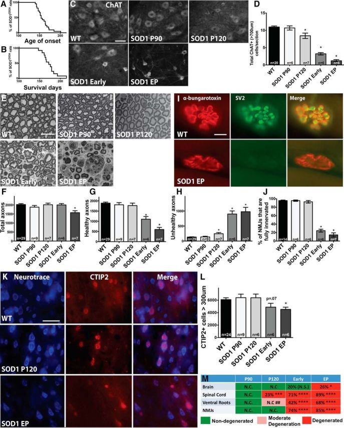
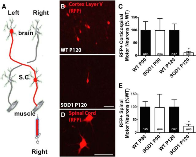
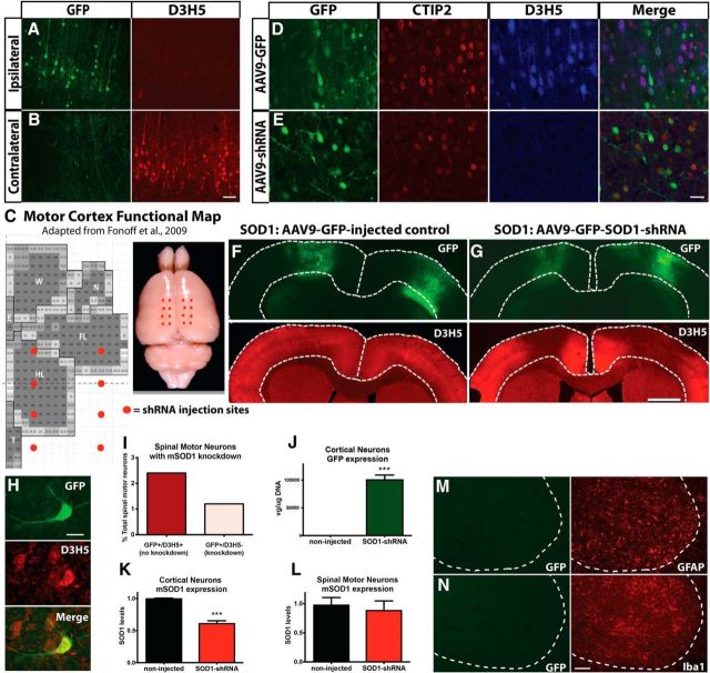
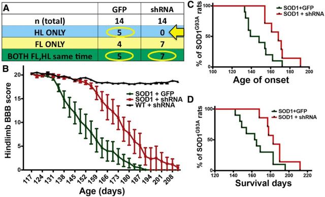
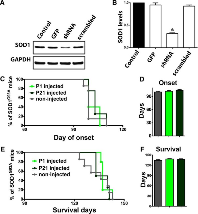
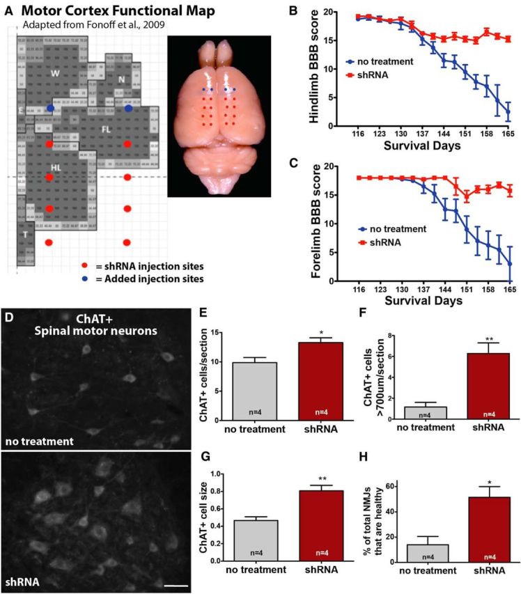
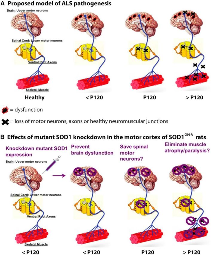
Sporadic amyotrophic lateral sclerosis (ALS) is a fatal disease with unknown etiology, characterized by a progressive loss of motor neurons leading to paralysis and death typically within 3-5 years of onset. Recently, there has been remarkable progress in understanding inherited forms of ALS in which well defined mutations are known to cause the disease. Rodent models in which the superoxide dismutase-1 (SOD1) mutation is overexpressed recapitulate hallmark signs of ALS in patients. Early anatomical changes in mouse models of fALS are seen in the neuromuscular junctions (NMJs) and lower motor neurons, and selective reduction of toxic mutant SOD1 in the spinal cord and muscle of these models has beneficial effects. Therefore, much of ALS research has focused on spinal motor neuron and NMJ aspects of the disease. Here we show that, in the SOD1(G93A) rat model of ALS, spinal motor neuron loss occurs presymptomatically and before degeneration of ventral root axons and denervation of NMJs. Although overt cell death of corticospinal motor neurons does not occur until disease endpoint, we wanted to establish whether the upper motor neuron might still play a critical role in disease progression. Surprisingly, the knockdown of mutant SOD1 in only the motor cortex of presymptomatic SOD1(G93A) rats through targeted delivery of AAV9-SOD1-shRNA resulted in a significant delay of disease onset, expansion of lifespan, enhanced survival of spinal motor neurons, and maintenance of NMJs. This datum suggests an early dysfunction and thus an important role of the upper motor neuron in this animal model of ALS and perhaps patients with the disease.

Keywords: ALS; RNAi; SOD1; amyotrophic lateral sclerosis; motor neuron disease; neurodegenerative disorder.


Figures

Figure 1.
Figure 1.
Spinal motor neurons are lost presymptomatically, before corticospinal motor neuron loss and before significant axon and muscle degeneration in the SOD1G93A rat. P90 and P120 were designated “presymptomatic” time points based on Kaplan–Meier analyses showing that disease onset for rats in this study ranged from 140 to 201 d, with a median of 154 d (n = 21) (A) and survival ranged from 147 to 218 d, with a median of 172 d (n = 13) (B). C, Immunostaining for ChAT+ spinal motor neurons in the lumbar spinal cord at P90, P120, early symptomatic, and endpoint (EP) showed qualitative changes between WT rats and SOD1G93A rats over time. D, Quantification revealed a significant reduction of large (>700 μm) ChAT+ spinal motor neurons starting at presymptomatic time point P120. E, Osmium tetroxide and toludine blue staining provided qualitative analysis of (L5) ventral root axons in WT versus SOD1G93A rats over time. Stereological quantification revealed that the number of total axons was not significantly reduced until rats reach endpoint (F), whereas the number of healthy axons is first significantly reduced when rats appear symptomatic (G) and a slight yet significant increase in degenerating (“unhealthy”) axons is seen starting at P120 (H). I, Immunostaining for α-bungarotoxin and SV2 in the muscle of WT versus SOD1G93A rats. J, Quantification revealed that NMJs do not become significantly denervated until the early symptomatic stages, after spinal motor neuron loss. K, Immunostaining of motor cortex layer V sections using CTIP2 that specifically stains nuclei of motor neurons, and Neurotrace, a fluorescent Nissl stain, in WT and SOD1G93A rats. L, Quantification revealed that the number of large corticospinal motor neurons was declined at early symptomatic stages and was significantly reduced at disease endpoint. Scale bars: C, K, 50 μm; E, I, 10 μm. *p < 0.05; error bars indicate SEM. M, Summary of the time course of degeneration in SOD1G93A rats showing the percentage of corticospinal motor neuron loss in the brain, spinal motor neuron loss, loss of healthy ventral root axons, and degenerated NMJs: *p < 0.05, **p < 0.001, ***p < 0.0001, ## indicates moderate degeneration designated attributable to a slight yet significant increase in numbers of unhealthy axons. N.C., No change; N.S., not significant.
Figure 2.
Figure 2.
The brain–muscle circuitry is disrupted in presymptomatic rats at P120. A, The brain-to-muscle pathway was retrogradely labeled by injecting RFP-tagged pseudorabies virus into the right gastrocnemius. B, Image of RFP-labeled corticospinal motor neurons at P120 in WT and SOD1G93A rats. C, Quantification of RFP-labeled motor neurons in the left motor cortex revealed a significant reduction in SOD1G93A rats at P120, but not at P90, relative to WT controls. D, Image of RFP-labeled spinal motor neurons at P120 in a WT rat. E, Quantification of RFP-labeled motor neurons in the right lumbar spinal cord revealed a significant reduction in SOD1G93A rats at P120, but not at P90, relative to WT controls. Scale bars, 75 μm. *p < 0.05; error bars indicate SEM.
Figure 3.
Figure 3.
Injection of AAV9–SOD1–shRNA in the SOD1G93A rat brain induces local suppression of mutant, misfolded SOD1 protein. A, A single injection of AAV9–SOD1–shRNA into the motor cortex of SOD1G93A rats induces local GFP expression (shown by histology) and suppression of mutant SOD1 (shown by immunohistochemistry using the D3H5 antibody in red that recognizes misfolded SOD1 protein) in these cells. B, Because AAV9–SOD1–shRNA does not cross into the contralateral side of the brain, there is no GFP expression and mutant SOD1 protein is still produced. C, Motor cortex functional map shows the location of AAV9 injection sites. Eight injections per hemisphere (total of 16 injections) were administered to target the posterior/hindlimb cortex. FL, Forelimb; HL, Hindlimb; W, whisker; N, neck; T, tail representation within the motor cortex. Immunostaining for GFP (green), CTIP2 (red), and D3H5 (blue) in coronal brain sections shows that a large number of CTIP2+ corticospinal motor neurons were virally transduced and that mutant SOD1 (D3H5) was still highly expressed in cells infected with AAV9–GFP control virus (D) but was knocked down in cells infected with AAV9–SOD1–shRNA (E). Low-power coronal brain images revealed that AAV9–GFP control-injected SOD1G93A rats display robust GFP expression and a remaining abundant expression of misfolded, mutant SOD1 protein in the motor cortex (F), whereas AAV9–SOD1–shRNA-injected SOD1G93A rats show robust GFP expression with a corresponding significant 40% reduction of misfolded SOD1 protein (G). H, Immunostaining using antibodies against GFP and D3H5 in the spinal cord revealed that very few (3.6%) total spinal motor neurons expressed GFP (and therefore AAV9–SOD1–shRNA), and the majority of these GFP+ cells did not show an abundant level of mutant SOD1 knockdown, resulting in only ∼1.2% of all spinal motor neurons with reduced levels of mutant protein (right, pink bar) (I). J, qRT-PCR analysis of laser-captured motor neurons from the motor cortex of SOD1G93A rats injected with AAV9–SOD1–shRNA revealed significant amounts of GFP vector expression and a corresponding significant reduction of mutant SOD1 expression compared with non-injected SOD1G93A rats (K). L, Similar qRT-PCR analysis of laser-captured motor neurons from the spinal cord revealed no loss of mutant SOD1 expression in these spinal motor neurons, suggesting a lack of vector expression. Immunostaining in the spinal cord after delivery of AAV9 to the motor cortex revealed that <1% of GFAP+ astrocytes (M) and Iba1+ microglia (N) were anterogradely transduced with AAV9 and therefore GFP+. Scale bars: A, B, M, N, 100 μm; D, E, H, 50 μm; F, G, 750 μm. ***p < 0.001; error bars indicate SEM.
Figure 4.
Figure 4.
Local suppression of mutant, misfolded SOD1 protein in the posterior motor cortex of SOD1G93A rat brain leads to improved hindlimb but not forelimb function, delayed disease onset, and an extension of survival. A, SOD1G93A rats administered AAV9–GFP control virus (n = 14) showed an expected variation in disease onset location in solely forelimb (n = 4; FL ONLY), solely hindlimb (n = 5; HL ONLY), or both (n = 5; BOTH FL,HL same time). In contrast, no SOD1G93A rats injected with AAV9–SOD1–shRNA developed exclusive hindlimb onset (arrow, A). This was likely attributable to the targeting field that knocked down mutant SOD1 primarily in the posterior/hindlimb region, while providing incomplete targeting in the anterior/forelimb motor cortex. In light of the targeting strategy, rats in both groups that developed exclusive forelimb onset were excluded from analysis. B, BBB hindlimb analysis showed that SOD1G93A rats administered AAV9–SOD1–shRNA had significant behavioral improvements relative to AAV9–GFP control-injected SOD1G93A rats. In addition, knockdown of mutant SOD1 expression in the motor cortex of SOD1G93A rats resulted in delayed hindlimb onset by ∼24 d (C) and extended survival time by ∼20 d (D).
Figure 5.
Figure 5.
AAV9–scrambled–shRNA has no effect on human SOD1 protein levels and does not improve motor function or survival. A, AAV9–scrambled–shRNA or AAV9–SOD1–shRNA were transfected into HEK293 cells, and 72 h later, cell lysates were collected for analysis by Western blot. Unlike AAV9–SOD1–shRNA-treated cells, AAV9–scrambled–shRNA treatment did not reduce human SOD1 levels compared with control conditions. B, Quantification of Western blot analysis of human SOD1 knockdown showed that AAV9–SOD1–shRNA-treated cells, but not AAV9–scrambled–shRNA-treated cells, showed a significant reduction (*p < 0.05) compared with control conditions. C–F, SOD1G93A mice were treated with a single intravenous injection of AAV9–scrambled–shRNA at P1 (n = 5; light green) or P21 (n = 4; dark green) and were monitored throughout the disease course compared with non-injected control mice (n = 7; gray). AAV9–scrambled–shRNA treatment did not significantly delay the disease onset (C, D) or increase survival (E, F).
Figure 6.
Figure 6.
Local suppression of mutant SOD1 in both the anterior and posterior motor cortices of SOD1G93A rats leads to improved forelimb and hindlimb function, enhanced spinal motor neuron survival, and increased muscle innervation. A, Previous experiments were repeated, with two additional AAV9 injection sites per hemisphere to the anterior motor cortex to target more of the forelimb region (total = 20 injections). This time, BBB analysis showed that both hindlimb (B) and forelimb (C) motor function were significantly improved in AAV9–SOD1–shRNA-injected rats relative to control rats. D, Immunostaining showed that SOD1G93A rats treated with AAV9–SOD1–shRNA in the motor cortex had preserved ChAT+ motor neurons in the spinal cord compared with non-treated SOD1G93A rats. Quantification confirmed that the enhanced survival was significant for both total ChAT+ spinal motor neurons (E) and large (>700 μm) spinal motor neurons (F). G, In addition, the average cell body size (μm2) of the spinal motor neurons was significantly larger in AAV9–SOD1–shRNA-treated SOD1G93A rats relative to nontreated control SOD1G93A rats. H, Finally, knockdown of mutant SOD1 in the brain also lead to increased muscle innervation relative to non-treated controls. Scale bar, 50 μm. *p < 0.05, **p < 0.01; error bars indicate SEM. FL, Forelimb; HL, hindlimb; W, whisker; N, neck; T, tail representation within the motor cortex.
Figure 7.
Figure 7.
Summary of the proposed model of ALS disease pathology and effects of mutant SOD1 knockdown in the brain. A, Proposed model of ALS disease progression whereby dysfunction (red/black symbols) but not cell death (black × symbols) of corticospinal motor neurons occurs at early, presymptomatic time points leading to presymptomatic loss of spinal motor neurons, which then results in the degeneration of ventral root axons and denervation of NMJs and concomitant appearance of disease symptoms. B, Consequences of knocking down mutant SOD1 in the brain based on model above (A), whereby the reduction of mutant SOD1 eliminates the dysfunction (purple symbol) of corticospinal motor neurons, which delays disease progression by preventing cell loss in the spinal cord and consequently preventing axon degeneration and NMJ denervation.

References

    1. Arlotta P, Molyneaux BJ, Chen J, Inoue J, Kominami R, Macklis JD. Neuronal subtype-specific genes that control corticospinal motor neuron development in vivo. Neuron. 2005;45:207–221. doi: 10.1016/j.neuron.2004.12.036. - DOI - PubMed
    1. Basso DM, Beattie MS, Bresnahan JC. A sensitive and reliable locomotor rating scale for open field testing in rats. J Neurotrauma. 1995;12:1–21. doi: 10.1089/neu.1995.12.1. - DOI - PubMed
    1. Berry JD, Cudkowicz ME. New considerations in the design of clinical trials for amyotrophic lateral sclerosis. Clin Investig (Lond) 2011;1:1375–1389. doi: 10.4155/cli.11.127. - DOI - PMC - PubMed
    1. Bilsland LG, Sahai E, Kelly G, Golding M, Greensmith L, Schiavo G. Deficits in axonal transport precede ALS symptoms in vivo. Proc Natl Acad Sci U S A. 2010;107:20523–20528. doi: 10.1073/pnas.1006869107. - DOI - PMC - PubMed
    1. Brockington A, Heath PR, Holden H, Kasher P, Bender FL, Claes F, Lambrechts D, Sendtner M, Carmeliet P, Shaw PJ. Downregulation of genes with a function in axon outgrowth and synapse formation in motor neurones of the VEGFdelta/delta mouse model of amyotrophic lateral sclerosis. BMC Genomics. 2010;11:203. doi: 10.1186/1471-2164-11-203. - DOI - PMC - PubMed

Publication types

MeSH terms